
При посиланні на статтю з нашого журналу
необхідно вказувати:
- назву журналу
українською мовою - Медичні перспективи
або транслітерацією - Medicni perspektivi
- DOI статті



Key words: artificial intelligence, item difficulty, item discrimination, large language models, medical education, meta-analysis, psychometrics, single best answer
Ключові слова: штучний інтелект, складність завдань, розрізнення завдань, великі мовні моделі, медична освіта, метааналіз, психометрія, єдина найкраща відповідь
Abstract
Single Best Answer Questions (SBAs) are essential and resource-intensive assessment tools in health professions education. Artificial Intelligence (AI), such as large language models (LLMs), can automate the creation of SBAs; however, evidence comparing the quality of AI-generated and human-created items is still dispersed. The purpose of the study is to compare the psychometric quality, measured by difficulty and discrimination indices, of AI-generated SBAs with those authored by humans in health professions education. The current study followed PRISMA guidelines. The search was conducted on Scopus, PubMed, and Google Scholar. Studies published through April 25th, 2025, and those that directly compared AI- and human-generated SBAs and reported the mean, standard deviation, and sample size for both difficulty and discrimination indices were included. Two reviewers independently extracted the data. Standardized mean differences (SMDs) were calculated and combined using random-effects models (Jamovi MAJOR module, version 2.6.44-06 March 2025). Heterogeneity and publication bias were assessed. Four studies met the inclusion criteria, providing eight comparison outcomes (4 for difficulty, 4 for discrimination). The combined analysis of both outcomes revealed no statistically significant difference, overall (SMD= -0.084, 95% CI: -0.65 to 0.49, p=0.773); however, the heterogeneity was very high (I²=92.7%). Separate analyses revealed that AI-generated questions were significantly easier than human-generated questions (SMD= +0.541, 95% CI: 0.17 to 0.91, p=0.004; I²=62.3%). Conversely, human-authored questions demonstrated significantly higher discrimination indices than AI-generated questions (SMD= -0.701, 95% CI: -1.33 to -0.08, p=0.028; I²=86.2%). No evidence of publication bias was found. AI-generated items tend to be easier, potentially aiding accessibility, whereas human-authored items currently exhibit superior discriminatory power, which is crucial for robust assessment. High heterogeneity underscores context dependency.
Реферат
Якість тестових завдань з однією найкращою відповіддю, згенерованих ШІ та людиною: систематичний огляд і метааналіз. Шаббір Мухаммад, Бенді Алтаф, Мехбуб Бушра, Махбуб Усман, Ліакат Амбрін, Аль-Маршад Ферас, Адам Сіті Хадіджа. Тестові завдання з єдиною найкращою відповіддю (Single Best Answer Questions, SBA) є важливими та ресурсоємними інструментами оцінювання в освіті фахівців сфери охорони здоров’я. Штучний інтелект (ШІ), зокрема великі мовні моделі (LLMs), може автоматизувати створення завдань типу SBA; однак дані щодо порівняння якості завдань, створених ШІ та людиною, наразі залишаються розрізненими. Метою дослідження було порівняти психометричну якість, виміряну за індексами складності та дискримінації, SBA, згенерованих ШІ, із завданнями, автором яких є людина, у сфері освіти медичних працівників. Дослідження виконано відповідно до рекомендацій PRISMA. Пошук літератури здійснювався в базах Scopus, PubMed та Google Scholar. До аналізу включалися дослідження, опубліковані до 25 квітня 2025 року, які безпосередньо порівнювали SBA, створені ШІ та людиною, і повідомляли середнє значення, стандартне відхилення та обсяг вибірки для індексів складності та дискримінації. Двоє рецензентів незалежно здійснювали вилучення даних. Стандартизовані середні різниці (standardized mean differences, SMDs) були розраховані та об’єднані з використанням моделей випадкових ефектів (модуль Jamovi MAJOR, версія 2.6.44-06 березня 2025 року). Також оцінювали гетерогенність та упередження публікації. Чотири дослідження відповідали критеріям включення, надавши вісім порівняльних результатів (4 для складності, 4 для дискримінації). Сукупний аналіз обох показників не виявив статистично значущої різниці загалом (SMD= -0,084; 95% ДІ: -0,65 до 0,49; p=0,773); однак гетерогенність була дуже високою (I²=92,7%). Окремі аналізи показали, що завдання, згенеровані ШІ, були статистично значуще простішими, ніж створені людьми (SMD= +0,541; 95% ДІ: 0,17-0,91; p=0,004; I²=62,3%). Водночас завдання, авторами яких були люди, демонстрували значно вищі індекси дискримінації порівняно з ШІ (SMD= -0,701; 95% ДІ: -1,33 до -0,08; p=0,028; I²=86,2%). Ознак публікаційного зміщення не виявлено. Завдання, створені ШІ, як правило, є простішими, що може підвищувати доступність оцінювання, тоді як завдання, створені людиною, наразі демонструють вищу дискримінативну здатність, що є критично важливим для надійного оцінювання. Висока гетерогенність підкреслює залежність результатів від контексту.
In the era of competency-based medical education, assessment plays a central role in guiding learning, evaluating progress, and ensuring that students attain the required competencies for clinical practice [1]. Among various assessment tools, single-best-answer (SBA) questions remain a cornerstone due to their objectivity, efficiency, and ability to cover diverse knowledge areas. When SBAs are designed around clinical scenarios, they promote critical thinking and decision-making by mimicking real-world medical situations [2, 3, 4]
However, the consistent development of high-quality SBAs is a resource-intensive process that requires subject matter expertise, psychometric sensitivity, and iterative validation. In this context, ChatGPT is considered a valuable tool for automating the generation of exam questions through generative AI [5, 6].
Recent studies have evaluated the psychometric performance and content quality of AI-generated SBAs compared to human-authored ones. While ChatGPT demonstrates efficiency and scalability in question generation, concerns remain regarding its accuracy, contextual alignment, and discriminatory ability [7, 8, 9]. Some studies report comparable item difficulty between AI- and human-generated questions [6], but consistently lower discrimination indices for AI items, which raises questions about their validity in differentiating learner performance [5].
Moreover, qualitative assessments have highlighted content-specific issues in AI-generated SBAs, such as factual inaccuracies, inappropriate difficulty levels, and culturally insensitive phrasing [10, 11]. These findings underscore the necessity of human oversight and robust quality assurance processes before AI-generated items can be integrated into high-stakes assessments [12].
The Purpose of the current systematic review and meta-analysis is to evaluate and synthesize existing evidence comparing the psychometric properties of AI-generated and human-created SBA questions in medical education contexts. Specifically, it addresses differences in item difficulty and discrimination, and examines the implications for assessment quality, validity, and utility in medical curricula. By quantitatively pooling data on these key psychometric properties where possible, we seek to provide a consolidated understanding of the relative strengths and weaknesses of current AI capabilities in SBA generation compared to traditional human authorship.
MATERIALS AND METHODS OF RESEARCH
The study was conducted as a systematic review and meta-analysis to compare the psychometric analysis of single-best-answer (SBA) questions generated by artificial intelligence (AI) with those written by human educators. The protocol included the review question, search strategy, problem description, intervention, comparison, outcome, and the end time point (PICOT), as well as inclusion criteria, types of studies to be included, data extraction, risk of bias, and strategies for evidence synthesis. The PICOT description was, Problem: Single best answer questions (SBAs) in health profession education; Intervention: SBAs generated by generative AI; Comparison: SBAs generated by faculty members; Outcome: Quality assessment of both set of questions through different psychometric parameters (discrimination and difficulty index); time frame: Studies published on the subject till 25th of April 2025 were included. The literature search was conducted from April 01, 2025, to April 25th, 2025, with an iterative process taken to get the best results.
Our focus was on two well-established item-level psychometric parameters: the "difficulty index” and the "discrimination index." These metrics are essential for evaluating the quality and effectiveness of assessment items. The difficulty index reflects the percentage of students who correctly answered a question, whereas the discrimination index assesses how well an item can distinguish between students who perform well and those who perform poorly. The Preferred Reporting Items for Systematic Reviews and Meta-Analyses (PRISMA) 2020 guidelines (https://www.prisma-statement.org/) were followed to ensure a structured, transparent, and replicable methodological approach.
A thorough literature review was conducted up to April 25, 2025, utilizing two primary electronic databases (Scopus, PubMed) and one search engine (Google Scholar). The search strategy was designed using Boolean operators and a combination of relevant keywords such as 'multiple choice questions', 'single best answer', 'AI-generated', 'ChatGPT', 'human-authored', and 'health professions education'. The references were managed by importing all citations into Rayyan AI (Rayyan: AI-Powered Systematic Review Management Platform). Duplicate entries were automatically removed. A blinded screening was carried out by two independent reviewers for title, abstract, and full-text relevance. Disagreements were settled through consensus, with a third reviewer available for arbitration if needed. Inter-rater reliability was measured using Cohen’s Kappa, which was calculated to be 0.81, signifying substantial agreement.
The inclusion criteria consisted of studies that directly compared SBA questions generated by AI models (e.g., ChatGPT) with those written by human experts. Furthermore, the studies included in the analysis had to report both difficulty and discrimination indices for questions generated by AI and humans. They also needed to provide adequate statistical data, such as the mean, standard deviation (SD), and sample size (N) for each group. Studies were excluded if they provided only qualitative evaluations, such as expert opinions, lacked detailed quantitative psychometric analysis, or were review articles, commentaries, or non-original research. Thus, only studies that offered strong, directly comparable data were included in the meta-analysis. Eligible studies had to be published in English and be accessible in full text.
To evaluate the quality of the included studies, the MERSQI instrument was used, which assesses various facets, including study design, sampling procedures, data collection, validity support, analytical approaches, and results. The studies scored from moderate to high, reflecting solid psychometric qualities and robust educational evaluations.
Two reviewers independently conducted data extraction using a standardized form. For each study, data elements extracted included author(s), year of publication, total number of items analyzed, the version of the AI model used, outcome type (difficulty or discrimination), group classification (AI vs. human), and the reported means and standard deviations of the relevant indices. Where necessary, difficulty index values presented as percentages (0 to 100) were converted to proportions (0.00 to 1.00) to ensure consistency across all studies. When standard deviations were not directly provided, they were approximated using the range-based method.: SD = (Max – Min) ÷ 4, which is commonly accepted in educational and psychometric research when only minimum and maximum values are provided.
Data Synthesis and Statistical Analysis
The MAJOR module in the Jamovi statistical software platform, version 2.6.44 for Windows (Jamovi desktop – Jamovi), was used for meta-analysis. The analysis compared the psychometric quality of AI- and human-generated SBA questions using standardized mean differences (SMDs). A separate random-effects model was applied to both the difficulty index and the discrimination index because of their distinct interpretations and psychometric significance. Additionally, a combined analysis was conducted by aggregating all available data from both parameters. The a priori selection of a random-effects model was made to address anticipated heterogeneity stemming from factors such as diverse study populations, AI models, quality of item writing, and educational disciplines.
Assessment of Heterogeneity and Publication Bias
The I² metric, Tau², and Cochran’s Q test were utilized to assess heterogeneity in results among the different studies. Additionally, 95% prediction intervals were calculated to assess the expected range of effects in future studies. In addition to mitigating the risk of publication bias, several diagnostic tools were employed. Funnel plots (Figs) were visually examined for asymmetry, and Egger’s regression test was performed to identify small-study effects. Additionally, the trim-and-fill technique was applied to estimate the potential number of unpublished studies, while the fail-safe N was computed to evaluate the stability of the findings. Influential data points and outliers were detected using studentized residuals and Cook’s distances. All outcomes are presented with 95% confidence intervals, with significance determined at p<0.05.
Risk of bias
A comprehensive evaluation of bias risk was conducted to uphold the credibility and internal validity of this meta-analysis. The Medical Education Research Study Quality Instrument (MERSQI) served as the primary tool for this purpose. MERSQI is an established and extensively utilized tool for evaluating the methodological rigor of research in medical education, focusing on six key areas: study design, sampling methods, data types, validity of evaluation tools, data analysis techniques, and results. Each domain contributes to a cumulative score ranging from 5 to 18, allowing classification of studies into low, moderate, or high methodological quality.
In this review, two reviewers independently evaluated all four studies using the MERSQI criteria. Two studies were rated as moderate quality (score =13), while the remaining two were classified as moderate to high quality (score =14). Discrepancies in ratings were resolved through consensus and, when necessary, by consulting a third reviewer.
RESULTS AND DISCUSSION
Study Inclusion and Overview
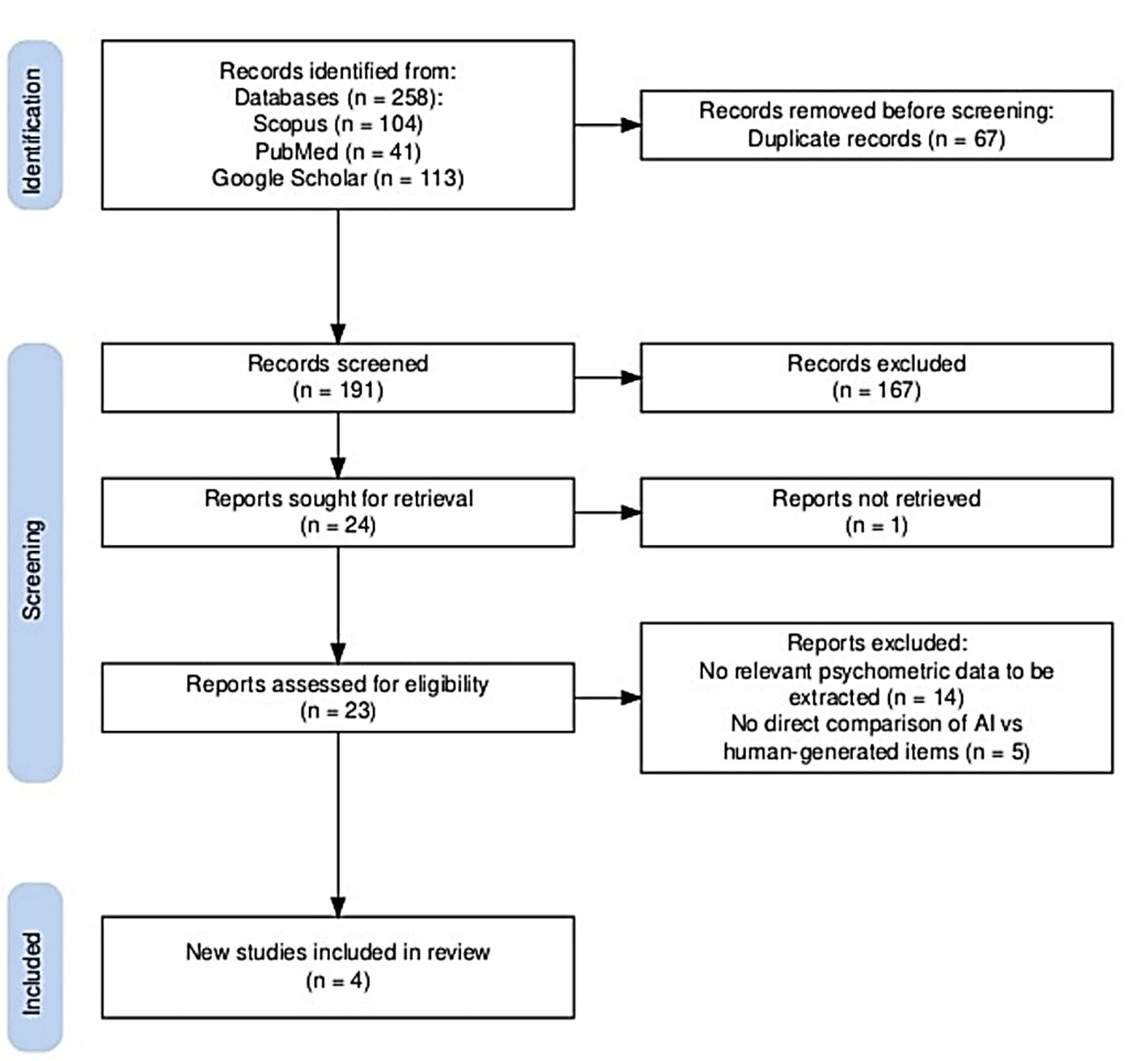
Among the 23 articles reviewed for eligibility, only four fulfilled all the inclusion criteria and were consequently included in the final meta-analysis (Fig. 1).
Fig. 1. Selection of studies. PRISMA diagram for selection of studies ↓
These four studies compared AI-generated and human-authored single best answer (SBA) questions in terms of two key psychometric parameters: difficulty index and discrimination index. Each study's quality was assessed using the MERSQI instrument [13]. Two of the selected studies were categorized as providing moderate to high-quality evidence. In contrast, the remaining two were classified as having a moderate level of evidence based on the MERSQI scores. Although the study mentioned different designs, the reviewers classified them as comparative quasi-experimental or crossover designs (e.g., comparing AI- vs human-generated questions). Furthermore, the study outcomes included objective psychometric parameters (e.g., difficulty index, discrimination index), which were taken into consideration in the MERSQI scoring system. Table 1 provides an overview of the characteristics of each study.
Author, year Study design Study setting Comparison AI Model Participants Outcomes Measured *Evidence level Law et al. (2025) [8] Comparative quasi-experimental or cross-over designs Hong Kong, PEEM exam (mock exam for AI authored questions followed by real exam AI vs Human-authored MCQs ChaGPT-4o Medical doctors Difficulty Index, Discrimination Index Moderate Points (13) Chauhan et al. (2025) [14] Comparative quasi-experimental or cross-over designs India, Physiology course exam (formative) AI vs Human-authored SBAs ChaGPT-4o MBBS students Difficulty Index, Discrimination Index Moderate–High Points (14) Laupichler et al. (2024) [6] Comparative quasi-experimental or cross-over designs Germany, Neurophysiology course exam (formative) AI vs Human-authored questions ChatGPT-3.5 Medical students Difficulty Index, Discrimination Index Moderate Points (13) Ahmed et al. (2025) [5] Comparative quasi-experimental or cross-over designs Scottish Graduate-Entry Medicine (ScotGEM) program (formative) AI vs Human-authored MCQs ChaGPT-4 Undergraduate medical students Difficulty Index, Discrimination Index Moderate–High Points (14) Note. * – quality of evidence determined using the MERSQI tool. Table 1. Characteristics of studies reporting Human versus AI in SBA(s) generation ↓
with human authored questions)
Each study reported the necessary summary statistics (mean, standard deviation, and sample size) for both AI and human groups, allowing for standardized mean difference (SMD) calculations (Table 2).
Combined Outcome Analysis
When combining all eight comparisons (across four studies and two outcomes), the overall standardized mean difference between questions generated by AI and humans was approximately –0.084 (with a 95% confidence interval ranging from -0.65 to +0.48). This result was not statistically significant (p=0.769), indicating that there was no clear overall benefit observed for either group across both outcomes (Table 3).
Estimate se Z p CI Lower Bound CI Upper Bound Intercept -0.0847 0.289 -0.293 0.769 -0.650 0.481 Tau² Estimator: Restricted Maximum-Likelihood Heterogeneity Statistics Tau Tau² I² H² R² df Q p 0.780 0.6083 (SE=0.3558) 93.01% 14.302 . 7.000 78.307 <0.001Table 3. Summary of combined variables (K8) ↓
The analysis revealed significant heterogeneity with an I² of 93.0%, reflecting differences in outcomes across various studies. The predicted range was from -1.72 to 1.55, implying that in future research, either AI-generated or human-generated questions could be deemed more effective, depending on the circumstances. No outliers were detected, and there was no evidence of publication bias, as indicated by an Egger’s test p-value of 0.685 and the trim-and-fill method not adding any studies (Fig. 2).
The heterogeneity (I²=93.0%), was further assessed by meta-regression with SMD’s of each study as the dependent variable and AI model version (GPT-3.5 vs. GPT-4 variants), Country of study (e.g., UK, India, Germany, Hong Kong) and Sample size (n) as moderator variables. The model's Residual Heterogeneity (τ²) was reduced by ~18%, indicating a partial explanation of variability, but not a complete one. The results of the regression analysis showed that none of the moderator variables had a significant impact on the observed effect sizes. However, there was a non-significant trend suggesting that questions generated by GPT-4 might have lower discrimination indices as compared to GPT-3.5 (β= -0.36, p=0.09). Country and sample size did not significantly moderate the effect size in either outcome.
The outcome measure employed in the analysis was the standardized mean difference. A random-effects model was applied to the data, with heterogeneity (τ²) estimated using the restricted maximum likelihood method. Additionally, tau², the Q-test for heterogeneity, and the I² statistic were reported. If any heterogeneity is detected (tau² >0, regardless of the Q-test results), a prediction interval for the true outcomes is provided. Studentized residuals and Cook's distances were employed to detect possible outliers and influential data points in the analysis. Outliers were defined as studies where the studentized residuals surpassed the critical value corresponding to the 100x(1-0.05/(2xk)) percentile of the standard normal distribution, with a Bonferroni adjustment to account for multiple comparisons. Influential studies were identified as those with Cook's distances exceeding the median plus six times the interquartile range across all Cook's distances. To evaluate funnel plot asymmetry, both the rank correlation test and a regression analysis were used, with the standard error of observed outcomes serving as a predictor.
The analysis involved four different studies and eight variables. The range of standardized mean differences observed was from -1.4567 to 1.1101, with approximately half of the estimates being negative. Using a random-effects model, the calculated mean standardized difference was -0.0837 with a 95% confidence interval between -0.6505 and 0.4802. This indicates that there was no significant difference from zero in the overall outcome, as reflected by a z-value of -0.2934 and a p-value of 0.7692. The Q-test suggests the presence of heterogeneity among the true effects, with a Q statistic of 78.3074 (degrees of freedom =7), p-value less than 0.0001, tau² =0.6083, I² =93.0081%. The predicted interval for true effects at 95% confidence spans from -1.7142 to 1.5452, illustrating that despite the average estimate being negative, individual studies could yield positive results. Residual analysis revealed no outliers, as none exceeded ±2.7344 in the studentized residuals. Cook's distance analysis confirmed that no studies exerted undue influence on the model. Additionally, statistical tests for funnel plot asymmetry, including the rank correlation and regression tests, showed no significant bias, with p-values of 0.3988 and 0.4936, respectively (Fig. 3).
Fig. 2. Combined funnel plot ↓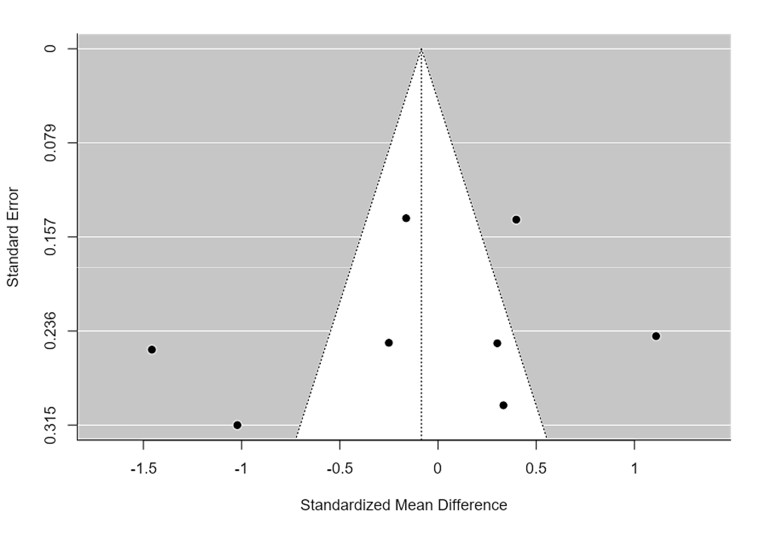
Test Name value p Fail-Safe N* 0.000 0.304 Begg and Mazumdar Rank Correlation -0.286 0.399 Egger's Regression -0.685 0.494 Trim and Fill Number of Studies 0.000 . Note. * – fail-safe N Calculation Using the Rosenthal Approach. Publication bias assessment ↓
Fig. 3. Combined forest plot ↓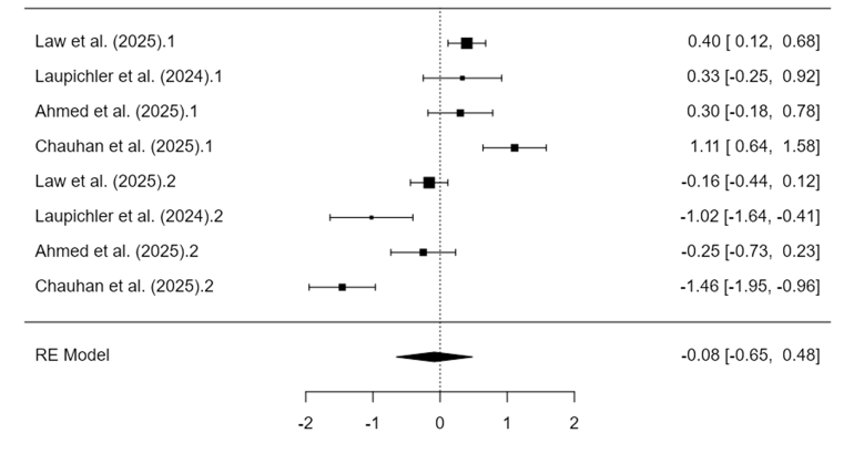
Individual outcome analysis
I. Difficulty Index
This analysis, focusing on the difficulty index (k=4), showed that questions generated by AI were significantly easier than those created by humans. The combined standardized mean difference was +0.534 (95% CI: 0.16-0.90; p=0.048), reflecting a moderate effect size that favors AI in terms of ease of use. This result exhibited moderate heterogeneity (I² =63.8%) and a prediction interval ranging from -0.15 to +1.23, suggesting that in some contexts, the difficulty level might be similar (Table 4).
According to Chauhan and colleagues' study, a potential outlier was found [14] having a studentized residual beyond the ±2 threshold; however, influence diagnostics did not suggest distortion of the overall results. No publication bias was found in this subgroup (Egger’s p=0.134) (Fig. 4).
Estimate se Z p CI Lower Bound CI Upper Bound Intercept 0.534 0.187 2.86 0.004 0.168 0.900 Random effect model (K4); Tau² Estimator: Restricted Maximum-Likelihood Heterogeneity statistics Tau Tau² I² H² R² df Q p 0.294 0.0867 (SE=0.1138 ) 63.87% 2.768 . 3.000 7.918 0.048Table 4. Analysis of difficulty index (k=4) ↓
The study utilized the standardized mean difference to measure outcomes. A random-effects approach was employed for data analysis. The heterogeneity, represented by tau², was estimated through the restricted maximum-likelihood method. Additionally, the report presents the Q-test for heterogeneity, originally developed by Cochran in 1954, along with the I² statistic. If any heterogeneity is detected (i.e., tau² >0, regardless of the Q-test results), a prediction interval for the true outcomes is also provided. To identify potential outliers or influential studies, the analysis uses Studentized residuals and Cook's distances. In the analysis, studies are flagged as potential outliers if their Studentized residuals exceed the 100×(1-0.05/(2×k)) percentile of the standard normal distribution, with a Bonferroni correction applied at a two-sided alpha level of 0.05 to account for the total number of studies (k). Additionally, studies with Cook's distances greater than the median plus six times the interquartile range are considered to have a notable influence on the findings. To assess funnel plot symmetry, both the rank correlation test and the regression test are utilized, with the regression test incorporating the standard error of the observed outcomes as a predictor (Fig. 4).
Fig. 4. Individual funnel plot (Difficulty Index Meta-Analysis) ↓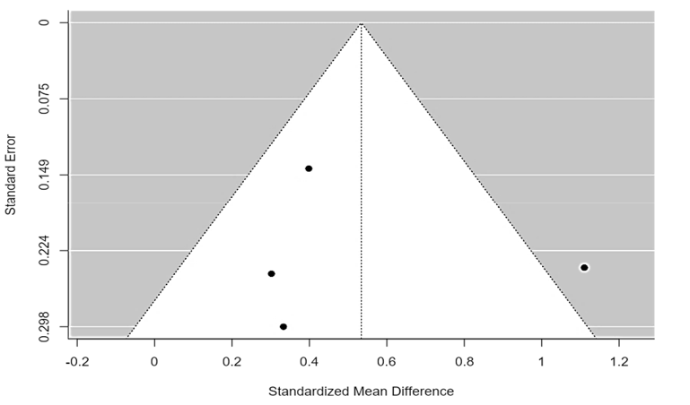
Test Name value p Fail-Safe N* 32.000 <.001 Begg and Mazumdar Rank Correlation 0.333 0.750 Egger's Regression -0.080 0.936 Trim and Fill Number of Studies 1.000 . Note. * – fail-safe N Calculation Using the Rosenthal Approach. Publication bias assessment ↓
A total of four studies were included in the analysis. The standardized mean differences observed ranged from 0.3018 to 1.1101, with all estimates being positive (100%). The overall average standardized mean difference, calculated using a random-effects model, was approximately 0.5341, with a 95% confidence interval from 0.1685 to 0.8996. This indicates that the overall outcome significantly deviates from zero (z=2.8632, p=0.0042). The heterogeneity test using the Q statistic suggested variability among the true effects (Q(3)=7.9179, p=0.0477, tau² =0.0867, I² =63.87%). The 95% prediction interval for the true effects spans from -0.1492 to 1.2173, meaning that despite a positive average effect, individual study outcomes could fall outside or below this range. Residual analysis identified one study (Chauhan et al., 2025) [14] as a potential outlier, exceeding the threshold value of ±2.4977. Cook's distance analysis revealed no single study exerted undue influence. Tests for publication bias, including rank correlation and regression methods, did not indicate asymmetry in the funnel plot (p=0.7500 and p=0.9363, respectively) (Fig. 5).
Fig. 5. Individual forest plot (Difficulty Index Meta-Analysis) ↓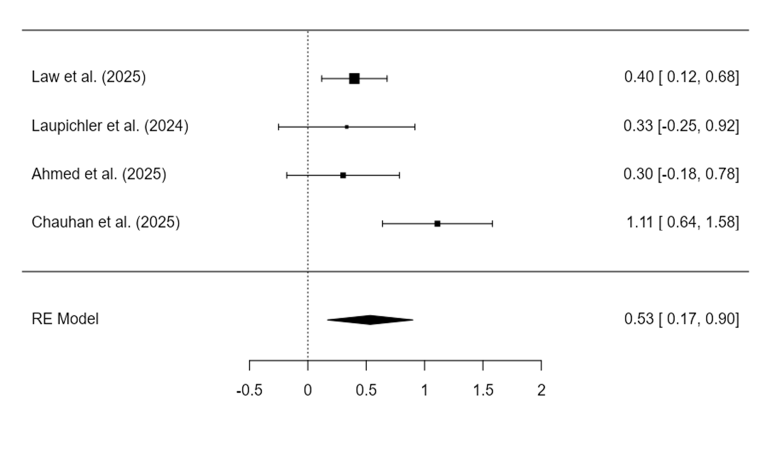
II. Discriminatory Index Meta-Analysis
The analysis of the discrimination index (k=4) revealed that human-authored questions were significantly more discriminative than AI-generated ones. The combined standardized mean difference was -0.699, with a 95% confidence interval ranging from -1.32 to 0.07, and a p-value of 0.027. This indicates a significant effect size favoring items created by humans. This result was accompanied by high heterogeneity (I² =86.98%) and a prediction interval from -2.01 to +0.61, suggesting variability in the magnitude and direction of effect across studies (Table. 5).
The analysis revealed no outliers or overly influential studies. Additionally, funnel plot tests did not indicate any publication bias, with Egger’s test yielding a p-value of 0.383 (Fig. 6).
Estimate se Z p CI Lower Bound CI Upper Bound Intercept -0.699 0.316 -2.21 0.027 -1.320 -0.079 Random effect model (K4); Tau² Estimator: Restricted Maximum-Likelihood Heterogeneity statistics Tau Tau² I² H² R² df Q p 0.585 0.3419 (SE= 0.3271) 86.98% 7.679 . 3.000 23.928 <0.001Table 5. Analysis of discriminatory index (k=4) ↓
In the analysis, the outcome measure selected was the standardized mean difference. The data were analyzed using a random-effects model, with heterogeneity (tau²) estimated via the restricted maximum-likelihood approach. The report presents the tau² estimate, the Q-test for heterogeneity, and the I² statistic. When heterogeneity is detected (tau² >0), a prediction interval for the true effects is also provided, regardless of the Q-test outcome. To identify outliers and influential studies, studentized residuals and Cook's distances were calculated. A study is considered a potential outlier if its studentized residual surpasses the percentile corresponding to 100×(1-0.05/(2×k)) of a standard normal distribution, applying a Bonferroni correction with a two-sided alpha of 0.05 based on the total number of studies (k) included. Studies with Cook's distance exceeding the median plus six times the interquartile range are classified as influential. The assessment of funnel plot asymmetry involved both the rank correlation test and the regression test, with the latter estimating asymmetry based on the standard error of the outcomes.
Fig. 6. Individual funnel plot (Discriminatory Index Meta-Analysis) ↓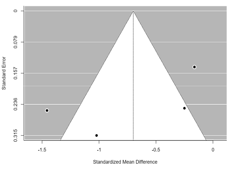
Test Name value p Fail-Safe N* 43.000 <0.001 Begg and Mazumdar Rank Correlation -0.667 0.333 Egger's Regression -1.154 0.249 Trim and Fill Number of Studies 0.000 . Note. * – fail-safe N Calculation Using the Rosenthal Approach. Publication bias assessment ↓
In a review of four studies, the standardized mean differences ranged from -1.4567 to -0.1623, with all estimates being negative, accounting for 100% of the cases. The average standardized mean difference, estimated using a random-effects model, was -0.6994 with a 95% confidence interval of
-1.3196 to -0.0791 (Fig. 7). This indicates a significant deviation from zero (z= -2.2100, p=0.0271). Heterogeneity among the true effect sizes was confirmed by the Q-test (Q(3) =23.9276, p<0.0001, tau² =0.3419, I² =86.9772%). A 95% prediction interval suggested that the true effects could range from -2.0024 to 0.6037, implying that while the average effect is negative, some individual studies might observe positive outcomes. Residual analysis showed no outliers, as none exceeded the threshold of ±2.4977, and Cook's distances indicated no studies were disproportionately influential. Tests for publication bias, including both rank correlation and regression methods, yielded p-values of 0.3333 and 0.2487 respectively, suggesting no evidence of funnel plot asymmetry.
Fig. 7. Individual forest plot (Discriminatory Index Meta-Analysis) ↓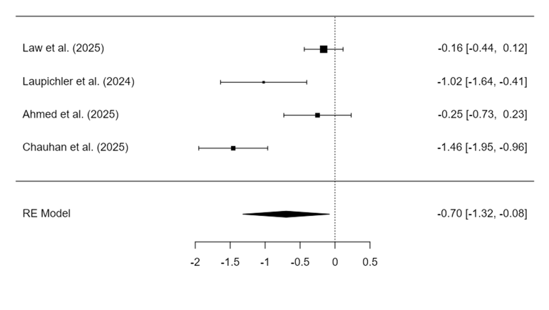
Overview of Findings
This meta-analysis critically evaluated the psychometric quality of single-best-answer (SBA) questions generated by artificial intelligence (AI) versus those authored by human experts in health professions education. The study is based on the difficulty and discrimination indices, providing data-driven insights into the comparability and potential applications of AI-generated assessments.
The analysis collectively showed no meaningful statistical difference between outputs created by artificial intelligence and those produced by humans, with a pooled standardized mean difference (SMD) of -0.084 (95% CI: -0.65 to 0.48), which was not statistically significant (p=0.769). However, the result must be interpreted considering the very high heterogeneity observed (I² =93.0%). Such variability suggests substantial differences in item construction methodologies, AI model versions, prompting techniques, or educational domains across studies. The wide prediction interval (-1.72 to +1.55) further emphasizes that the comparative effectiveness of AI versus human-authored questions is context-sensitive.
AI advantage in item difficulty
With regards to ease of answering questions, it was found that questions generated by using AI were significantly easier than human-authored ones (SMD=+0.534 (95% CI: 0.16-0.90; p=0.048)). Prior studies [15, 16, 17] suggest that AI tools like ChatGPT tend to construct items with simpler syntax and fewer ambiguities. Such linguistic clarity can reduce construct-irrelevant variance, thereby enhancing fairness, particularly for non-native English speakers [18].
Easier questions may also facilitate learning, particularly in formative assessments and early stages of training. Moreover, in high-stakes assessments, difficulty is not inherently valuable unless it is coupled with appropriate discrimination [19, 20]. Therefore, the utility of AI-generated questions should not be dismissed simply because they are easier. Rather, they could support scaffolding strategies in assessment design where foundational knowledge precedes more complex testing.
Human Superiority in Discrimination
Human-authored questions showed significantly higher discrimination indices than AI-generated ones (SMD= -0.699 (95% CI: -1.32 - 0.07; p=0.027), indicating a better ability to differentiate among high- and low-performing examinees. This is a well-documented advantage, supported by studies that show expert-written items to integrate clinical judgment, pedagogical intent, and cognitive load management more effectively.
The lower discrimination of AI-generated items could stem from several factors. First, AI lacks the contextual experience to generate refined distractors. Second, many studies did not specify prompts targeting higher-order cognitive domains. Third, without alignment to curriculum outcomes, AI may generate fact-based recall items that are less effective in evaluating deeper understanding. These limitations can be mitigated through prompt engineering, AI fine-tuning, and structured human-AI collaboration models.
Heterogeneity and Its Implications
High heterogeneity in the combined and discrimination outcomes (I² >85%) suggests inconsistency in study methods, question topics, and analytical approaches. Some studies relied on simulated assessments, while others used real student performance data; some utilized GPT-3.5, while others employed GPT-4 or hybrid prompts. This diversity reflects both the evolving nature of AI tools and the varied ways in which they are deployed in educational settings.
This heterogeneity (I² =93.01%), was further assessed by meta-regression with SMD’s of each study as the dependent variable and AI model version (GPT-3.5 vs. GPT-4 variants), Country of study (e.g., UK, India, Germany, Hong Kong) and Sample size (n) as moderator variables. The model Residual Heterogeneity (τ²): reduced by ~18%, indicating partial explanation of variability, but not complete. The analysis of the regression results indicated that none of the moderator variables significantly influenced the observed outcome. However, the AI model version revealed a non-significant trend, suggesting that GPT-4-generated questions may exhibit lower discrimination indices compared to GPT-3.5 (β= -0.36, p=0.09). Country and sample size did not significantly moderate the effect size in either outcome. These findings suggest that heterogeneity may stem from unmeasured factors such as prompt design, educational context, domain specificity, or question vetting processes. Future studies should standardize these parameters to facilitate clearer interpretation and reduce unexplained variance (Supp. 2).
Rather than undermining the analysis, this heterogeneity highlights a key strength: it reflects real-world variability. Meta-analyses that synthesize such diverse evidence provide stakeholders with a better understanding of the potential range of outcomes across various use cases. Future studies should strive to standardize psychometric reporting and stratify results by AI version, domain, and prompt quality.
Educational Implications
Contrary to the view that AI-generated questions are not suitable for high-stakes exams, the findings suggest that their current limitations lie in discrimination rather than difficulty or content accuracy. Provided that AI-generated items are reviewed and modified by experts, they can be effectively integrated into high-stakes assessments. In fact, some AI-generated items in included studies met or exceeded standard psychometric thresholds [8, 14].
Moreover, AI tools offer scalability, consistency, and speed in item generation – qualities that are highly valuable in large-scale assessments. With proper training datasets and expert oversight, AI could evolve into a reliable source for high-quality questions. Therefore, the current findings support a hybrid assessment model where AI serves as a productive starting point and human experts refine and validate the output.
Limitations
The limitations include a small number of included studies (four), which reduces the statistical power and generalizability of the results. Significant heterogeneity was observed, particularly for the discrimination index (I² >85%), indicating substantial variability across different study contexts, AI models, and prompting techniques that could not be fully explored with meta-regression. Furthermore, the rapid evolution of AI technology means these findings reflect the capabilities of models used in the included studies, which may differ from the most current versions.
Future Directions
Based on the findings and limitations identified in this review, future research should prioritize several key areas. Firstly, systematic investigations are needed to determine how variations in AI models (e.g., GPT-4 vs. specialized models), prompt engineering strategies targeting specific cognitive levels (like Bloom's taxonomy), and subject matter domains influence the psychometric quality, particularly the discrimination index, of generated SBAs. Secondly, research should focus on developing and rigorously evaluating hybrid human-AI co-authorship workflows, assessing their impact on efficiency, item quality, and the mitigation of AI weaknesses, such as poor distractor generation. Finally, studies exploring the effectiveness of fine-tuning large language models specifically on high-quality assessment item data could lead to AI tools intrinsically better suited for creating psychometrically sound questions.
CONCLUSION
While the combined analysis pooling difficulty and discrimination outcomes yielded no statistically significant overall difference, this result is overshadowed by substantial heterogeneity and masks crucial distinctions revealed in separate analyses.
- creating high-quality single-best-answer questions in medical education is a resource-intensive process.
- while Artificial intelligence generated questions are easier and useful for formative assessments, human-written questions have higher discrimination, making them better for high-stakes exams.
- results vary widely due to factors such as Artificial intelligence version and setting, highlighting the need for standardization and human oversight.
Acknowledgement. The authors thank the Deanship of Scientific Research (DSR) at Shaqra University for their continuous research support.
Contributors:
Shabbir Muhammad – conceptualization, data curation, formal analysis, investigation, methodology, resources, software, validation, visualization, writing – original draft, writing – review and editing.
Bandy Altaf – conceptualization, data curation, formal analysis, investigation, methodology, writing – original draft.
Mehboob Bushra – conceptualization, data curation, formal analysis, investigation, methodology, visualization, writing – original draft, writing – review and editing.
Mahboob Usman – methodology, project administration, resources, supervision, writing – review and editing.
Liaqat Ambreen – formal analysis, data curation, reviewed the manuscript.
Almarshad Feras – formal analysis, data curation, reviewed the manuscript.
Adam Siti Khadijah – supervision, validation, writing – review and editing.
Funding. This research received no external funding.
Conflict of interests. The authors declare no conflict of interest.
REFERENCES
Key words: neuro-glio-capillary system, cerebral stability, pediatric respiratory infections, systemic inflammation, neurovascular unit, astrocyte metabolism, cerebral vulnerability
Ключові слова: нейрогліокапілярна система, церебральна стабільність, педіатричні респіраторні інфекції, системне запалення, нейроваскулярна одиниця, метаболізм астроцитів, церебральна вразливість
Abstract
Acute respiratory infections in children are accompanied by systemic inflammatory, metabolic, and microcirculatory disturbances that may affect the developing brain even without focal neurological symptoms. During critical neurodevelopmental stages, brain function depends on stable oxygenation, perfusion, and energy metabolism, increasing the vulnerability of the neuro-glio-capillary system to combined inflammatory and hypoxic stress. The aim of this study was to substantiate a systemic model of cerebral stability, identify threshold markers of its alteration, and develop the Neuro-Glio-Capillary System Risk Score (NGCS-RS) for early detection of cerebral vulnerability. A retrospective-prospective study included 1,243 children aged 1 month to 18 years. Clinical and laboratory parameters were analyzed throughout the infection course. Inclusion criteria were confirmed viral or bacterial respiratory infections; children with chronic neurological, endocrine, or metabolic disorders were excluded. Participants were divided into viral infection, bacterial infection, and healthy control groups. Statistical validation involved correlation analysis and prognostic modeling. A critical threshold of alteration was identified: C-reactive protein ≥8 mg/L, body temperature ≥39°C, and oxygen saturation ≤90%. Risk scores differed significantly between groups, with the highest values in bacterial infections. Receiver operating characteristic analysis showed excellent accuracy (AUC 0.87). A cutoff of 10 points yielded 85.3% sensitivity and 83.7% specificity. Respiratory infections may be associated with threshold-dependent potential cerebral instability via neuro-glio-capillary dysregulation. The reference standard for validation comprised independent clinical outcomes not included in NGCS-RS scoring: ICU admission, hospital stay >7 days, and/or objective neurological complications. The revised AUC was 0.87 (95% CI: 0.82-0.92), with sensitivity 85.3% and specificity 83.7%, reflecting robust but clinically realistic discriminative performance. The proposed scale may be used for preliminary risk stratification of children at elevated risk of severe course and potential cerebral vulnerability, requiring intensive monitoring.
Реферат
Фебрильні респіраторні інфекції в дітей: оцінка клінічної тяжкості та церебральної вразливості за допомогою шкали NGCS-RS. Бондаренко Я.Д., Каук О.І., Різниченко О.К., Черкашина Л.В., Говбах І.О. Гострі респіраторні інфекції в дітей супроводжуються системними запальними, метаболічними та мікроциркуляторними порушеннями, що можуть впливати на мозок, який розвивається, навіть за відсутності вогнищевих неврологічних симптомів. Під час критичних етапів нейророзвитку функція мозку залежить від стабільної оксигенації, перфузії та енергетичного метаболізму, що підвищує вразливість нейрогліокапілярної системи до поєднаного запального та гіпоксичного стресу. Метою цього дослідження було обґрунтувати системну модель церебральної стабільності, визначити порогові маркери її зміни та розробити шкалу оцінки ризику нейрогліокапілярної системи (NGCS-RS) для оцінювання клінічної тяжкості та церебральної вразливості в дітей з фебрильними респіраторними інфекціями. Ретроспективно-проспективне дослідження включало 1243 дитини віком від 1 місяця до 18 років. Клінічні та лабораторні параметри аналізувалися протягом усього перебігу інфекції. Критеріями включення були підтверджені вірусні або бактеріальні респіраторні інфекції; діти з хронічними неврологічними, ендокринними або метаболічними порушеннями були виключені. Учасників було розподілено на групи вірусної інфекції, бактеріальної інфекції та здорового контролю. Статистична валідація включала кореляційний аналіз і прогностичне моделювання. Було визначено критичний поріг для зміни функції: С-реактивний білок ≥8 мг/л, температура тіла ≥39°С та сатурація кисню ≤90%. Значення ризикового бала достовірно відрізнялися між групами, з найвищими показниками при бактеріальних інфекціях. Аналіз ROC-кривих продемонстрував відмінну точність (AUC 0,87). Порогове значення 10 балів забезпечувало чутливість 85,3% та специфічність 83,7%. Респіраторні інфекції можуть індукувати порогозалежну церебральну нестабільність через нейрогліокапілярну дизрегуляцію. Еталонний стандарт для валідації включав незалежні клінічні результати: госпіталізацію до ВРІТ, перебування в стаціонарі >7 днів та/або об'єктивні неврологічні ускладнення. Переглянутий AUC становив 0,87 (95% ДІ: 0,82-0,92), чутливість 85,3% та специфічність 83,7%. Запропонована шкала може бути використана для попередньої стратифікації дітей з підвищеним ризиком тяжкого перебігу та потенційної церебральної вразливості, що потребують інтенсивного моніторингу.
Respiratory febrile infections in children trigger a systemic inflammatory response involving immune, metabolic, and microvascular mechanisms of the CNS [1-6]. The developing brain is highly dependent on aerobic oxidative phosphorylation, has limited macroergic substrate reserves, and is sensitive to changes in cerebral perfusion and oxygenation [2, 3], making even moderate systemic disturbances functionally significant [3, 4, 6]. Respiratory inflammation generates a cytokine profile dominated by IL-1β, IL-6, and TNF-α, modulating cellular metabolism, vascular tone, and neuroglial reactivity via NF-κB and JAK/STAT pathways [5]. Concurrently, infection-associated hyperthermia, ventilation disturbances, and dehydration reduce tissue oxygen delivery and activate HIF-1α-mediated hypoxic signaling [6, 7, 8], shifting energy metabolism toward a glycolytic profile [9].
Lactate accumulation during systemic inflammation and relative hypoxia reflects a targeted metabolic shift at the pyruvate node of the Embden–Meyerhof-Parnas pathway, initiated by HIF-1α activation and proinflammatory cascades (IL-6–STAT3, TNF-α–NF-κB), upregulating hexokinase-2, phosphofructokinase-1, and LDH-A [10, 11]. Concurrent PDH kinase–mediated phosphorylation of the pyruvate dehydrogenase E1 subunit limits acetyl-CoA entry into the TCA cycle, shifting metabolism to a glycolytic mode [11]. The elevated lactate–pyruvate ratio signals energy compensation with limited stability reserve, with lactate modulating intracellular pH, neuronal excitability, and microvascular tone [11, 12]. Systemic cytokinemia remodels tight junction proteins (claudin-5, occludin), upregulates aquaporin-4 in perivascular astrocytes, and may destabilize the glycocalyx [7], while TLR4-dependent microglial activation and oxidative stress may further lower the blood-brain barrier permeability threshold [5, 7, 8], consistent with the pivotal role of microglia in neurovascular integrity [5-12].
Together, systemic inflammation, hypoxia-induced metabolic reprogramming, and blood-brain barrier dysfunction may collectively threaten cerebral homeostasis in pediatric respiratory infections. Darweesh et al. [8] demonstrated that viral infections systematically reprogram host cell metabolism, including in neurovascular unit cells, suggesting a biologically plausible mechanism of potential transient cerebral alteration. Threshold-dependent cerebral stability – where subthreshold stressors synergistically cause functional decompensation – remains poorly characterized in children. The proposed neuro-glio-capillary model and Neuro-glio-capillary system risk score (NGCS-RS) scale shift assessment from descriptive to systems-level risk stratification, translating BBB dynamics, astrocytic metabolism, and microvascular dysfunction into a clinically applicable tool using routine parameters. The study covers: (1) pathophysiological characterization of neuro-glio-capillary integration under inflammatory stress; (2) identification of critical thresholds (C-reactive protein (CRP), temperature, SpO₂, hydration) marking the compensation–subcompensation transition; and (3) development and validation of a multi-domain cerebral risk screening tool.
The aim of the study was to identify the critical thresholds of systemic load that trigger neuro-glio-capillary alteration and asthenovegetative symptoms in children with febrile respiratory infections, and to develop the NGCS-RS scale for early risk stratification and preventive monitoring.
MATERIALS AND METHODS OF RESEARCH
The presented retrospective-prospective study was conducted between 2023 and 2026 at the Municipal Non-Profit Enterprise "City Children's Polyclinic No. 15" of the Kharkiv City Council. Data collected retrospectively from anonymized medical records for 2023-2025 did not require prior ethics approval under institutional policy for de-identified data. The prospective data collection phase (2026) was approved by the Institutional Bioethics Committee (Protocol No. 2, dated February 04, 2026). The study was conducted in accordance with the Declaration of Helsinki. The study design and reporting conform to the STROBE (Strengthening the Reporting of Observational Studies in Epidemiology) guidelines. Written informed consent was mandatory and obtained from parents or legal guardians of all children enrolled prospectively prior to any clinical or laboratory data collection, whereas retrospective data from the 2023-2025 period were analyzed in a strictly anonymized form in accordance with applicable institutional regulations. The cohort included 1,243 children aged 1 month to 18 years (mean 7.1±2.8 years; 52% boys). Inclusion criteria: age under 18, confirmed febrile ARI (viral or bacterial) [13, 14], and written consent. Exclusion criteria: chronic neurological, metabolic, or endocrine disorders, structural cerebral anomalies, acute neuroinfections, or antibacterial/corticosteroid use within 24 hours prior to examination. Group allocation was based solely on etiological diagnosis, established independently of NGCS-RS scoring. Group A (bacterial, n=526) was confirmed by positive bacterial cultures, radiological pneumonia evidence, and/or procalcitonin >0.5 ng/mL [15, 16]. Group B (viral, n=579) – by positive multiplex PCR for respiratory pathogens and/or procalcitonin <0.25 ng/mL with negative cultures. Severity parameters (CRP, temperature, SpO₂) were measured after group assignment and were not used as grouping criteria [6, 16, 17, 18]. Group C (control, n=138) comprised healthy children with normative laboratory values. Age distribution: infants (6.0%, n=75), early childhood (6.8%, n=85), preschool (14.7%, n=183), younger school age (34.2%, n=425), middle school age (28.4%, n=353), and adolescents (9.9%, n=122). Bacterial infection was confirmed by positive bacterial culture (blood, nasopharynx, or sputum), radiological consolidation consistent with pneumonia, and/or procalcitonin ≥0.5 ng/mL. Viral infection was confirmed by positive multiplex RT-PCR [19] for respiratory viruses, procalcitonin <0.25 ng/mL, and absence of bacterial growth. Cases not meeting clear criteria for either category were excluded.
Score weights (0-3 points) were derived from beta-coefficients and ORs of predictors significant at p<0.10 in univariable analysis and entered into a multivariable logistic regression model, model fit was confirmed by the Hosmer-Lemeshow test (p=0.831) [20]. Normality was assessed by the Shapiro-Wilk test [21]; data are presented as Me (Q1; Q3) or M±SD. Group comparisons used the Kruskal-Wallis test [22] with Dunn's post-hoc correction [23]; categorical variables were analyzed by Pearson's χ². Scale reliability was evaluated using Cronbach's alpha and ICC. Logistic regression modelling followed standard procedures [24]; the Hosmer-Lemeshow test [20] assessed calibration. ROC analysis [25] determined the optimal cut-off, AUC, sensitivity, and specificity. Bootstrap resampling (1,000 iterations) was used for internal validation [26]. K-fold cross-validation (k=10) was applied to assess model generalizability [27], achieving 10-fold CV AUC=0.86±0.03. Statistical significance was set at p<0.05. Statistical analyses were performed using JASP (version 0.18.3, University of Amsterdam), distributed under the GNU General Public License (GPLv3). Univariable and multivariable logistic regression analyses were performed for each predictor (CRP, body temperature, SpO₂, mucous membranes/skin turgor, hematocrit, and astheno-vegetative symptoms). Score weights spanning from 0 to 3 points were derived from the multivariable logistic regression model. It is important to note that the logistic regression analysis presented in Table 5 represents an auxiliary exploratory analysis demonstrating the general statistical significance of systemic predictors; it does NOT constitute a direct mathematical derivation of the final practical clinical scale (Table 2). To avoid methodological circularity, objective neurological complications – seizures and impaired consciousness (GCS ≤14) – are exclusively defined as primary outcome endpoints and are NOT incorporated as predictors in the logistic regression model or as scoring items in the clinical scale. The practical clinical scale (Table 2) incorporates only early non-specific asthenovegetative symptoms (somnolence, hyporeactivity, irritability, apathy) as the neurovegetative domain. Both sub-components of the Hydration-Hemorheology domain – mucous membrane status/skin turgor and hematocrit – are entered as independent predictors in the regression model (Table 5), mirroring their separate representation in the clinical scale (Table 2), thereby enabling individual β-coefficients to inform the contribution of each sub-component. The mathematical transformation algorithm involved dividing the multivariable β-coefficients of each statistically significant predictor (p<0.05) by the smallest significant β-coefficient value obtained in the model (β-min =0.36), which served as the baseline unit. The resulting raw quotients were subsequently rounded to the nearest whole integer (0, 1, 2, or 3) to establish intuitive, clinically practical integer weights. Collinearity among predictors was assessed using the Variance Inflation Factor (VIF) [28]; VIF values were: CRP =1.43, body temperature =1.38, SpO₂ =1.51, mucous membranes/skin turgor =1.24, hematocrit =1.31, astheno-vegetative symptoms =1.34 (all VIF <2.0, indicating acceptable collinearity). Patients with missing data on any predictor were excluded by listwise deletion; the final analytic sample in the logistic regression model comprised n=1,187 (95.5% of the original cohort; n=56 excluded due to incomplete SpO₂ or hematocrit measurements).
RESULTS AND DISCUSSION
Threshold model of cerebral stability. Integrative analysis of clinical and biochemical parameters identified a critical factor combination associated with the transition from compensated neuro-glio-capillary regulation to cerebral subcompensation: simultaneous hyperthermia >39.0°C, CRP ≥8 mg/L, and SpO₂ <90% [29, 30] Based on these findings, three levels of neuro-glio-capillary functional resilience were defined, reflecting sequential phases of adaptation, subcompensation, and decompensation of cerebral homeostasis (Table 1).
Stability Level Criteria Physiological Status Probability of a clinical profile linked to possible cerebral vulnerability I. Normostable CRP<3 mg/L, SpO₂ >95%, Intact BBB, normal neuroglial metabolism <10% II. Subcompensated CRP 3–8 mg/L, SpO₂ 91–94%, T 38.5–39.0 °C Reversible gliocapillary alteration, asthenovegetative symptoms 40-60% III. Decompensated CRP≥8 mg/L, SpO₂ ≤90%, Potential BBB compromise, clinically inferred microcirculatory instability, transient neurodysfunction [31] 80-95%Table 1. Cerebral Stability levels ↓
T<38.5°C
T≥39.0°C
Overproduction of proinflammatory cytokines (IL-6, TNF-α) induces microglial activation, increased blood–brain barrier permeability, and local microcirculatory disorders [6, 7, 32]. This dysfunction of the neurovascular unit leads to transient general cerebral symptoms without focal neurological deficits.
NGCS-RS (Basic Clinical Screening Version). Based on these pathophysiological patterns, the NGCS-RS Basic scale (Neuro-Glio-Capillary System Risk Score) was developed for the assessment of clinical severity and preliminary risk stratification of cerebral vulnerability in children with febrile respiratory infections. Intended for primary care and outpatient settings, it provides quantitative cerebral risk assessment without specialized diagnostics, integrating six key predictors – systemic inflammatory load, thermo-metabolic stress, oxygenation status, hydration–hemorheological balance (mucous membranes/skin turgor and hematocrit), and neurovegetative functional status (astheno-vegetative symptoms) – into a multiparametric scoring model for patient stratification and timely clinical decision-making.
Pathophysiological essence of the criteria.
C-reactive protein serves as a molecular indicator of blood-brain barrier permeability. Its elevation activates the IL-6-TNF-α axis, remodeling tight interendothelial junctions (claudin-5, occludin) and upregulating aquaporin-4 in astrocytes, forming a permeability "window" for peripheral cytokines – triggering microglial activation, microedema, and reduced cognitive energy efficiency [6, 7, 32, 33].
Body temperature elevation. The cytokine-induced pyrogenic cascade drives metabolic reprogramming of glial cells, shifting from oxidative phosphorylation to glycolysis. This metabolic protection state limits neuronal activity, manifesting clinically as asthenia, somnolence, and psychomotor slowing [6, 7].
Impaired oxygenation. Decreased saturation triggers hypoxic-ischemic decompensation via HIF-1α activation and VEGF induction, increasing microvascular permeability. A vicious cycle ensues: reduced oxygenation enhances microglial activation and cytokine production, which deepens hypoxia – a critical risk point for loss of cerebral homeostasis [6, 7, 32, 33].
Dehydration. Cytokine-dependent vascular hydrodynamic restructuring causes fluid redistribution and hematocrit changes, impairing capillary transport of oxygen and glucose and reducing perivascular exchange efficiency. Astrocytes lose adequate buffering capacity for K⁺, NH₄⁺, and glutamate, producing ionic instability and subclinical excitotoxicity – clinically expressed as irritability, impaired attention, and cognitive dysfunction. Combined with hematocrit-dependent restructuring, dehydration represents a key mechanism of transition into neuro-glio-capillary instability [6, 7]. The obtained data are presented in Table 2.
NGCS-RS-Basic score interpretation follows the risk levels in Table 3, reflecting sequential transition from preserved compensation to subcompensation and critical decompensation of cerebral regulation. Each level corresponds to a defined clinical profile and recommended management strategy – from observation to urgent intervention.
Group A (n=526): Bacterial infections. Characterized by intense systemic inflammatory response with fever, desaturation, dehydration, and systemic intoxication. The mean NGCS-RS score was 11.8±2.6 (95% CI: 11.6-12.0; median 12.0; IQR 10.0-14.0), corresponding to the high cerebral risk zone. No patient fell within the low-risk zone; 14.4% (n=83) reached the critical level (≥14 points), with a mean age of 3.8±2.1 years, of whom 87.2% required hospitalization with intensive monitoring. These findings suggest that bacterial respiratory infections may be a powerful trigger of neuro-glio-capillary alteration.
Domain Parameter 0 points 1 point 2 points 3 points Systemic Inflammatory Load CRP (mg/L) <5 5-7 8-12 >12 Thermo-Metabolic Stress T (°C) <38.0 38.0-38.5 38.6-39.0 >39.0 Oxygenation SpO₂ (%) ≥96 94-95 91-93 ≤90 Hydration–Hemorheology Mucous membranes and skin turgor Moist mucosa, normal skin turgor Mildly dry mucosa Dry mucosa, reduced skin turgor Severely dry mucosa, marked skin turgor reduction, decreased urine output Hematocrit (Hct, %) 36-42 33-35 or 43-45 30-32 or 46-50 <30 or >50 Neurovegetative Functional Status Astheno-vegetative symptoms Normal activity, no symptoms Reduced activity, easy fatigability Somnolence, hypo-reactivity, irritability Marked lethargy, apathy, or pronounced psychomotor instabilityTable 2. Components and Scoring of the NGCS-RS-Basic Scale ↓
Group B (n=579): Uncomplicated viral infections. Characterized by a mild systemic inflammatory response, stable oxygenation, and preserved or minimally altered hydration status. The mean NGCS-RS score for this cohort was 3.2±1.6 points (95% CI: 3.1-3.3; median 3.0; IQR 2.0-4.0), which strictly corresponds to the low cerebral risk zone (0-5 points). The clinical-laboratory distribution demonstrated that 91.2% (n=528) of patients fell within the low-risk stratum, 6.7% (n=39) were classified into the moderate-risk zone, and only 2.1% (n=12) exhibited a high-risk profile. No patient with uncomplicated viral infection reached the critical risk threshold (≥14 points). Hospitalization with intensive monitoring or day-care observation was required for only 2.3% (n=13) of children in this group, primarily due to non-neurological, age-related feeding difficulties or transient hyperthermia. These precise statistical indicators confirm that uncomplicated viral respiratory pathogens predominantly preserve native neuro-glio-capillary compensation mechanisms and possess a significantly lower potential for inducing systemic-cerebral instability compared to bacterial infections.
Score Risk Level Clinical Interpretation Recommendations 0-5 Low Preserved cerebral stability; no systemic maladaptation Outpatient care; reassessment in 24–48 h 6-9 Moderate Early neuro-glio-capillary changes; emerging metabolic and microcirculatory stress Clinical and neurological screening; monitor SpO₂, temperature, hydration 10-13 High Potential cerebral instability; neurovegetative dysfunction and clinically inferred microcirculatory instability Hospital/day-care observation; monitor SpO₂, CRP, hematocrit, consciousness; correct hydration ≥14 Critical High risk of neuro-glio-capillary failure; impaired microcirculation, evolving cerebral vulnerability risk Emergency care; oxygen therapy, fluid resuscitation, continuous monitoring, urgent neurological consultTable 3. Stratification of Risk Levels and Clinical Management Guidelines ↓
Group C (n=138). Clinically healthy children with normal laboratory parameters, body temperature, saturation, and hydration. The mean NGCS-RS score was 0.4±0.6 (median 0.0; IQR 0.0-1.0), corresponding to the low-risk zone (0-5 points), confirming preserved cerebral stability in the absence of systemic inflammatory load. Near-zero scores across all domains with minimal physiological fluctuations confirm high NGCS-RS specificity in differentiating normality from cerebral risk. Domain-specific data are presented in Table 4.
Parameter Group A Score Group B Score Group C Score CRP (mg/L) 42.7±28.3 8-12: 24.6%; >12: 75.4% 2.4±0.7 4.1±1.8 <5: 68.4%; 5–7: 31.6% 0.6±0.5 0.3±0.2 <5: 100% 0.0±0.0 T (°C) 39.3±0.6 38.6-39.0: 18.1%; 2.6±0.6 38.2±0.4 <38.0: 24.3%; 0.8±0.6 36.6±0.3 0.0±0.0 SpO₂ (%) 91.2±2.8 91-93: 52.4%; ≤90: 47.6% 2.1±0.9 96.8±1.2 ≥96: 82.1%; 94-95: 17.9% 0.3±0.5 98.4±0.8 0.0±0.0 Hydration status Mild: 31.2%; moderate: 48.5%; severe: 20.3% 1.8±0.9 Normal: 73.8%; mild dryness: 26.2% 0.4±0.6 Normal: 100% 0.0±0.0 Hct (%) 41.8±5.4 36-42: 42.3%; 43-45 or 33-35: 38.6%; >45 or <33: 19.1% 1.2±1.1 38.4±2.1 36-42: 91.3%; 33-35 or 43-45: 8.7% 0.2±0.4 38.9±1.8 0.0±0.2 Asthenovegetative symptoms Reduced activity: 14.7%; somnolence: 70.9%; lethargy: 14.4% 2.3±0.7 Normal: 47.2%; mild fatigue: 44.6%; somnolence: 8.2% 0.9±0.8 Normal: 68.9%; mild fatigue: 31.1% 0.4±0.6Table 4. NGCS-RS-Basic Structure Across Groups ↓
>39.0: 81.9%
38.0-38.5: 58.2%;
38.6-39.0: 17.5%
<38.0: 100%
≥96: 100%
36-42: 94.4%;
33-35 or
43-45: 5.6%
Validation of the NGCS-RS scale. The reference standard comprised independent clinical outcomes not included in NGCS-RS scoring: ICU admission, hospital stay >7 days, and/or objective neurological complications (seizures, impaired consciousness GCS ≤14). High risk was identified in 443 patients (35.6%): Group A – 431 (81.9%), Group B – 12 (2.1%), Group C – 0. Among patients categorized under the high-risk and critical strata (total n=443) who experienced adverse clinical outcomes, objective acute neurological complications were explicitly documented in 21.0% (n=93) of cases. Specifically, manifest febrile seizures (including complex and prolonged episodes) occurred in 13.1% (n=58) of these high-risk patients, while a transient or sustained alteration of consciousness with a Glasgow Coma Scale score of GCS ≤14 was verified in 7.9% (n=35) of individuals. This granular event distribution confirms the scale's sensitivity toward acute cerebral vulnerability rather than generic systemic single-organ failure. ROC analysis yielded AUC =0.87 (95% CI: 0.82-0.92); at the optimal threshold ≥10 points: sensitivity 85.3%, specificity 83.7%, PPV 84.1%, NPV 85.0%, accuracy 84.5%. Hosmer-Lemeshow calibration confirmed predicted-observed agreement (χ² =4.27, p=0.831; slope 0.98 [0.92-1.04]; Brier score 0.043). AUC remained stable across age subgroups (p=0.724) and sexes (p=0.412). Internal consistency: Cronbach’s α =0.710 (0.682-0.736); inter-rater reliability: ICC =0.986 (0.973-0.993), κ=0.916 (0.845-0.987). Construct validity: F=3353, p<0.001, Cohen’s d=4.13 (A vs B); correlations with fever duration (r=0.742), hospitalization (r=0.816), leukocytes (r=0.581), procalcitonin (r=0.693), pSOFA (r=0.724; all p<0.001). Hospitalization rates by risk stratum: low 1.3%, moderate 27.7%, high 80.2%, critical 97.6%; OR per point 2.08 (p<0.001), hospitalization outcome sub-analysis: AUC=0.967; R² =0.648 for length of stay. Score dynamics declined from 12.4±2.1 at admission to 3.2±1.8 at discharge (F=342.7, p<0.001, η² =0.648; SRM = -2.71, MCID =2.4). For the primary outcome (composite adverse events: ICU admission, hospital stay >7 days, and/or neurological complications), bootstrap AUC=0.88 (0.83-0.93). The predictive model for the primary composite outcome demonstrated a highly realistic and stable discriminative capability with an overall AUC of 0.87 (95% CI: 0.82-0.92), which remained consistent during internal validation protocols, while an R² =0.648 was observed for the length of hospital stay. NGCS-RS outperformed pSOFA: ΔAUC = +0.053, NRI=34.6%, IDI=0.128 (all p<0.001), with equivalent performance across infection localizations (p=0.538).
Among all enrolled children (N=1,243), neurological events were recorded in 7.5% of cases (n=93). Specifically, seizures occurred in 58 children (4.7%), and impaired consciousness (GCS ≤14) was documented in 35 children (2.8%). A sub-group ROC analysis for the composite neurological outcome (seizures and/or GCS ≤14) yielded AUC=0.84 (95% CI: 0.79-0.89), confirming the discriminative capacity of the NGCS-RS scale for cerebral vulnerability specifically.
Predictor Univariate OR (95% CI) p Multivariate β Multivariate OR p Score (0-3) CRP (mg/L) 3.2 (2.1-4.8) <0.001 1.12 3.1 (2.1-4.5) <0.001 3 Body temperature (°C) 2.1 (1.5-3.0) <0.001 0.74 2.1 (1.5-3.0) <0.001 2 SpO₂ (%) 1.9 (1.4-2.7) <0.001 0.71 2.0 (1.4-2.9) <0.001 2 Mucous membranes/Skin turgor 1.7 (1.2-2.4) 0.002 0.39 1.5 (1.1-2.1) 0.021 1 Hematocrit (Hct, %) 1.5 (1.1-2.0) 0.012 0.36 1.4 (1.0-2.0) 0.036 1 Astheno-vegetative symptoms 2.3 (1.6-3.3) <0.001 0.68 2.0 (1.4-2.8) <0.001 2 Notes: score weights derived by dividing each multivariate β-coefficient by the minimum significant β (0.36) and rounding to the nearest integer (0-3). Mucous membranes/skin turgor (β = 0.39) and hematocrit (β = 0.36) each carry weight 1 individually; their combined additive contribution within the Hydration-Hemorheology domain mirrors their dual representation in the clinical scale (Table 2). Model calibration: Hosmer-Lemeshow test χ² =4.27, p=0.831; AUC=0.87 (95% CI: 0.82-0.92); Brier score = 0.043; Nagelkerke R² = 0.612; sensitivity 85.3%, specificity 83.7% at threshold ≥10 points. Table 5. Multivariable Logistic Regression Analysis and Derivation of NGCS-RS Score Weights ↓
(95% CI)
Limitations include absent external validation, composite surrogate criterion, domain subjectivity, single-center design, and exclusion of critically ill patients. Multicenter prospective studies with independent validation, objective biomarkers, and long-term neurodevelopmental follow-up are required before widespread clinical implementation.
The results should be interpreted within a systemic view of the brain as an open metabolic–microvascular system with limited adaptive reserve. In childhood, active ontogenetic maturation with high dependence on perfusion, oxygenation, and energy substrate stability renders this system vulnerable to disproportionate functional shifts under even moderate systemic inflammation. Bondarenko et al. (2025) [6] confirmed a clear correlation between inflammatory intensity and neurological manifestations: at CRP 1-7 mg/L, mild symptoms predominated (lethargy 73%, tachycardia 40%) with preserved consciousness and SpO₂ >95%, whereas at CRP ≥8 mg/L, disturbances of consciousness (33%), desaturation to 88-90% (27%), motor disturbances (20%), and tachycardia (80%) were observed, with a critical temperature threshold of ~39.0°C combined with dehydration (33%) [6]. Takahashi et al. (2022) demonstrated that astrocytes induce vasodilation or vasoconstriction depending on oxygenation status: under hypoxia, increased lactate production leads to prostaglandin E2–mediated vasodilation, whereas under sufficient oxygenation 20-HETE induces vasoconstriction; loss of astrocytic support causes neurovascular unit dysfunction underlying numerous neurological disorders [34].
The combination of hyperthermia, elevated CRP, and reduced SpO₂ forms a unified threshold state within which cerebral metabolism and microcirculation are reorganized. Song et al. (2016) demonstrated a similar threshold effect: systemic inflammation or short-term hypoxia alone did not induce cerebral edema, whereas their combination did so through synergistic astrocyte and microglial activation, blood-brain barrier disruption, and reduced Na⁺/K⁺-ATPase activity, confirming the concept of a critical threshold formed by multiple interacting stressors [35]. This state is characterized by glial transition to a glycolytic compensatory mode, reduced astrocytic metabolic support of neurons, and increased capillary sensitivity to cytokine-mediated influences. Pamies et al. (2021) demonstrated that neuroinflammatory response to TNFα and IL1β is accompanied by increased glycolysis and lactate release, with upregulation of GLUT1, MCT4, and PKM2, increased basal glycolytic rate, and simultaneous decrease in respiration and ATP production – consistent with metabolic reprogramming of astrocytes during inflammation. [36] Thus, systemic inflammation acts as both a trigger and modulator of cerebral resilience, shifting the neuro-glio-capillary unit toward metabolic vulnerability.
The proposed threshold model differs from linear "infection severity – neurological severity" concepts: clinically minor infections may cause latent decreases in cerebral resilience without focal symptoms, supporting the view of the brain as a target organ of systemic inflammation. The NGCS-RS scale formalizes these interactions into an integral risk indicator whose value lies in the systemic combination of parameters, enabling identification of the subcompensation phase critical for preventive intervention.
Pathophysiological mechanisms discussed above, including potential BBB compromise, astrocytic metabolic reprogramming, and microglial activation, are presented as hypothetical frameworks consistent with our previous research [5, 7, 20]. These mechanisms have not been directly measured in the current cohort and should be interpreted as biologically plausible explanations for the observed clinical findings, requiring confirmation by future studies incorporating direct neurological outcome measures, neuroimaging, and cerebrospinal fluid analysis.
CONCLUSION
1. Febrile respiratory infections in children can alter the threshold function of the neuro-glio-capillary complex under the combined influence of inflammatory, thermo-metabolic, and hypoxic loads.
2. Asthenovegetative symptoms during infection may be pathophysiologically associated with potential metabolic reprogramming of glial cells and reduced cerebral energy stability, consistent with the proposed neuro-glio-capillary model.
3. Critical threshold biomarkers were identified: C-reactive protein ≥8 mg/L, body temperature ≥39°C, and SpO₂ ≤90%, the simultaneous exceeding of which indicates functional overload of cerebral regulatory systems.
4. Bacterial respiratory infections are associated with a significantly higher clinical load and an increased risk of potential cerebral vulnerability compared to uncomplicated viral infections, whose clinical profiles demonstrate the predominant preservation of compensatory physiological mechanisms.
5. The neuro-glio-capillary system risk score scale may be used for preliminary stratification of children with an elevated risk of severe course and potential cerebral vulnerability, supporting differentiated monitoring and pathogenesis-oriented decision-making in outpatient practice.
Acknowledgements. This research did not receive any outside support, including financial support.
Contributors:
Bondarenko Ya.D. – conceptualization, methodology, software, formal analysis, investigation, data curation, writing – original draft, visualization, funding acquisition, validation;
Kauk O.I. – investigation, data curation, writing – review & editing, methodology, project administration, validation, resources, supervision;
Riznychenko O.K. – writing – review & editing, validation;
Cherkashyna L.V. – writing – review & editing;
Hovbakh I.O. – writing – review & editing.
Funding. This research received no external funding. The authors declare that no financial support was provided by any organization or institution for this study.
Conflict of interests. The authors declare no conflict of interest.
REFERENCES
1. Okuyan O, Elgormus Y, Dumur S, Sayili U, Uzun H. New generation of systemic inflammatory markers for respiratory syncytial virus infection in children. Viruses. 2023;15(6):1245. doi: https://doi.org/10.3390/v15061245
2. Zhou HH, Qian KL. Comparison of the systemic inflammatory response index and the systemic immune-inflammatory index in pediatric community-acquired pneumonia caused by respiratory syncytial virus and Mycoplasma pneumoniae. Front Pediatr. 2025;13:1694856. doi: https://doi.org/10.3389/fped.2025.1694856
3. Oyarzábal A, Musokhranova U, Barros LF, García-Cazorla A. Energy metabolism in childhood neurodevelopmental disorders. EBioMedicine. 2021;69:103474. doi: https://doi.org/10.1016/j.ebiom.2021.103474
4. Kombe AJ, Fotoohabadi L, Gerasimova Y, Nanduri R, Lama Tamang P, Kandala M, et al. The role of inflammation in the pathogenesis of viral respiratory infections. Microorganisms. 2024;12(12):2526. doi: https://doi.org/10.3390/microorganisms12122526
5. Mora VP, Kalergis AM, Bohmwald K. Neurological impact of respiratory viruses: insights into glial cell responses in the central nervous system. Microorganisms. 2024;12(8):1713. doi: https://doi.org/10.3390/microorganisms12081713
6. Bondarenko YaD, Kauk OI, Stetsenko SO, Pliten OM. Neuro-glio-capillary dysfunction in children with respiratory infections: early clinical markers and the role of outpatient screening. Psychiatry Neurol Med Psychol. 2025;12(4(30)):449-71. doi: https://doi.org/10.26565/2312-5675-2025-30-03
7. Bondarenko YD, Kauk OI, Stetsenko SO, Rykhlik SV. Changes in the neuro-glial-vascular interface in metabolic intoxications in children (based on acetonemic syndrome and hyperammonemia). Ukr Neurosurg J. 2025;31(4):3-10. doi: https://doi.org/10.25305/unj.331349
8. Darweesh M, Mohammadi S, Rahmati M, Al-Hamadani M, Al-Harrasi A. Metabolic reprogramming in viral infections: the interplay of glucose metabolism and immune responses. Front Immunol. 2025;16:1578202. doi: https://doi.org/10.3389/fimmu.2025.1578202
9. Wigfield SM, Winter SC, Giatromanolaki A, Taylor J, Koukourakis ML, Harris AL. PDK-1 regulates lactate production in hypoxia and is associated with poor prognosis in head and neck squamous cancer. Br J Cancer. 2008;98(12):1975-84. doi: https://doi.org/10.1038/sj.bjc.6604356
10. Zhang X, Peng L, Kuang S, Wang T, Wu W, Zuo S, et al. Lactate accumulation from HIF-1α-mediated PMN-MDSC glycolysis restricts brain injury after acute hypoxia in neonates. J Neuroinflammation. 2025;22(1):59. doi: https://doi.org/10.1186/s12974-025-03385-8
11. Li X, Yang Y, Zhang B, et al. Lactate metabolism in human health and disease. Signal Transduct Target Ther. 2022;7:305. doi: https://doi.org/10.1038/s41392-022-01151-3
12. Ronaldson PT, Davis TP. Regulation of blood-brain barrier integrity by microglia in health and disease: a therapeutic opportunity. J Cereb Blood Flow Metab. 2020;40(1 Suppl):S6-S24. doi: https://doi.org/10.1177/0271678X20951995
13. Li Y, Min L, Zhang X. Usefulness of procalcitonin (PCT), C-reactive protein (CRP), and white blood cell (WBC) levels in the differential diagnosis of acute bacterial, viral, and mycoplasmal respiratory tract infections in children. BMC Pulm Med. 2021;21(1):386. doi: https://doi.org/10.1186/s12890-021-01756-4
14. Nijman RG, Moll HA, Smit FJ, Gervaix A, Weerkamp F, Vergouwe Y, et al. C-reactive protein, procalcitonin and the lab-score for detecting serious bacterial infections in febrile children at the emergency department: a prospective observational study. Pediatr Infect Dis J. 2014;33(11):e273-e279. doi: https://doi.org/10.1097/INF.0000000000000466
15. Ratageri VH, Panigatti P, Mukherjee A, Das RR, Goyal JP, Bhat JI, et al. Role of procalcitonin in diagnosis of community acquired pneumonia in children. BMC Pediatr. 2022;22(1):217. doi: https://doi.org/10.1186/s12887-022-03286-2
16. Stockmann C, Ampofo K, Killpack J, Williams DJ, Edwards KM, Grijalva CG, et al. Procalcitonin accurately identifies hospitalized children with low risk of bacterial community-acquired pneumonia. J Pediatric Infect Dis Soc. 2018;7(1):46-53. doi: https://doi.org/10.1093/jpids/piw091
17. Tissières P, Esteban Torné E, Hübner J, Randolph AG, Rey Galán C, Weiss SL. Use of procalcitonin in therapeutic decisions in the pediatric intensive care unit. Ann Intensive Care. 2025;15(1):55. doi: https://doi.org/10.1186/s13613-025-01470-y
18. Krause JC, Panning M, Hengel H, Henneke P. The role of multiplex PCR in respiratory tract infections in children. Dtsch Arztebl Int. 2014;111(38):639-45. doi: https://doi.org/10.3238/arztebl.2014.0639
19. Boondouylan T, Nuwong W, Horthongkham N, et al. Performance evaluation of TaqMan Array Card real-time PCR for multi-pathogen detection in acute undifferentiated febrile illness. BMC Infect Dis. 2026;26:332. doi: https://doi.org/10.1186/s12879-026-12639-6
20. Surjanovic N, Loughin TM. Improving the Hosmer-Lemeshow goodness-of-fit test in large models with replicated Bernoulli trials. J Appl Stat. 2023;51(7):1399-411. doi: https://doi.org/10.1080/02664763.2023.2272223
21. Kamath A, Poojari S, Varsha K. Assessing the robustness of normality tests under varying skewness and kurtosis: a practical checklist for public health researchers. BMC Med Res Methodol. 2025;25(1):206. doi: https://doi.org/10.1186/s12874-025-02641-y
22. Clark JSC, Kulig P, Podsiadło K, Rydzewska K, Arabski K, Białecka M, et al. Empirical investigations into Kruskal-Wallis power studies utilizing Bernstein fits, simulations and medical study datasets. Sci Rep. 2023;13(1):2352. doi: https://doi.org/10.1038/s41598-023-29308-2
23. Dinno A. Nonparametric pairwise multiple comparisons in independent groups using Dunn's test. Stata J. 2015;15(1):292-300. doi: https://doi.org/10.1177/1536867X1501500117
24. Austin PC, Leckie G. Extending the median odds ratio (MOR), the interval odds ratio (IOR), and the proportion of opposed odds ratios (POOR) for use with 3-level multilevel logistic regression models. Stat Med. 2026;45(8-9):e70558. doi: https://doi.org/10.1002/sim.70558
25. DeLong ER, DeLong DM, Clarke-Pearson DL. Comparing the areas under two or more correlated receiver operating characteristic curves: a nonparametric approach. Biometrics. 1988;44(3):837-45. PMID: 3203132
26. Efron B, Tibshirani RJ. An introduction to the bootstrap. New York: Chapman & Hall; 1994. 456 p. doi: https://doi.org/10.1201/9780429246593
27. Oh J, Shin YE. A surrogate-calibrated updating method for logistic regression with missing covariates. Stat Med. 2026;45(6-7):e70489. doi: https://doi.org/10.1002/sim.70489
28. Kim JH. Multicollinearity and misleading statistical results. Korean J Anesthesiol. 2019;72(6):558-69. doi: https://doi.org/10.4097/kja.19087
29. Bernardi L, Bossù G, Dal Canto G, Giannì G, Esposito S. Biomarkers for serious bacterial infections in febrile children. Biomolecules. 2024;14(1):97. doi: https://doi.org/10.3390/biom14010097
30. Ginsburg AS, Van Cleve WC, Thompson MI, English M. Oxygen and pulse oximetry in childhood pneumonia: a survey of healthcare providers in resource-limited settings. J Trop Pediatr. 2012;58(5):389-93. doi: https://doi.org/10.1093/tropej/fmr103
31. Rochfort KD, Collins LE, McLoughlin A, Cummins PM. Tumour necrosis factor-α-mediated disruption of cerebrovascular endothelial barrier integrity in vitro involves the production of proinflammatory interleukin-6. J Neurochem. 2016;136(3):564-72. doi: https://doi.org/10.1111/jnc.13408
32. Gryka-Marton M, Grabowska AD, Szukiewicz D. Breaking the barrier: the role of proinflammatory cytokines in BBB dysfunction. Int J Mol Sci. 2025;26(8):3532. doi: https://doi.org/10.3390/ijms26083532
33. Siegmund M, Pagel J, Scholz T, Rupp J, Härtel C, Lauten M. Pro-inflammatory cytokine ratios determine the clinical course of febrile neutropenia in children receiving chemotherapy. Mol Cell Pediatr. 2020;7(1):5. doi: https://doi.org/10.1186/s40348-020-00097-2
34. Takahashi S. Metabolic contribution and cerebral blood flow regulation by astrocytes in the neurovascular unit. Cells. 2022;11(5):813. doi: https://doi.org/10.3390/cells11050813
35. Song TT, Bi YH, Gao YQ, Huang R, Hao K, Xu G, et al. Systemic pro-inflammatory response facilitates the development of cerebral edema during short hypoxia. J Neuroinflammation. 2016;13(1):63. doi: https://doi.org/10.1186/s12974-016-0528-4
36. Pamies D, Sartori C, Schvartz D, González-Ruiz V, Pellerin L, Nunes C, et al. Neuroinflammatory response to TNFα and IL1β cytokines is accompanied by an increase in glycolysis in human astrocytes in vitro. Int J Mol Sci. 2021;22(8):4065. doi: https://doi.org/10.3390/ijms22084065
Ключові слова: трихофітія, тактика лікування, трихоскопія, зміни дерми, підлітки (дівчата)
Key words: trichophytosis, treatment tactics, trichoscopy, dermis changes, adolescents (girls)
Реферат
Епідеміологія трихофітії (стригучого лишаю) в підлітків має унікальні особливості через гормональну перебудову організму, яка часто трансформує гостру дитячу інфекцію у хронічну форму. Впровадження інноваційних підходів до лікування трихофітії в підлітків є актуальним та естетично важливим напрямком сучасної дерматології. Метою роботи стала оцінка трихоскопічної характеристики ураження волосистої частини голови в підлітків з трихофітією на тлі комплексної терапії (гризеофульвін + токоферол +аскорбінова кислота + крем з рівнем захисту від ультрафіолетового випромінювання SF-50). Спостерігалось 30 підлітків, які страждали на трихофітію в період з 2023 до 2025 р. Пацієнти були у віці від 13 до 17 років, середній вік – 15,1±1,8 року. Пацієнтів було розподілено на дві рівнозначні за чисельністю групи (по 15 осіб): основна група (комплексна поетапна терапія) та група порівняння (стандартна антимікотична терапія). За допомогою поляризаційної дерматоскопії визначався зовнішній вигляд шкіри голови та характер її змін, для чого оцінювалося не менше 3-х полів зору в осередку ураження. Для виявлення додаткових ознак захворювання використовувався імерсійний метод дерматоскопії на ділянці ураженої шкіри 3-х полів зору в осередку ураження. Трихоскопічне дослідження проводилося як детальний огляд під час дерматоскопії. На початковому етапі дослідження обидві підгрупи характеризувалися високою частотою більшості патологічних трихоскопічних ознак, що свідчило про виражену активність запально-деструктивного процесу у волосяних фолікулах. Зокрема, зламані стрижні волосся, чорні крапки та перифолікулярні лусочки реєструвалися в 95% пацієнток обох підгруп, тоді як жовті крапки, феномен «мильних пухирів», еритема та точкові судини (червоні крапки) – практично у всіх обстежених, що відображало поєднання інтенсивного запального процесу, активної фолікулярної деструкції та порушень мікроциркуляції, характерних для високої активності трихофітії. Моніторинг пацієнтів через 3 місяців виявив стійке покращення клінічної картини в основній підгрупі. Частота виявлення зламаних стрижнів волосся, чорних та жовтих точок знизилася до 35% (p<0,001 для всіх показників), а феномен «мильних пухирів» визначався лише у 25% випадків (p<0,001). Еритема та перифолікулярне лущення повністю регресували (0%; p<0,001). Застосований метод лікування продемонстрував виражену позитивну динаміку вже через 3 місяці, що підтверджується значним зниженням ключових маркерів захворювання та повним регресом запальних явищ (еритеми та лущення до 0%; p<0,001). Ознаки активного патологічного процесу (чорні/жовті точки, феномен «мильних пухирів») повністю нівелювалися в 100% підлітків першої підгрупи.
Abstract
Innovative treatment of trichophytosis in adolescents. Zakharov S.V., Makarenko O.V. The epidemiology of trichophytosis (ringworm) in adolescents has unique features due to hormonal changes in the body, which often transforms an acute childhood infection into a chronic form. The introduction of innovative approaches to the treatment of trichophytosis in adolescents is an urgent and aesthetically important direction of modern dermatology. Purpose of the work – to assess the trichoscopic characteristics of scalp lesions in adolescents with trichophytosis against the background of complex therapy (griseofulvin + tocopherol + ascorbic acid + cream with a level of UV protection SF-50). 30 adolescents suffering from trichophytosis were observed in the period from 2023 to 2025. Patients were aged 13 to 17 years, the average age was 15.1±1.8 years. Patients were allocated to two equal groups (15 people each): the main group (complex step-by-step therapy) and the comparison group (standard antimycotic therapy). Using polarization dermatoscopy, the appearance of the scalp and the nature of its changes were determined, for which at least 3 fields of view in the lesion were evaluated. To identify additional signs of the disease, the immersion method of dermatoscopy was used on the affected skin area in 3 fields of view in the lesion. Trichoscopic examination was performed as a detailed examination during dermatoscopy. At the initial stage of the study, both subgroups were characterized by a high frequency of most pathological trichoscopic signs, which indicated a pronounced activity of the inflammatory-destructive process in the hair follicles. In particular, broken hair shafts, black dots and perifollicular scales were recorded in 95% of patients in both subgroups, while yellow dots, the phenomenon of "soap bubbles", erythema and pinpoint vessels (red dots) were recorded in almost all examined, which reflected the combination of an intense inflammatory process, active follicular destruction and microcirculation disorders characteristic of high activity of trichophytosis. Monitoring of patients after 3 months revealed a stable improvement in the clinical picture in the main subgroup. The frequency of detection of broken hair shafts, black and yellow dots decreased to 35% (p<0.001 for all indicators), and the phenomenon of "soap bubbles" was determined only in 25% of cases (p<0.001). Erythema and perifollicular scaling completely regressed (0%; p<0.001). The applied treatment method demonstrated a pronounced positive dynamics after 3 months, which is confirmed by a significant decrease in key markers of the disease and complete regression of inflammatory phenomena (erythema and peeling to 0%; p<0.001). Signs of an active pathological process (black/yellow dots, the phenomenon of "soap bubbles") were completely eliminated in 100% of adolescents of the first subgroup.
Відомо, що трихофітія (дерматофітія шкіри голови, tinea capitis) – це інфекційне захворювання, спричинене грибами роду Trichophyton. Гриби проникають у верхні шари шкіри та волосяні фолікули, викликаючи запалення, пошкодження волосся і, в деяких випадках, їх повне випадіння на уражених ділянках. Хвороба більше часто зустрічається в підлітків, але може уражати й дорослих.
Основні види грибів, що викликають трихофітію: Trichophyton tonsurans: часто зустрічається у дітей; Trichophyton mentagrophytes: викликає більш виражене запалення; Trichophyton verrucosum: частіше передається від тварин.
Захворювання є висококонтагіозним (заразним) грибковим ураженням шкіри та волосся. Початкова стадія характеризується появою невеликих ділянок, що лущаться, на шкірі голови, які часто розцінюють як сухість шкіри або лупу. Хронічна трихофітія волосистої частини голови, у свою чергу, проявляється невеликими зонами лущення та «чорними цятками» (волосся, що обламалося на рівні шкіри). Осередки малопомітні, що ускладнює своєчасну ізоляцію підлітка [1, 2].
Епідеміологія трихофітії (стригучого лишаю) в підлітків має унікальні особливості через гормональну перебудову організму, яка часто трансформує гостру дитячу інфекцію у хронічну форму. У той час як у дітей молодшого віку переважає гостра поверхнева форма, у підлітковому віці (особливо в дівчат) нелікований грибок адаптується до нових біохімічних умов шкіри [3]. Підлітковий вік є критичним переломним моментом для перебігу інфекції:
- у хлопчиків-підлітків у період статевого дозрівання змінюється склад шкірного сала (збільшується концентрація фунгістатичних жирних кислот). Це часто призводить до самовилікування від поверхневої форми;
- у дівчаток-підлітків на тлі ендокринних змін (дисфункція яєчників, дефіцит естрогенів або щитоподібної залози) поверхнева трихофітія часто переходить у хронічну форму. Такі дівчата стають головним епідеміологічним резервуаром інфекції, заражаючи в майбутньому своїх дітей [4, 5].
Гризеофульвін – основний базовий протигрибковий антибіотик спрямованої дії проти дерматофітів. Гризеофульвін є антибіотиком, що продукується пліснявим грибком Penicillinum nigricans, фунгістатичним засобом, активним відносно різних дерматоміцетів (трихофітонів, мікроспорумів, епідермофітонів) [6, 7]. Серед додаткових засобів для відновлення порушень окиснено-відновлених порушень дерми зазвичай призначають вітамін Е (токоферол) – це потужний антиоксидант, який допомагає зменшити запалення, зняти свербіж і прискорити регенерацію тканин. Отже, розробка та впровадження інноваційного терапевтичного комплексу для лікування трихофітії є перспективним та актуальним завданням.
Нами розроблена програма комплексної терапії трихофітії в підлітків, котра направлена на іррадіацію грибкового ураження та зменшення проявів свербежу за допомогою використання токоферолу та засобів захисту від ультрафіолету з рівнем SF-50 місцевого використання.
Мета – оцінити трихоскопічні характеристики ураження волосистої частини голови в підлітків з трихофітією на тлі комплексної терапії (гризеофульвін + токоферол + аскорбінова кислота + крем з рівнем захисту від ультрафіолетового випромінювання SF-50).
МАТЕРІАЛИ ТА МЕТОДИ ДОСЛІДЖЕНЬ
Спостерігалось 30 підлітків (дівчат), які страждали на трихофітію в період з 2023 до 2025 р. Пацієнти були у віці від 13 до 17 років, середній вік – 15,1±1,8 року. Пацієнтів було розподілено на дві рівнозначні за чисельністю групи (по 15 осіб): основна група (комплексна поетапна терапія) та група порівняння (стандартна антимікотична терапія).
Діагноз трихофітія (за МКХ-10 – B35.0) встановлювався на підставі проведення клінічних, анамнестичних, лабораторних (клінічний та біохімічний аналізи крові), інструментальних (трихоскопія та дерматоскопія) досліджень.
Комплексна поетапна терапія: гризеофульвін 10 днів + токоферол + аскорбінова кислота та засіб з догляду з рівнем захисту від ультрафіолету SF-50. Комплексний підхід передбачав послідовний вплив на основні патогенетичні ланки захворювання, а саме: запалення та свербіж. Стандартна терапія складалась з гризеофульвіну та токоферолу.
За допомогою поляризаційної дерматоскопії визначався зовнішній вигляд шкіри голови та характер її змін, для чого оцінювалося не менше 3-х полів зору в осередку ураження. Для виявлення додаткових ознак захворювання використовувався імерсійний метод дерматоскопії на ділянці ураженої шкіри 3-х полів зору в осередку ураження. Трихоскопічне дослідження проводилося як детальний огляд під час дерматоскопії.
Дослідження схвалено комісією з питань біомедичної етики ДДМУ (протокол № 34 від 16.01.2026 р.) та проведено відповідно до принципів біоетики, викладених у Гельсінській декларації «Етичні принципи медичних досліджень за участю людини у якості об’єкта дослідження» та «Загальній декларації про біоетику та права людини (ЮНЕСКО)». Згоду на участь у дослідженні підписували батьки або опікуни підлітків.
Статистичну обробку результатів дослідження проводили з використанням програмних продуктів Microsoft Excel (https://www.microsoft.com/microsoft-365/free-office-online-for-the-web) та
R-середовища (версія 4.3.1; https://www.r-project.org) із застосуванням стандартних медико-статистичних методів. Якісні (категоріальні) змінні наводили у вигляді абсолютних (n) та відносних (%) частот. Для описання змінних використовувалися кількість спостережень (n), мінімальні та максимальні значення (мін – макс), кількісні дані представлені у вигляді середнього арифметичного і стандартного відхилення. Якісні дані – у форматі n (%) [8].
РЕЗУЛЬТАТИ ТА ЇХ ОБГОВОРЕННЯ
Порівняльний аналіз трихоскопічних показників у пацієнтів з трихофітією виявив значні відмінності в терапевтичній відповіді залежно від вибраної тактики лікування. Подані в таблиці дані відображають динаміку трихоскопічних ознак у ході активної терапії та подальшого довгострокового моніторингу. Це дозволяє комплексно оцінити як пряму ефективність лікування, так і стабільність досягнутих результатів у середньостроковій та віддаленій перспективі.
На початковому етапі дослідження обидві підгрупи характеризувалися високою частотою більшості патологічних трихоскопічних ознак, що свідчило про виражену активність запально-деструктивного процесу у волосяних фолікулах. Зокрема, зламані стрижні волосся, чорні крапки та перифолікулярні лусочки реєструвалися в 95% пацієнтів обох підгруп, тоді як жовті крапки, феномен «мильних пухирів», еритема та точкові судини (червоні крапки) – практично у всіх обстежених, що відображало поєднання інтенсивного запального процесу, активної фолікулярної деструкції та порушень мікроциркуляції, характерних для високої активності трихофітії (табл.).
Ознака 1 підгрупа – комплексна терапія 2 підгрупа – базова терапія протимікотичними ЛЗ (n2=15) до терапії через через через до терапії через через через Зламані стрижні волосся 16/88 14/72* 6/24*** 1/5*** 20/100 15/75* 15/75* 15/75* Чорні крапки 17/88 13/70* 5/25*** 0/0*** 20/100 15/7* 15/75* 15/75* Жовті крапки 19/100 19/48*** 4/25*** 0/0*** 20/100 10/50*** 12/60** 15/75* «Мильні пухирі» 19/100 9/48*** 6/30*** 0/0*** 19/95 10/50** 15/75 15/75 Порожні фолікулярні отвори 18/83 12/68 12/60 7/35* 19/95 15/ 75 14/85 14/85 Шкірні заглиблення 12/60 15/75 12/65 12/65 17/85 14/85 14/85 14/85 Політрихія 12/60 13/65 12/65 12/65 16/80 16/80 16/80 16/80 Еритема 20/100 6/30*** 0/0*** 0/0*** 20/100 6/30*** 12/60** 18/90 Примітки: * – p<0,05, ** – p<0,01, *** – p<0,001 за критерієм Мак-Немара (відмінності статистично значущі порівняно з показником до лікування у відповідній підгрупі). Таблиця. Порівняння динамічних змін трихоскопічних ознак у підгрупах дослідження (n/ %) ↓
(n1=15)
2 тижні
3 місяці
6 місяців
2 тижні
3 місяці
6 місяців
У пацієнтів 1-ї підгрупи, які отримували комплексну терапію, вже через 2 тижні лікування відзначалася виражена позитивна динаміка більшості запальних і деструктивних трихоскопічних маркерів. Частота зламаних стрижнів волосся зменшилася з 95% до 70% (p<0,05), чорних крапок – з 95% до 70% (p<0,05), тоді як жовтих крапок та проявів феномену «мильних пухирів» – удвічі, до 50% (p<0,001).
У пацієнтів 2-ї підгрупи, які отримували базову антимікотичну терапію з токоферолом, на 2-му тижні лікування також спостерігалося зменшення частоти окремих запальних трихоскопічних ознак, однак цей ефект мав переважно транзиторний характер. Зокрема, частота зламаних стрижнів волосся та чорних крапок зменшувалася зі 100% до 78% (p<0,05), жовтих крапок – до 45% (p<0,001), еритеми – до 28% (p<0,001), а точкових судин – до 40% (p<0,001).
Моніторинг пацієнтів через 3 місяці виявив стійке покращення клінічної картини в основній підгрупі. Частота виявлення зламаних стрижнів волосся, чорних та жовтих точок знизилася до 35% (p<0,001 для всіх показників), а феномен «мильних бульбашок» визначався лише у 25% випадків (p<0,001). Еритема та перифолікулярне лущення повністю регресували (0%; p<0,001).
Найбільш показовими були результати віддаленого спостереження. У пацієнтів першої підгрупи через 6 місяців практично всі ознаки активної трихофітії були відсутні. Зламані стрижні волосся реєструвалися лише в 3% пацієнтів, чорні та жовті крапки, феномен «мильних пухирів» не виявлялися в жодному випадку (p<0,001). Значно зменшилася частота фіброзних змін – аморфні білі ділянки визначалися лише в 10% випадків, що свідчить про формування стійкої клініко-трихоскопічної ремісії.
Провідне діагностичне значення на поточному етапі мало зіставлення частоти реєстрації перифолікулярних лусочок та точкових судин (червоних крапок) як маркерів інтенсивності екскудації та мікроциркуляторних розладів. Через пів року лікування в основній підгрупі зафіксовано практично повний регрес запальних проявів: перифолікулярне лущення повністю нівелювалося (0%), а точкові геморагії візуалізувалися лише в 5% спостережень. У референтній групі такі панікулітні ознаки персистували в 30% та 60% випадків відповідно (Fisher’s Exact Test (FET), p<0,001). Отримані дані верифікують патогенетичну перевагу розробленої терапевтичної схеми в купіруванні перифолікулярної інфільтрації та нормалізації судинного русла, що мінімізує ризик подальшого прогресування деструктивного процесу.
Оцінювання фіброзних змін виявило виражену міжпідгрупову дивергенцію щодо частоти реєстрації аморфних білих ділянок. Зокрема, через пів року спостереження цей трихоскопічний маркер діагностували лише в 10% осіб основної підгрупи проти 35% у контрольній (FET, p<0,001), що демонструє високу антифібротичну ефективність комплексної схеми та стабілізацію архітектоніки волосяних фолікулів. Натомість такі сформовані структурні дефекти, як шкірні заглиблення («кишені») та політрихія, не виявили динаміки під впливом терапії (p>0,05), підтверджуючи свою резистентність до медикаментозного лікування.
Результати трихоскопічного дослідження продемонстрували надзвичайно високу інформативність цього методу для диференційної діагностики. Практично всі специфічні трихоскопічні ознаки були статистично достовірно асоційовані з трихофітією (p<0,0001). Зокрема, зламані стрижні волосся, чорні крапки та жовті крапки виявлялися в 97,5-100% пацієнтів з трихофітією.
Загалом, отримані результати засвідчують, що трихофітія в підлітків супроводжуються зокрема й психоемоційними та соціальними наслідками. Виявлені клінічні та трихоскопічні маркери мають високу діагностичну та прогностичну значущість, що обґрунтовує доцільність їх включення до комплексного алгоритму ранньої діагностики та персоналізованого лікування пацієнтів з трихофітією.
ВИСНОВКИ
1. Застосований метод лікування продемонстрував виражену позитивну динаміку вже через 3 місяці, що підтверджується значним зниженням ключових маркерів захворювання та повним регресом запальних явищ (еритеми та лущення до 0%; p<0,001).
2. Ознаки активного патологічного процесу (чорні/жовті точки, феномен «мильних пухирів») повністю нівелювалися в 100% підлітків першої підгрупи.
3. Отримані дані верифікують патогенетичну перевагу розробленої терапевтичної схеми в купіруванні перифолікулярної інфільтрації та нормалізації судинного русла, що мінімізує ризик подальшого прогресування деструктивного процесу.
Перспективи подальших досліджень. Подальший ретельний аналіз зібраних даних міг би сприяти виявленню можливих кореляцій між вибором конкретних лікарських засобів і клінічними результатами лікування, що відкриває нові можливості для подальших досліджень, спрямованих на оптимізацію лікувальних протоколів. Також необхідно проаналізувати профілактичні заходи щодо трихофітії серед дітей та підлітків.
Внески авторів:
Захаров С.В. – ведення та консультування проєкту, адміністрування проєкту, концептуалізація, формальний аналіз, дослідження;
Макаренко О.В. – концептуалізація, ресурси, ведення, методологія, візуалізація, написання – рецензування та редагування.
Фінансування. Виконання роботи проводиться в рамках НДР кафедри шкірних та венеричних хвороб: «Порушення адаптаційних механізмів при дерматозах і інфекціях, що передаються статевим шляхом, і методи їх корекції» (№ держреєстрації 0100U000395, 01.01.2023 р. – 31.12.2026 р.) та НДР кафедри соціальної медицини, громадського здоров’я та управління охороною здоров’я з теми: «Наукове обґрунтування стратегій збереження та відновлення громадського здоров’я через вплив на детермінанти ефективності системи охорони здоров’я» (№ держреєстрації 0123U104849, 01.01.2024 р. – 31.12.2027 р.).
Конфлікт інтересів. Автори заявляють про відсутність конфлікту інтересів.
REFERENCES
1. Vargas-Navia N, Ayala Monroy GA, Franco Rúa C, Malagón Caicedo JP, Rojas Hernández JP. [Tinea capitis in children]. Rev Chil Pediatr. 2020 Oct;91(5):773-83. Spanish. doi: https://doi.org/10.32641/rchped.vi91i5.1345
2. Zhi H, Shen H, Zhong Y, Sang B, Lv W, Li Q, et al. Tinea capitis in children: A single-institution retrospective review from 2011 to 2019. Mycoses. 2021 May;64(5):550-4. doi: https://doi.org/10.1111/myc.13243
3. Serbina I, Khobzei K, Liadova T, Litus O, Vozianova S, Andrashko Yu, et al. [Clinical recommendations from the Ukrainian Society for Hair Research. Diagnosis and treatment of alopecia areata]. Aktualni problemy suchasnoi medytsyny. 2023;12:45-67. Ukrainian. doi: https://doi.org/10.26565/2617-409X-2023-12-06
4. Biran R, Zlotogorski A, Ramot Y. The genetics of alopecia areata: new approaches, new findings, new treatments. J Dermatol Sci. 2015;78(1):11-20. doi: https://doi.org/10.1016/j.jdermsci.2015.01.004
5. Montes de Oca-Loyola ML, Lumbán Ramírez P, Gómez-Daza F, Bonifaz A. An Overview of Trichobacteriosis (Trichomycosis): An Underdiagnosed Disease. Cureus. 2023 Sep 25;15(9):e45964. doi: https://doi.org/10.7759/cureus.45964
6. Leung AKC, Hon KL, Leong KF, Barankin B, Lam JM. Tinea Capitis: An Updated Review. Recent Pat Inflamm Allergy Drug Discov. 2020;14(1):58-68. doi: https://doi.org/10.2174/1872213X14666200106145624
7. Khurana A, Sharath S, Sardana K, Chowdhary A. Clinico-mycological and therapeutic updates on cutaneous dermatophytic infections in the era of Trichophyton indotineae. J Am Acad Dermatol. 2024 Aug;91(2):315-23. doi: https://doi.org/10.1016/j.jaad.2024.03.024
8. Hruzieva TS, Lekhan VM, Ohniev VA, Haliienko LI, Kriachkova LV, Palamar BI, et al. [Biostatistics]. 2020. 384 p. Ukrainian.
Key words: hyperglycemia, inflammation, interleukin-6, peripheral blood mononuclear cells, type-2 diabetes mellitus
Ключові слова: гіперглікемія, запалення, інтерлейкін-6, мононуклеарні клітини периферичної крові, цукровий діабет 2 типу
Abstract
Type 2 diabetes mellitus is a significant risk factor for dysregulated inflammatory responses during Severe Acute Respiratory Syndrome Coronavirus-2 (SARS-CoV-2) infection. Corticosteroids, commonly used as anti-inflammatory agents in diabetic patients, often cause hyperglycemia, making infections and inflammation more difficult to control. These conditions highlight the need for laboratory models that can support preliminary screening of anti-inflammatory drug candidates under elevated-glucose conditions. This exploratory study aimed to establish a human peripheral blood mononuclear cell (PBMC) inflammatory model induced by SARS-CoV-2 and lipopolysaccharide in the presence of high glucose and to assess the modulatory activity of Phaleria macrocarpa fruit ethanol extract on interleukin-6 (IL-6) mRNA expression under combined stimulation. This in vitro study used a human PBMC model induced by the SARS-CoV-2 spike protein and lipopolysaccharide under elevated glucose conditions. PBMCs were isolated from venous blood of a limited number of healthy adult volunteers. The concentrations of glucose, SARS-CoV-2 spike protein, and lipopolysaccharides used in PBMC cultures were selected for their ability to increase IL-6 mRNA expression under the tested conditions. Non-cytotoxic concentrations of Phaleria macrocarpa fruit ethanol extract were identified by maintaining cell viability of at least 80 percent. Combined stimulation with the SARS-CoV-2 spike protein, lipopolysaccharide, and high glucose increased IL-6 mRNA expression. Treatment with Phaleria macrocarpa fruit ethanol extract was comparable with dexamethasone in reducing IL-6 mRNA expression under the combined stimulation condition. These preliminary findings suggest that Phaleria macrocarpa suppresses IL-6 mRNA expression in response to combined stimulation in this in vitro PBMC model. However, our findings should be interpreted as exploratory due to the small number of the PBMC donor pool. Further studies using a larger, sex-balanced donor population, broader cytokine panels, and chemical standardization are required.
Реферат
Пошукове дослідження впливу екстракту плодів Phaleria macrocarpa на експресію мРНК інтерлейкіну-6 у мононуклеарних клітинах периферичної крові людини, індукованих spike-білком SARS-CoV-2 та ліпополісахаридом в умовах підвищеного рівня глюкози in vitro. Нурхасанах А.Г., Луїса М., Ангелина М., Естунінгтіяс А. Цукровий діабет 2-го типу є значущим фактором ризику розвитку порушених запальних реакцій під час інфекції, спричиненої вірусом Severe Acute Respiratory Syndrome Coronavirus-2 (SARS-CoV-2). Кортикостероїди, які широко застосовуються як протизапальні засоби в пацієнтів з діабетом, часто викликають гіперглікемію, що ускладнює контроль інфекційних і запальних процесів. Ці обставини зумовлюють потребу у створенні лабораторних моделей для попереднього скринінгу потенційних протизапальних лікарських засобів в умовах підвищеної концентрації глюкози. Метою цього пошукового дослідження було створення запальної моделі на основі мононуклеарних клітин периферичної крові людини (peripheral blood mononuclear cell, PBMCs), індукованої SARS-CoV-2 та ліпополісахаридом за умов високого рівня глюкози, а також оцінювання модулювальної активності етанольного екстракту плодів Phaleria macrocarpa щодо експресії мРНК (матрична РНК) інтерлейкіну-6 (interleukin-6, IL-6) за комбінованої стимуляції. У дослідженні in vitro використовували модель людських PBMCs, стимульованих spike-білком SARS-CoV-2 та ліпополісахаридом в умовах підвищеної концентрації глюкози. PBMCs виділяли з венозної крові обмеженої кількості здорових дорослих добровольців. Концентрації глюкози, spike-білка SARS-CoV-2 та ліпополісахариду для культивування PBMCs підбирали на основі їхньої здатності підвищувати експресію мРНК IL-6 за досліджуваних умов. Нетоксичні концентрації етанольного екстракту плодів Phaleria macrocarpa визначали шляхом забезпечення життєздатності клітин на рівні не менше 80%. Комбінована стимуляція spike-білком SARS-CoV-2, ліпополісахаридом та високою концентрацією глюкози спричиняла підвищення експресії мРНК IL-6. Вплив етанольного екстракту плодів Phaleria macrocarpa щодо зниження експресії мРНК IL-6 за умов комбінованої стимуляції був зіставним з ефектом дексаметазону. Отримані попередні результати свідчать, що Phaleria macrocarpa пригнічує експресію мРНК IL-6 у відповідь на комбіновану стимуляцію в цій моделі PBMCs in vitro. Водночас результати слід розглядати як пошукові через невелику кількість донорів PBMCs. Для підтвердження отриманих даних необхідні подальші дослідження із залученням більшої та гендерно збалансованої популяції донорів, ширших панелей цитокінів і проведенням хімічної стандартизації екстракту.
Individuals with type 2 diabetes mellitus (T2DM) face a higher risk of developing severe coronavirus disease 2019 (COVID-19) and have higher mortality rates compared to non-diabetic individuals. Although the exact mechanisms underlying greater severity in diabetic patients are not fully understood, both conditions involve dysregulated immune and inflammatory responses [1, 2]. A key feature of severe COVID-19 is the cytokine storm, which causes vascular hyperpermeability, multiorgan failure, and death. In severe cases, there is an overproduction of proinflammatory cytokines and chemokines, along with a limited induction of interferons (IFN-α, IFN-β, and IFN-γ). Increased interferon production activates nuclear factor kappa light chain enhancer of activated B cells (NF-κB), a crucial mediator of inflammatory responses. When activated, NF-κB translocates to the nucleus, leading to increased production of proinflammatory molecules, including interleukin-6 (IL-6). IL-6 is a key biomarker of COVID-19-related cytokine storms and is inversely associated with immune function impairment [3, 4].
Current therapeutic strategies recommended by the World Health Organization (WHO) for managing hyperinflammation include corticosteroids such as dexamethasone; however, their effectiveness remains limited [5, 6]. Additionally, severe COVID-19 and its steroid-based treatments can negatively impact diabetes management by worsening hyperglycemia through increased insulin resistance and impaired β-cell secretory function. In turn, uncontrolled hyperglycemia may further increase COVID-19 severity [7]. High-dose dexamethasone is also associated with significant adverse effects [7, 8, 9]. These limitations highlight the need for complementary strategies that can reduce hyperinflammation while minimizing risks in patients with metabolic comorbidities.
Natural products remain an important source of potential anti-inflammatory agents [10, 11]. One known natural product is Phaleria macrocarpa fruit, which has been reported to possess various bioactive properties, including antioxidant, antidiabetic, antiviral, anti-inflammatory, and immunomodulatory activities [12-15]. However, its anti-inflammatory activity has not been thoroughly evaluated in a human immune cell hyperinflammatory model with increased glucose exposure, a context relevant to diabetes-related vulnerability.
Previous mechanistic studies indicate that the Severe Acute Respiratory Syndrome Coronavirus 2 (SARS-CoV-2) spike protein can enhance inflammatory responses to lipopolysaccharide (LPS), a well-established toll-like receptor 4 (TLR4) ligand, thereby increasing proinflammatory cellular responses both in vitro and in vivo [16, 17, 18]. Evidence also suggests that higher glucose exposure may amplify immune cell inflammatory responses. Therefore, a peripheral blood mononuclear cell model that combines increased glucose exposure with stimulation by the spike protein and lipopolysaccharide could serve as a practical platform for early screening of potential anti-inflammatory interventions [19, 20]. Given its known pharmacological properties, P. macrocarpa was selected for preliminary evaluation in this model, with IL-6 mRNA expression used as the primary inflammatory marker.
This study aimed to establish an exploratory model of inflammatory peripheral blood mononuclear cells (PBMCs) under high-glucose exposure by sequential stimulation with SARS-CoV-2 spike protein and lipopolysaccharide, and to evaluate whether an ethanol extract of Phaleria macrocarpa fruit modulates the induced IL-6 mRNA response.
MATERIALS AND METHODS OF RESEARCH
This study adhered to the Declaration of Helsinki and was approved by the Institutional Ethics Committee of the Faculty of Medicine, Universitas Indonesia (No. KET-1415/UN2.F1/ETIK/PPM.00.02/2025). Written informed consent was obtained for participation and PBMC sample collection.
Plant collection and extract preparation
The dried fruit of Phaleria macrocarpa was obtained from a sample collected by the National Research and Innovation Agency (BRIN). The plant determination was done by the Traditional Medicine Raw Material Standardization Laboratory, BRIN (Voucher Number: 6449-244096-1).
Approximately 166.8 g of the powdered sample was sequentially extracted by maceration in 70% ethanol for 4 days at ambient temperature, with periodic shaking. The extract was then concentrated under reduced pressure using a rotary evaporator (Buchi Laboratorium-Technik AG, Flawil, Switzerland) at 45°C until all solvent was removed, yielding the dried crude extract.
Phytochemical analysis
The bioactive compounds of the Phaleria macrocarpa fruit extract were analyzed using phytochemical screening. The test procedures involved the examination of alkaloids, phenols, flavonoids, saponins, tannins, and terpenoids [21, 22].
High-performance liquid chromatography (HPLC) analysis
HPLC analysis was performed to confirm the presence of phalerin and mangiferin as markers for Phaleria macrocarpa fruit ethanol extracts. The study was conducted using a Waters HPLC 2695 Alliance system equipped with a photodiode array detector (2998). Chromatographic separation was achieved on a Symmetry C18 column. Sample solutions were filtered through a 0.22 μm membrane filter and injected into the column in a 30 μL volume. For phalerin, isocratic separation was performed using acetonitrile and water (63:35, v/v) as the mobile phase at 35°C, a flow rate of 0.9 mL/min, and detection at 220 nm. Mangiferin analysis was conducted using a mobile phase consisting of methanol and 0.5% formic acid (30:70, v/v). The chromatographic conditions were as follows: column temperature, 40°C; flow rate, 1.0 mL/min; and detection at 257 nm [18, 19].
PBMC isolation
Human peripheral blood mononuclear cells were isolated from venous blood of healthy adult volunteers. Eligibility criteria for donors were healthy men or women aged 18-45 years, blood group O, who received at least two doses of the COVID-19 vaccine. Individuals with confirmed diabetes mellitus were excluded. PBMCs were obtained from two healthy female donors. Three experimental replicates were performed and should be interpreted as technical replicates. A blood volume of 15 mL was collected from each donor at each collection.
Whole blood was diluted 1:1 with cold Dulbecco's phosphate-buffered saline (DPBS, Gibco, Cat No. 14190-144) and layered over an equal volume of density-gradient medium (Ficoll-PaqueTM PLUS, Cytiva, Cat No. 17144003) [13]. Samples were centrifuged at 2,500 rpm for 30 minutes. The PBMC layer was collected, washed twice with cold Dulbecco's phosphate-buffered saline (DPBS, and centrifuged at 1,200 rpm for an additional 10 minutes at room temperature. The isolated pellets were resuspended in 1 mL of cold DPBS. Cell count was determined using a hemocytometer, and viability was assessed by trypan blue exclusion. Samples with >90% viability were selected for downstream experiments [23].
PBMC culture, glucose conditioning, and hyperinflammatory stimulation
PBMCs were cultured in complete medium consisting of RPMI-1640 (Gibco, Cat No. 11875-093) supplemented with 10% heat-inactivated fetal bovine serum (Sigma-Aldrich, Cat No. F9665) and 1% penicillin-streptomycin (Sigma-Aldrich, Cat No. P4333). Cells were incubated for 2 h at 37°C in a humidified 5% CO2 atmosphere. Afterward, adherent cells were separated from the non-adherent ones [24]. Adherent PBMCs were seeded at a density of 1.5×10⁶ cells per well in high-glucose medium and incubated overnight.
PBMCs were cultured in high-glucose medium conditioned for 2 hours by supplementing the culture media with glucose at 5.5, 15, 25, or 30 mM. The resulting glucose concentrations in culture supernatants were measured spectrophotometrically at 500 nm using the Glucose GOD FS assay (DiaSys, Cat. No. 1 2500 99 83 021). Based on these measurements and the corresponding IL-6 mRNA response, 15 mM glucose supplementation was selected for subsequent experiments.
To induce a hyperinflammatory condition, adherent PBMCs were subsequently stimulated with SARS-CoV-2 spike protein (RayBiotech, Cat No. 30-01101-10) for 2 hours, followed by lipopolysaccharide (LPS) (Sigma-Aldrich, Cat No. L2880) at 10 ng/mL for 24 hours. Spike protein exposure was maintained during the LPS phase. An overview of the study workflow is presented in Figure 1.
Treatment with Phaleria macrocarpa extract
Adherent PBMCs were treated with varying concentrations of Phaleria macrocarpa fruit ethanol extract (12.5, 25, and 50 mg/mL) or dexamethasone (50 mg/mL) for 24 hours. Dexamethasone (Infalabs, B0120206) served as a positive anti-inflammatory control. Vehicle controls contained dimethylsulfoxide (Sigma-Aldrich, Cat No. D2650) at the same final concentrations as treatment groups. Phaleria macrocarpa fruit extracts or dexamethasone were added after sequential PBMC stimulation with SARS-CoV-2 spike protein and lipopolysaccharides (Fig. 1).
Interleukin-6 mRNA gene expression
For gene expression analysis, cells were harvested after 24 h, and total RNA was isolated using the Quick-RNA™ Miniprep Plus Kit (Zymo Research, Cat No. R1058). Total RNA was reverse-transcribed into cDNA using the ReverTra AceÒ RT-qPCR Master Mix with gDNA remover (Toyobo, Cat No. FSQ-301), following the manufacturer's instructions. Relative mRNA expression was quantified using the comparative method, with b-actin serving as the internal reference gene. Real-time PCR was performed using the SensiFAST SYBRÒ No-ROX (Biolane, Cat No. BIO-98005) on a MiniOpticon instrument (Bio-Rad). Primer sequence used for Interleukin-6 (IL-6): Forward: CACTCACCTCTTCAGAACGAAT and Reverse: GCTGCTTTCACACATGTTACTC. As a housekeeping gene, β-actin was used, with primer sequences: Forward: ACAGGATGCAGAAGGAGATTAC; Reverse: ATAGAGCCACCAATCCACAC. The data consisted of cycle threshold (CT) values automatically generated by the software. Relative mRNA expression levels were calculated using the Livak (2^DDCt) method [25].
Data analyses
Data are presented as the median and interquartile range (IQR). Differences between groups were tested using the Kruskal-Wallis test, followed by the Mann-Whitney U test. A p-value less than 0.05 was considered significant at the 95% confidence level. Because of the limited number of PBMCs used in this experiment, the analysis was considered preliminary. The experiment was conducted in three replicates and served as a technical control. All inferential statistics should be interpreted with caution and regarded as exploratory. Plots were created using GraphPad Prism version 10.1.1 (GraphPad Software, San Diego, CA, USA).
Fig. 1. Overview of the study workflow: (a) Blood samples were collected from patients using standard venipuncture procedures; (b) PBMCs were isolated by density-gradient centrifugation using Ficoll-Paque; and (c) isolated PBMCs were cultured and sequentially stimulated to establish an exploratory inflammatory model. Cells were first cultured under high-glucose conditions, then exposed to the SARS-CoV-2 spike protein and lipopolysaccharide (LPS). PBMCs were then treated with Phaleria macrocarpa fruit extract for 24 h. After treatment, total RNA was isolated for gene expression analysis ↓
RESULTS AND DISCUSSION
Phytochemical profile and extraction yield
The ethanol extraction of 166.8 g of Phaleria macrocarpa fruit powder yielded 56.7 g of dried extract, corresponding to an extraction yield of 34%. Qualitative phytochemical screening (Table 1) indicated the presence of alkaloids, phenols, flavonoids, saponins, tannins, and triterpenoids. This broad phytochemical profile is consistent with the reported pharmacological potential of P. macrocarpa and supports its evaluation in anti-inflammatory assays in various conditions [26, 27, 28].
References in previous studies indicate that phalerin and mangiferin are the two main components and markers of Phaleria macrocarpa extracts [29, 30, 31]. Therefore, we conducted a targeted HPLC analysis using the reference compounds phalerin and mangiferin. The results showed that the Phaleria macrocarpa fruit ethanol extract contained phalerin as the main compound at 3710 ng/mL (17.04%) and mangiferin, a bioactive compound with antidiabetic properties, at 82.17 ng/mL (2.93%). Studies have shown that Phaleria macrocarpa extracts rich in phalerin and mangiferin exert antidiabetic and anti-inflammatory activities [27, 30, 31, 32], thereby substantiating their use in hyperinflammatory conditions aggravated by hyperglycemia.
Types of qualitative tests Results Alkaloids + Phenols + Flavonoids + Saponins + Tannins + Triterpenoids + Note. (+) shows the presence of the phytochemical constituent. Table 1. Phytochemical test results of Phaleria macrocarpa fruit ethanol extract ↓
Selection of a high-glucose culture condition
Glucose concentrations in culture supernatants were evaluated under the specified glucose conditions and are presented in Table 2. Among the four glucose-supplementation conditions tested, the 15 mM glucose-supplemented medium yielded a supernatant glucose concentration of 358 mg/dL, which corresponds to the hyperglycemic range. When peripheral blood mononuclear cells were incubated for 24 hours under 15 mM glucose-supplemented conditions, IL-6 mRNA expression increased by about 1.80-fold compared with control (Fig. 2, a). Accordingly, 15 mM glucose was selected for subsequent experiments because it produced a measurable IL-6 mRNA response and represented exposure above the physiological reference of 5.5 mM.
Concentrations of glucose supplementation Supernatant glucose concentrations after 24-hour treatment in mM in mg/dL 5.5 14.15 257.23 15 19.73 358.60 25 28.82 524.04 30 32.10 578.41Table 2. Glucose concentrations in PBMC culture media after supplementation with varying glucose concentrations ↓
in culture media (mM)
Proinflammatory response to SARS-CoV-2 spike protein and lipopolysaccharides
In the present study, IL-6 mRNA expression was assessed as a marker of the acute proinflammatory response. Stimulation of PBMCs with the SARS-CoV-2 spike protein at 12.5 ng/mL for 24 hours resulted in about an 18-fold increase in IL-6 expression compared with the control group (Fig. 2, b). Thus, the SARS-CoV-2 spike protein at 12.5 ng/mL was selected for sequential stimulation because it elicited the highest IL-6 response in this step.
Mechanistically, the SARS-CoV-2 spike protein activates proinflammatory signaling in immune cells. A study by Olajide et al. showed that high–dose SARS-CoV-2 S1 spike protein significantly increases secretion of TNF-α, IL-6, IL-1, and IL-8, along with upregulation of NF-κB signaling pathways [33]. Similar increases in IL-1 and IL-6 expression have been observed in PBMCs from patients with immune-mediated hearing loss (IMHL) stimulated with the SARS-CoV-2 spike protein at 12 ng/mL [34]. Prior ex vivo studies also indicate that the spike protein acts synergistically with low-dose LPS to enhance TNF- and IL-1 production [35]. Additionally, in silico studies have reported that the SARS-CoV-2 spike protein can bind not only to the angiotensin-converting enzyme 2 receptor but also to TLR4, a key receptor for lipopolysaccharide, potentially intensifying downstream inflammatory signaling [36]. However, receptor engagement and downstream pathway activation were not directly assessed in the present study; therefore, these mechanisms should be interpreted as literature-supported hypotheses rather than confirmed findings.
To induce a hyperinflammatory state, lipopolysaccharide (LPS) was added to the culture medium to stimulate the innate immune response in PBMCs. LPS, a structural component of Gram-negative bacteria, a potent activator of the innate immune system and widely used in experimental models of inflammation because it mimics cytokine-mediated inflammatory responses. Upon interaction with innate immune cells, particularly monocytes and macrophages, LPS stimulates the production of proinflammatory cytokines and chemokines, thereby contributing to host defense mechanisms [37]. Toll-like receptor 4 (TLR4) is the primary receptor for LPS recognition and signal transduction. Binding of LPS to TLR4 activates NF-κB through recruitment and activation of MyD88, IL-1 receptor–associated kinase (IRAK), TNF receptor-associated factor 6 (TRAF6), and NADPH oxidase (Nox). NF-κB plays a central role in regulating the transcription of genes related to innate immunity and inflammatory responses [24].
Our results showed that PBMCs stimulated with LPS at 10 ng/mL had the greatest increase in IL-6 mRNA expression (Fig. 2, c). These findings align with previous studies showing that LPS stimulates the production of proinflammatory cytokines and chemokines in innate immune cells. Even low concentrations of LPS have been shown to induce IL-6 and TNF-α production [37].
* - p<0.05 between the two groups Fig. 2. Interleukin-6/actin mRNA expression stimulation with (a) high glucose; (b) SARS-CoV-2 spike protein; (c) lipopolysaccharides. Results are presented as median (interquartile range) ↓
Activity of dexamethasone or Phaleria macrocarpa fruit extract on IL-6 expression in lipopolysaccharide-induced conditions
In the lipopolysaccharide-induced model (10 ng/mL), dexamethasone reduced IL-6 mRNA expression, with the greatest reduction at 50 ng/mL (Fig. 3, a). This effect is consistent with the known ability of glucocorticoids to suppress inflammatory gene transcription [38]. However, glucocorticoid receptor signaling and nuclear factor-κB (NF-κB) activation were not directly measured in this experiment.
Treatment with Phaleria macrocarpa fruit extracts at several concentrations after LPS stimulation at 10 ng/mL generally reduced IL-6 mRNA expression compared with the stimulated control (Fig. 3, b). However, the pattern was not entirely consistent. This variability may be attributable to the complex and heterogeneous chemical composition of herbal extracts, in which observed effects may result from the combined interactions of multiple constituents [39]. Therefore, these findings should be interpreted as a preliminary indication of an IL-6 mRNA-suppressive signal, suggesting that Phaleria macrocarpa fruit extract contains components that may attenuate LPS-induced IL-6 upregulation.
* - p<0.05 between the two groups Fig. 3. Interleukin-6/b-actin mRNA expression after stimulations with (a) lipopolysaccharides 10 ng/mL and dexamethasone 50, 100, or 200 ng/mL; (b) lipopolysaccharides 10 ng/mL and Phaleria macrocarpa (PM) fruit extract 6.25, 12.5, 25, 50, or 100 mg/mL. Results are presented in median (interquartile range) ↓
Cell viability assessment and determination of non-cytotoxic conditions
To confirm that changes in inflammatory markers were unaffected by the treatment's cytotoxic effects, cell viability was evaluated under the designated inflammatory stimulation and treatment conditions. The stimulatory agents (glucose, SARS-CoV-2 spike protein, and lipopolysaccharides) and the treatment agents (dexamethasone and Phaleria macrocarpa fruit extract) were assessed for their effects on PBMC viability. None of the treatments showed a significant reduction in cell viability compared with the control cells, and the average cell viability remained above 80% (Fig. 4).
The results suggest that the stimulation treatments used in subsequent analysis were unlikely to account for the observed effects on IL-6, indicating that these effects were unlikely to result from a significant loss of viable cells.
Suppression of IL-6 mRNA expression by Phaleria macrocarpa extracts in the combined spike protein and lipopolysaccharide model under high-glucose conditions
Figure 5 addresses the central objective of establishing an inflammatory model that incorporates both infection-associated stimuli and a high-glucose condition, and then applying it to the early screening of drug candidates.Fig. 4. Percentage of viable PBMC after treatment with stimulatory or treatment agents. (a) stimulatory agents: glucose 15 mM, SARS-CoV-2 spike protein 12.5 ng/mL; lipopolysaccharide 10 ng/mL; combination of SARS-CoV-2 spike protein 12.5 ng/mL and LPS 10 ng/mL; or combination of glucose 15 mM, SARS-CoV-2 spike protein 12.5 ng/mL, and LPS 10 ng/mL; (b) treatment agents: dexamethasone 50 ng/mL, Phaleria macrocarpa (PM) fruit extract 50 mg/mL, or Phaleria macrocarpa (PM) fruit extract 50 mg/mL. Results are presented as median (interquartile range) ↓
Hyperglycemia is a common feature of poorly controlled diabetes, prompting investigation into how immune cells pre-exposed to elevated glucose levels respond to inflammatory stimuli. A previous study modeled this clinical context by pre-incubating PBMCs with graded glucose concentrations (5.5, 8, 16, and 24 mM) for 48 h, followed by stimulation with the TLR3 agonist poly(I: C) (20 µg/mL) for an additional 24 h. Hyperglycemia has been reported to increase proinflammatory cytokine production while impairing type I interferon generation and signaling, mechanisms that may exacerbate inflammatory responses and weaken antimicrobial defenses in diabetes [40].
Additionally, hyperglycemia has been reported to predispose monocytes/macrophages to a proinflammatory phenotype by increasing oxidative stress and enhancing NF-κB signaling, thereby intensifying responses to toll-like receptor 4 (TLR4) ligands, including lipopolysaccharide [40]. The combined use of high glucose, the SARS-CoV-2 spike protein, and lipopolysaccharide is therefore a biologically plausible experimental model of hyperglycemia, in which chronic metabolic stress may lower the threshold for cytokine production. However, the model should not be considered a full replication of diabetes-associated COVID-19 hyperinflammation.
Consistent with the above concept, increasing in vitro glucose concentrations from 5 to 25 mmol/L had only marginal effects on cytokine release after stimulation with M. tuberculosis lysate, LPS, or Candida albicans. In contrast, exposure to 40 mmol/L glucose markedly increased production of TNF-α, IL-1β, IL-6, and IL-10. Notably, IL-6 and IL-1β increased in a dose-dependent manner, whereas T-cell–derived cytokines showed greater variability. Collectively, these findings support the concept that substantial hyperglycemia can amplify inflammatory signaling in immune cells [41].
Stimulation with the SARS-CoV-2 spike protein in combination with LPS under high-glucose conditions resulted in elevated IL-6 expression compared with PBMCs maintained in control medium. These findings suggest that elevated glucose may enhance IL-6 mRNA responses to infectious or inflammatory stimuli in this experimental system. The observed response is consistent with the previous literature on spike protein-LPS interaction [42]. However, because nuclear factor kappa B (NF-kB), TLR4, MAPK, and related pathways were not directly measured, the mechanism cannot be confirmed by the present data. Treatment with the Phaleria macrocarpa fruit ethanol extract resulted in concentration-dependent suppression of IL-6 expression in the inflammatory PBMC model. This attenuation was comparable to that of dexamethasone, the positive control, thereby substantiating the preliminary data indicating that Phaleria macrocarpa fruit extract may inhibit IL-6 expression under the combined condition.
* - p<0.05 between the two groups Fig. 5. Effect of Phaleria macrocarpa fruit extract on IL-6/β-actin mRNA expression in PBMCs induced with SARS-CoV-2 spike protein and LPS under high-glucose conditions. Treatments: High glucose (15 mM); SARS+LPS: SARS-CoV-2 spike protein 12.5 ng/mL + lipopolysaccharides 10 ng/mL; High glu+SARS+LPS: glucose 15 mM + SARS-CoV-2 spike protein 12.5 ng/mL + lipopolysaccharides 10 ng/mL; High glu+SARS+LPS+PM 12.5: glucose 15 mM + SARS-CoV-2 spike protein 12.5 ng/mL + lipopolysaccharides 10 ng/mL + Phaleria macrocarpa fruit extract 12.5 µg/mL; High glu+SARS+LPS+PM 25: glucose 15 mM + SARS-CoV-2 spike protein 12.5 ng/mL + lipopolysaccharides 10 ng/mL + Phaleria macrocarpa fruit extract 25 µg/mL; High glu+SARS+LPS+PM 50: glucose 15 mM + SARS-CoV-2 spike protein 12.5 ng/mL + lipopolysaccharides 10 ng/mL + Phaleria macrocarpa fruit extract 50 µg/mL; High glu+SARS+LPS+Dex: glucose 15 mM + SARS-CoV-2 spike protein 12.5 ng/mL + lipopolysaccharides 10 ng/mL + dexamethasone 50 ng/mL. Results are presented as the median (interquartile range) ↓
Taken together, these results (Fig. 5) demonstrate that the ethanol extract reduced IL-6 mRNA expression induced by the SARS-CoV-2 spike protein and LPS under high-glucose conditions. This finding aligns with prior reports that P. macrocarpa contains bioactive constituents, such as alkaloids, flavonoids, phenolics, sterols, and terpenoids, which may inhibit IL-6 production and therefore possess potential anti-inflammatory activity relevant to inflammation associated with viral infections and hyperglycemia [43-46]. However, the present study did not identify which constituent was responsible for the observed effect and did not assess IL-6 at the protein level.
Our findings support the feasibility of a peripheral blood mononuclear cell inflammatory platform incorporating high-glucose exposure. SARS-CoV-2 spike protein and lipopolysaccharide, when combined with high-glucose exposure, enhanced inflammatory responses. Additionally, initial evidence suggests that Phaleria macrocarpa fruit ethanol extract may reduce IL-6 mRNA expression in this system. Dexamethasone, used as a control, produced a similar suppression of IL-6 mRNA expression, consistent with its known anti-inflammatory pharmacology [38]. However, comparisons with dexamethasone should be interpreted with caution because only IL-6 mRNA expression was measured, and no protein-level or pathway-specific validation was performed.
The present study remains an early-stage exploratory experiment because its primary endpoint is a single messenger of RNA marker. To improve mechanistic and translational relevance, subsequent studies should validate effects at the protein level, such as IL-6 concentrations in the supernatant; incorporate additional inflammatory mediators, including TNF-α, IL-1β, interferon responses, and oxidative stress markers; and improve the interpretability of extracts through analytical standardization and/or fractionation.
The fruit of Phaleria macrocarpa contains phenolic and flavonoid compounds, including mangiferin and phalerin, that have been reported to influence inflammatory signaling and oxidative stress in preclinical studies [12, 29, 31]. The current study did not identify a single active constituent. Therefore, any proposed involvement of upstream pathways, such as NF-κB or mitogen-activated protein kinase cascades, remains hypothetical and should be tested directly in future work using pathway-specific assays [47]. Chemical standardization, such as high-performance liquid chromatography profiling, will be crucial for ensuring repeatability and facilitating future translation.
From a translational standpoint, the use of primary human immune cells offers advantages over immortalized cell lines; however, inter-donor variability and limited biological replicates pose significant limitations. This study has several limitations. First, PBMCs were obtained from only two healthy female donors; therefore, donor-to-donor variability, sex-related immune differences, and generalizability could not be adequately assessed. Second, IL-6 mRNA expression was the sole inflammatory endpoint, and no other inflammatory markers, such as TNF-α, IL-1β, or interferons, were measured. Finally, the findings are based on an in vitro model and should not be interpreted as evidence of clinical efficacy in COVID-19, diabetes, or diabetes-associated hyperinflammation. Subsequent research should involve a broader, more heterogeneous donor population, protein-level multiplex cytokine assays, and an expanded endpoint panel and functional pathways.
CONCLUSIONS
1. This exploratory study established an in vitro inflammatory model using human peripheral blood mononuclear cells exposed to elevated glucose levels, subsequently stimulated with the Severe Acute Respiratory Syndrome Coronavirus-2 spike protein and lipopolysaccharides, with interleukin-6 mRNA expression as the primary marker of effect.
2. Phaleria macrocarpa fruit ethanol extract reduced the interleukin-6 mRNA expression at non-cytotoxic concentrations under the tested conditions. Due to the limited number of PBMC donors, this data should be considered preliminary.
3. A subsequent study using a larger, sex-balanced donor population, broader anti-inflammatory endpoints, and chemical standardization is needed before this cell-based platform can be applied to screen anti-inflammatory candidates under conditions of elevated glucose exposure.
Contributors:
Nurhasanah A.H. – data curation, formal analysis, writing original draft;
Louisa M. – conceptualization, methodology, supervision, review & editing;
Angelina M. – validation, supervision, review & editing;
Estuningtyas A. – validation, review & editing.
Funding. This research was funded by the Master Thesis Research Grant of the Ministry of Higher Education, Science and Technology, Republic of Indonesia 2025 (Contract no. PKS-597/UN2/RST/HKP.05.00/2025) and Program Center of the Health Research Organization, National Research and Innovation Agency (Decision of the Head of The Health Research Organization, BRIN, No. 72/III.9/HK/2025).
Conflict of interests. The authors declare no conflict of interest.
REFERENCES
1. Carbajal J, Ballon‑Salcedo C, Uribe‑Cavero L, Saravia G, Cuadros‑Aguilar S, Lopez M, et al. Risk factors associated with the mortality of COVID‑19 in patients with type 2 diabetes mellitus. World Acad Sci J. 2024;6:62. doi: https://doi.org/10.3892/wasj.2024.277
2. Sharma P, Behl T, Sharma N, Singh S, Grewal AS, Albarrati A, et al. COVID-19 and diabetes: Association intensify risk factors for morbidity and mortality. Biomed Pharmacother. 2022;151:113089. doi: https://doi.org/10.1016/j.biopha.2022.113089
3. Zhou Q, Zhang L, Dong Y, Wang Y, Zhang B, Zhou S, et al. The role of SARS-CoV-2-mediated NF-κB activation in COVID-19 patients. Hypertension Res. 2024;47:375-84. doi: https://doi.org/10.1038/s41440-023-01460-2
4. Hiti L, Markovič T, Lainscak M, Farkaš Lainščak J, Pal E, Mlinarič-Raščan I. The immunopathogenesis of a cytokine storm: The key mechanisms underlying severe COVID-19. Cytokine Growth Factor Rev. 2025;82:1-17. doi: https://doi.org/10.1016/j.cytogfr.2024.12.003
5. Sharun K, Tiwari R, Dhama J, Dhama K. Dexamethasone to combat cytokine storm in COVID-19: Clinical trials and preliminary evidence. Int J Surgery. 2020;82:179-81. doi: https://doi.org/10.1016/j.ijsu.2020.08.038
6. Tuba S, Aprianto MR. Effectivity of Dexamethasone Fighting Cytokine Storm in Severe COVID-19 Patients; Does it still reliable used for COVID-19? Formosa J Sci Tech. 2022;1:157-64. doi: https://doi.org/10.55927/fjst.v1i3.835
7. Landstra CP, de Koning EJP. COVID-19 and Diabetes: Understanding the Interrelationship and Risks for a Severe Course. Front Endocrinol (Lausanne). 2021 Jun 17;12:649525. doi: https://doi.org/10.3389/fendo.2021.649525
8. RECOVERY Collaborative Group. Dexamethasone in Hospitalized Patients with COVID-19. New Engl J Med. 2021;384:693-704. doi: https://doi.org/10.1056/NEJMoa2021436
9. Arcani R, Cauchois R, Suchon P, Jean R, Jarrot P, Gomes De Pinho Q, et al. Factors associated with dexamethasone efficacy in COVID‐19. A retrospective investigative cohort study. J Med Virol. 2022;94:3169-75. doi: https://doi.org/10.1002/jmv.27712
10. Ghasemian M, Owlia S, Owlia MB. Review of Anti-Inflammatory Herbal Medicines. Adv Pharmacol Sci. 2016;2016:1-11. doi: https://doi.org/10.1155/2016/9130979
11. Gupta M, Singh N, Gulati M, Gupta R, Sudhakar K, Kapoor B. Herbal bioactives in treatment of inflammation: An overview. South Afr J Botany. 2021;143:205-25. doi: https://doi.org/10.1016/j.sajb.2021.07.027
12. Hendra R, Ahmad S, Oskoueian E, Sukari A, Shukor MY. Antioxidant, Anti-inflammatory and Cytotoxicity of Phaleria macrocarpa (Boerl.) Scheff Fruit. BMC Complement Altern Med. 2011;11:110. doi: https://doi.org/10.1186/1472-6882-11-110
13. Ahmad R, Khairul Nizam Mazlan M, Firdaus Abdul Aziz A, Mohd Gazzali A, Amir Rawa MS, Wahab HA. Phaleria macrocarpa (Scheff.) Boerl.: An updated review of pharmacological effects, toxicity studies, and separation techniques. Saudi Pharm J. 2023;31:874-88. doi: https://doi.org/10.1016/j.jsps.2023.04.006
14. Lestari I, Lindarto D, Ilyas S, Widyawati T, Mustofa M, Jusuf N, et al. Effect of Phaleria Macrocarpa (Scheff.) Boerl Leaf Ethanol Extract on Serum IL-6 and TNF-α Levels in Diabetic Rats. Med Archiv. 2023;77:254. doi: https://doi.org/10.5455/medarh.2023.77.254-257
15. Heriatmo NL, Estuningtyas A, Soetikno V, Poerwaningsih EH, Kusmardi K. Attenuation of TNF-α and Iron Levels in Renal Hemosiderosis by Phaleria macrocarpa (Scheff.) Boerl Extract in a Rat Iron Overload Model. Indones J Pharm. 2024;35(3):481-90. doi: https://doi.org/10.22146/ijp.6811
16. Petruk G, Puthia M, Petrlova J, Samsudin F, Strömdahl A-C, Cerps S, et al. SARS-CoV-2 spike protein binds to bacterial lipopolysaccharide and boosts proinflammatory activity. J Mol Cell Biol. 2021;12:916-32. doi: https://doi.org/10.1093/jmcb/mjaa067
17. Hansur L, Louisa M, Ernawaty B, Wuyung PE, Zaini J, Fadillah F, et al. Histopathology assay of the lung after intratracheal injection of SARS-CoV-2 spike protein recombinant in mice: A preliminary study. AIP Conf Proc. 2024;3080:090002. doi: https://doi.org/10.1063/5.0199399
18. Hansur L, Louisa M, Wuyung PE, Fadilah F. Daphnoretin from Carthamus tinctorius as a Potential Inflammatory Inhibitor in COVID-19 by Binding to Toll-like Receptor-4: An in silico Molecular Docking Study. Open Access Maced J Med Sci. 2022;10:220-7. doi: https://doi.org/10.3889/oamjms.2022.7961
19. Melgaço JG, Azamor T, Silva AMV, Linhares JHR, dos Santos TP, Mendes YS, et al. Two-Step In Vitro Model to Evaluate the Cellular Immune Response to SARS-CoV-2. Cells. 2021;10:2206. doi: https://doi.org/10.3390/cells10092206
20. Nichols JH, Smith AM, Jonsson CB. The Intersection of SARS-CoV-2 and Diabetes. Microorganisms. 2025;13:1390. doi: https://doi.org/10.3390/microorganisms13061390
21. Abubakar AR, Haque M. Preparation of medicinal plants: Basic extraction and fractionation procedures for experimental purposes. J Pharm Bioallied Sci. 2020;12:1-10. doi: https://doi.org/10.4103/jpbs.JPBS_175_19
22. Rizki M, Harahap U, Sitorus P. Phytochemical Screening Of Phaleria Macrocarpa (Scheff.) Boerl.) And Antibacterial Activity Test Of Ethanol Extract Against Staphylococcus Aureus Bacteria. Int J Sci Tech Management. 2023;4:422-7. doi: https://doi.org/10.46729/ijstm.v4i2.781
23. Čolić M, Bekić M, Tomić S, Đokić J, Radojević D, Šavikin K, et al. Immunomodulatory Properties of Pomegranate Peel Extract in a Model of Human Peripheral Blood Mononuclear Cell Culture. Pharmaceutics. 2022;14:1140. doi: https://doi.org/10.3390/pharmaceutics14061140
24. Ngkelo A, Meja K, Yeadon M, Adcock I, Kirkham PA. LPS induced inflammatory responses in human peripheral blood mononuclear cells is mediated through NOX4 and G iα dependent PI-3kinase signalling. J Inflamm. 2012 Jan 12;9(1):1. doi: https://doi.org/10.1186/1476-9255-9-1
25. Livak K, Schmittgen T. Analysis of Relative Gene Expression Data Using Real-Time Quantitative PCR and the 2(-Delta Delta C(T)) Method. Methods. 2001;25:402-8. doi: https://doi.org/10.1006/meth.2001.1262
26. Agustin E, Insanu M, Mauludin R. Improvement in Pharmacological Activity of Mahkota Dewa (Phaleria macrocarpa (Schef. Boerl)) Seed Extracts in Nanoemulsion Dosage Form: In vitro and In vivo Studies. Pharm Nanotechnol. 2024;12:90-7. doi: https://doi.org/10.2174/2211738511666230602100045
27. Shofiah A, Christina YI, Dwijayanti DR, Soewondo A, Widodo N, Djati MS. Exploring the inflam_matory pathway modulation of Phaleria macrocarpa: evidence from in vitro and in silico studies. Pharmacia. 2025;72:1-14. doi: https://doi.org/10.3897/pharmacia.72.e153095
28. Rizki M, Harahap U, Sitorus P. Phytochemical Screening of Phaleria macrocarpa (Scheff.) Boerl.) and Antibacterial Activity Test of Ethanol Extract Against Staphylococcus aureus Bacteria. Int J Sci Technol Management. 2023;4:422-7. doi: https://doi.org/10.46729/ijstm.v4i2.781
29. Oshimi S, Zaima K, Matsuno Y, Hirasawa Y, Iizuka T, Studiawan H, et al. Studies on the constituents from the fruits of Phaleria macrocarpa. J Nat Med. 2008;62:207-10. doi: https://doi.org/10.1007/s11418-007-0209-9
30. Ali RB, Atangwho IJ, Kaur N, Abraika OS, Ahmad M, Mahmud R, et al. Bioassay-Guided Antidiabetic Study of Phaleria macrocarpa Fruit Extract. Molecules. 2012;17:4986-5002. doi: https://doi.org/10.3390/molecules17054986
31. Altaf R, Asmawi M Bin, Dewa A, Sadikun A, Umar M. Phytochemistry and medicinal properties of Phaleria macrocarpa (Scheff.) Boerl. extracts. Pharmacogn Rev. 2013;7:73. doi: https://doi.org/10.4103/0973-7847.112853
32. Lestari IC, Ghufron M, Herwiyanti S, Ari Sumiwi YA. The effects of ethanolic extract of Phaleria macrocarpa (Scheff.) Boerl leaf on macrophage phagocytic activity in diabetic rat model. J Med Sci. 2018;50:140-9. doi: https://doi.org/10.19106/JMedSci005002201802
33. Olajide OA, Iwuanyanwu VU, Lepiarz‐Raba I, Al‐Hindawi AA, Aderogba MA, Sharp HL, et al. Garcinia kola and garcinoic acid suppress SARS‐CoV‐2 spike glycoprotein S1‐induced hyper‐inflammation in human PBMCs through inhibition of NF‐κB activation. Phytother Res. 2021;35:6963-73. doi: https://doi.org/10.1002/ptr.7315
34. Pathak S, Tan N, Vambutas A. A pilot study on the effect of SARS-CoV-2 spike protein on IL-1β-mediated inflammation in peripheral blood immune cells from AIED patients. Mol Med. 2025;31:174. doi: https://doi.org/10.1186/s10020-025-01227-0
35. Tumpara S, Gründing AR, Sivaraman K, Wrenger S, Olejnicka B, Welte T, et al. Boosted Pro-Inflammatory Activity in Human PBMCs by Lipopolysaccharide and SARS-CoV-2 Spike Protein Is Regulated by α-1 Antitrypsin. Int J Mol Sci. 2021;22:7941. doi: https://doi.org/10.3390/ijms22157941
36. Bhattacharya M, Sharma AR, Mallick B, Sharma G, Lee S-S, Chakraborty C. Immunoinformatics approach to understand molecular interaction between multi-epitopic regions of SARS-CoV-2 spike-protein with TLR4/MD-2 complex. Infect Genet Evol. 2020;85:104587. doi: https://doi.org/10.1016/j.meegid.2020.104587
37. Chaiwut R, Kasinrerk W. Very low concentration of lipopolysaccharide can induce the production of various cytokines and chemokines in human primary monocytes. BMC Res Notes. 2022 Feb 10;15(1):42. doi: https://doi.org/10.1186/s13104-022-05941-4
38. Świerczek A, Jusko WJ. Pharmacokinetic/Pharmacodynamic Modeling of Dexamethasone Anti-Inflammatory and Immunomodulatory Effects in LPS-Challenged Rats: A Model for Cytokine Release Syndromes. J Pharmacol Exp Ther. 2023;384:455-72. doi: https://doi.org/10.1124/jpet.122.001477
39. Kiyomi A, Matsuda A, Nara M, Yamazaki K, Imai S, Sugiura M. Immunological Differences in Human Peripheral Blood Mononuclear Cells Treated with Traditional Japanese Herbal Medicines Hochuekkito, Juzentaihoto, and Ninjin’yoeito from Different Pharmaceutical Companies. Evidence-Based Compl Altern Med. 2021;2021:7605057. doi: https://doi.org/10.1155/2021/7605057
40. Hu R, Xia CQ, Butfiloski E, Clare-Salzler M. Effect of high glucose on cytokine production by human peripheral blood immune cells and type I interferon signaling in monocytes: Implications for the role of hyperglycemia in the diabetes inflammatory process and host defense against infection. Clin Immunol. 2018;195:139-48. doi: https://doi.org/10.1016/j.clim.2018.06.003
41. Lachmandas E, Vrieling F, Wilson LG, Joosten SA, Netea MG, Ottenhoff TH, et al. The effect of hyperglycaemia on in vitro cytokine production and macrophage infection with mycobacterium tuberculosis. PloS One. 2015 Feb 9;10(2):e0117941. doi: https://doi.org/10.1371/journal.pone.0117941
42. Petruk G, Puthia M, Petrlova J, Samsudin F, Strömdahl AC, Cerps S, et al. SARS-CoV-2 spike protein binds to bacterial lipopolysaccharide and boosts proinflammatory activity. J Mol Cell Biol. 2020;12:916-32. doi: https://doi.org/10.1093/jmcb/mjaa067
43. Ahmad R, Khairul Nizam Mazlan M, Firdaus Abdul Aziz A, Mohd Gazzali A, Amir Rawa MS, Wahab HA. Phaleria macrocarpa (Scheff.) Boerl.: An updated review of pharmacological effects, toxicity studies, and separation techniques. Saudi Pharm J. 2023;31:874-88. doi: https://doi.org/10.1016/j.jsps.2023.04.006
44. Lestari IC, Ghufron M, Herwiyanti S, Ari Sumiwi YA. The effects of ethanolic extract of Phaleria macrocarpa (Scheff.) Boerl leaf on macrophage phagocytic activity in diabetic rat model. J Med Sci. 2018;50:140-9. doi: https://doi.org/10.19106/jmedsci005002201802
45. Kusmardi K, Situmorang NY, Zuraidah E, Estuningtyas A, Tedjo A. The effect of mahkota dewa (Phaleria macrocarpa) leaf extract on the Mucin 1 expression in mice colonic epithelial cells induced by dextran sodium sulfate (DSS). Pharmacogn J. 2021;13:1509-15. doi: https://doi.org/10.5530/pj.2021.13.192
46. Ali R, Atangwho I, Kaur N, Abraika O. Bioassay-Guided Antidiabetic Study of Phaleria macrocarpa Fruit Extract. Molecules. 2012;17:4986-5002. doi: https://doi.org/10.3390/molecules17054986
47. Millrine D, Jenkins RH, Hughes STO, Jones SA. Making sense of IL‐6 signalling cues in pathophysiology. FEBS Lett. 2022;596:567-88. doi: https://doi.org/10.1002/1873-3468.14201
 UK
UK  EN
EN