

 UK
 EN



Вібава Дж.К.1
 Аюбі Н.2
Хераваті Л.3
Путро Б.Н.1
Сейсаррія В.М.1
Пратама Х.Г.1
Фебріанто Н.1
Фудін М.С.1
Курназ М.5
Ключові слова: малорухливий спосіб життя, фізичні вправи, тренування з обтяженнями, малоновий діальдегід, антиоксидант
Key words: sedentary life style, physical exercise, resistance training, malondialdehyde, antioxidant
Реферат
Підвищений окиснювальний стрес пов’язаний з малорухливим способом життя та відіграє певну роль у розвитку низки хронічних захворювань. Одним з важливих біомаркерів для оцінювання окиснювального ушкодження та перекисного окиснення ліпідів в організмі є малоновий діальдегід (МДА). Однією з можливих немедикаментозних стратегій зниження рівня окиснювального стресу є тренування з обтяженнями. Метою цього дослідження було визначити вплив тренувань з обтяженнями на рівень МДА як біомаркера окиснювального стресу. У цьому експериментальному дослідженні використовували дизайн із визначенням показників в експериментальній та контрольній групах до і після втручання. Учасниць відбирали методом випадкової вибірки, після чого їх розподілили на дві групи: група втручання отримувала програму тренувань з обтяженнями, тоді як учасниці контрольної групи не виконували жодної фізичної активності та перебували в положенні сидячи. У дослідженні взяли участь 22 здорові жінки з малорухливим способом життя віком від 18 до 26 років. Збір даних проводили протягом двох днів, починаючи зі збору інформації про характеристики учасниць. Перед виконанням фізичних вправ у них брали зразки крові для визначення вихідних (передтестових) показників. Після цього учасниці виконували розминку. Далі вони виконували тренування з обтяженнями відповідно до своєї групи. Програма включала тренування з обтяженнями до виснаження з максимальною інтенсивністю. Після завершення тренування з обтяженнями повторно брали зразки крові для визначення післятестових показників. Згідно з результатами дослідження, в експериментальній групі рівень МДА до втручання становив 1,81±0,58, а після втручання — 1,15±0,43. Це свідчить про те, що силове тренування з обтяженнями достовірно знижує рівень МДА в жінок, які ведуть малорухливий спосіб життя (p<0,05). У контрольній групі рівень МДА становив 1,81±0,71 до та 1,89±1,23 після втручання, що свідчить про відсутність статистично значущої різниці (p>0,05). Таким чином, можна стверджувати, що силове тренування з обтяженнями може знижувати рівень МДА — біомаркера окиснювального стресу. Тренування з обтяженнями можна рекомендувати як альтернативний вид фізичних вправ для людей, які ведуть малорухливий спосіб життя, з метою покращення стану здоров’я, зокрема шляхом зниження окиснювального стресу, який негативно впливає на здоров’я.
Abstract
Acute resistance training significantly reduces malondialdehyde levels in women with a sedentary lifestyle: a randomized controlled trial. Wibawa J.C., Ayubi N., Herawati L., Putro B.N., Sceisarriya V.M., Pratama H.G., Febrianto N., Fudin M.S., Kurnaz M. Increased oxidative stress is linked to a sedentary lifestyle and plays a role in the emergence of a number of chronic illnesses. One important biomarker for evaluating oxidative damage and lipid peroxidation in the body is malondialdehyde (MDA). One possible non-pharmacological strategy to lower oxidative stress levels is resistance exercise. This study sought to ascertain how resistance training affected MDA levels, a biomarker of oxidative stress. Experimental and control groups before and after were used in this experimental study. Individuals were selected through random sampling, and after their selection, two groups were formed: the treatment group received a resistance training intervention, and there was no activity in the control group, only sitting. The study involved twenty-two healthy women with a sedentary lifestyle. Study participants were between 18 and 26 years of age. Data collection took place over two days, beginning with gathering information about the subjects' characteristics. Before the physical exercises were performed, blood samples were collected to determine baseline (pre-test) values. After then, the participants warmed up. Then, the subjects performed resistance training according to their group. The exercises performed included resistance training to exhaustion at maximum intensity. After the resistance training, blood samples were taken as post-test data. According to the study results, the MDA level in the experimental group was 1.81±0.58 before the intervention and 1.15±0.43 after the intervention, this testifies that acute resistance training significantly reduces MDA levels in women who live a sedentary lifestyle (p<0.05). In the control group, the MDA level was 1.81±0.71 before and 1.89±1.23 after the intervention, indicating no statistically significant difference (p>0.05). Conclusions: Thus, it may be said that acute resistance training can reduce MDA levels, a biomarker of oxidative stress. Resistance training can be recommended as an alternative exercise for people with a sedentary lifestyle to improve their health, particularly by reducing oxidative stress, which has adverse health effects.
Sedentary lifestyles are getting more and more common these days. Long stretches of inactivity are indicative of a sedentary lifestyle, such as playing video games, using computers, watching television for extended periods, and spending a lot of time in front of other electronic devices. As technology advances and digital devices become more accessible in the modern era, this lifestyle is becoming more and more common [1]. The sort of work, hobbies, amenities and comforts, and habitual characteristics are the first variables that contribute to a sedentary lifestyle [2]. Additional effects of a sedentary lifestyle include a higher risk of cancer, metabolic illnesses brought on by screen time habits, and increased fat from a passive lifestyle [1]. Living a sedentary lifestyle can raise your risk of developing hypertension [3]. In addition, oxidative stress is increased by sedentary behavior [4].
When the body's ROS levels rise above its antioxidant capacity, a state known as oxidative stress occurs. This damages cellular constituents such proteins, lipid structures, and deoxyribonucleic acid (DNA), leading to pathophysiological illnesses [5]. A recent concern regarding the health benefits of regular exercise is the compatibility with oxidative stress and its consequences. Free radicals are molecules with a loose electron in their outer orbit having a propensity to react and oxidatively decay with cellular and macromolecular components. Oxidative processes caused by the synthesis of these molecules are referred to as oxidative stress [6]. One is the antioxidant system of the defense systems that living things and cells use to counteract these negative consequences [6]. This condition is also known as oxidative stress when the quantity of cellular oxidants produced exceeds the physiological capacity of the neutralizing organs.
One of the main byproducts of free radicals breaking down unsaturated fatty acids is malondialdehyde or MDA, created by a class of free radicals known as hydroxyl radicals that lead to lipid peroxidation. Lipid peroxidation generates compounds like malondialdehyde (MDA), and 8-hydroxydeoxyguanosine (8-OHdG) is used to quantify DNA damage [7]. One indicator of oxidative stress is MDA [8]. Blood MDA is currently a significant predictor of the degree of oxidative damage both in vivo and in vitro, according to a wealth of studies [9]. Previous research has shown that elevated blood MDA levels are linked to an increased risk of hypertension [9]. So this needs to be watched out for and alternative solutions to prevent the negative effects resulting from a sedentary lifestyle.
Many myths exist today regarding the negative health implications of weighted resistance training. Over the past few decades, scientific research has documented the advantages of consistent resistance exercise, demonstrating that resistance training offers a host of health advantages, such as enhanced bone density, better body composition, and stronger muscles [10]. Furthermore, there is growing evidence that regular resistance training lowers blood pressure, cholesterol, lipoprotein, and homocysteine levels, being risk factors for cardiovascular disease [11]. In the fourth decade of life, free radical production increases, while the activity of antioxidant enzymes such as glutathione peroxidase (GPX) and superoxide dismutase (SOD) decreases [12]. Reactive oxygen species (ROS) oxidative damage to lipid membranes and DNA through programmed cell death [12].
Exercise offers numerous health advantages, and prolonged exercise has positive effects such as boosting the capacity of the antioxidant system and reducing the generation of free radicals [13]. By postponing and reducing the negative effects of aging, resistance training (RT) is an effective non-pharmacological method to enhance one's quality of life while maintaining physical and mental health [14]. Resistance training has a significant impact on maintaining or improving muscle strength, mobility, muscle repair, and functional independence [15], it is essential for preserving independence and avoiding the aging-related weakness [16]. It has also been discovered that resistance exercise reduces oxidative stress. However, larger volume exercise may decrease compliance and increase fatigue or injury risk in fragile older persons, particularly those with cognitive impairment. Research on how resistance training affects oxidative stress levels is still limited. It is unclear how resistance training affects MDA levels. A thorough analysis of the response of resistance training to MDA is essential. Therefore, the aim of this study was to determine how resistance training affects MDA levels, a biomarker of oxidative stress.
MATERIALS AND METHODS OF RESEARCH
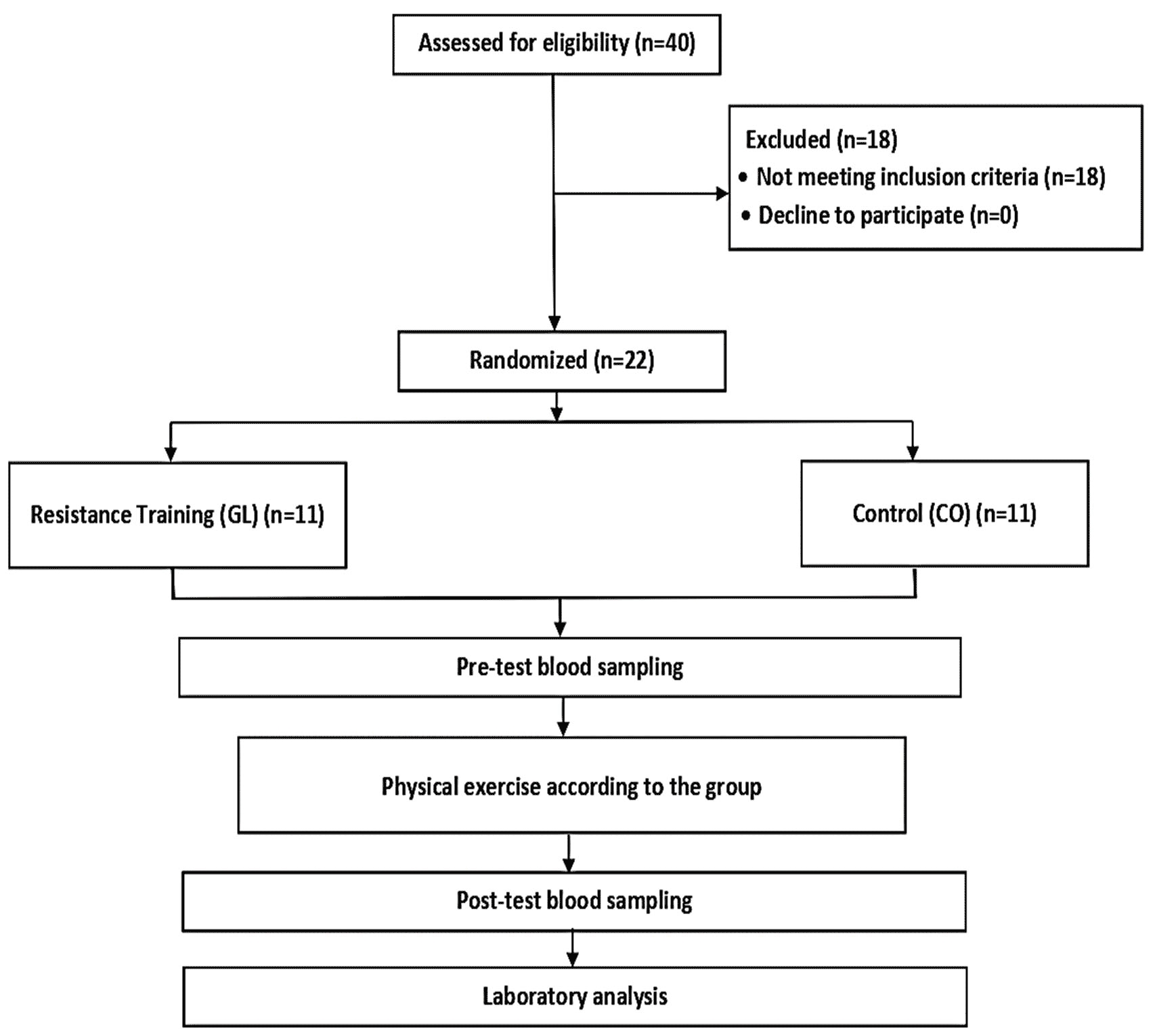
Twenty-two women were randomly selected and divided into two groups: a resistance training (GL) group (n=11) and a control (CO) group (n=11). The GL group underwent resistance training according to a program determined by the researcher, while the control group did nothing. Details of the training program will be further explained in the exercise intervention section below. Before conducting the study, all respondents were given informed consent and read the research activity plan. After that, they agreed to the research activities as respondents by filling out a consent form before the study. This study was conducted in July 2025.
The State Polytechnic of Health in Malang's ethical committee gave its approval for this study, which was carried out in compliance with the Declaration of Helsinki. This study included twenty-two healthy women (the subjects' characteristics are shown in Table 1). The researchers created inclusion and exclusion criteria to assess if the participants met the study requirements. Subjects have to be aged 18-26 years, with a normal body mass index (BMI), lead a sedentary lifestyle, do not smoke, have no history of cancer, cardiovascular disease, or musculoskeletal disorders, do not use dietary supplements or ergogenic aids, and be willing to take part in the study. According to the short form of the International Physical Activity Questionnaire (IPAQ), a sedentary lifestyle is classified as one that involves fewer than 600 MET-minutes/week (metabolic equivalent of task) of physical activity. Additionally, respondents were not required to engage in regular exercise. Participants under the age of eighteen were not permitted to participate in this study. Our study additionally eliminated respondents with hypertension who had abnormal blood pressure before exercise (systolic blood pressure ≥130 mmHg and/or diastolic blood pressure ≥85 mmHg). Subjects were also excluded if they were taking non-steroidal anti-inflammatory medicines (NSAIDs).
Exclusion criteria included reluctance to continue the prescribed exercise intervention (not attending the full exercise session), pregnancy, starting a special diet, as well as taking medicine. A formal consent form was read and signed by each chosen participant. This study involved 22 female volunteers who were randomly [17] assigned to two groups: a control group (CO) (n=11) and a resistance training group (GL) (n=11). The study was conducted at a fitness center in Tulungagung City, East Java Province. Furthermore, all participants were guided by certified professional fitness trainers during the exercise intervention (Fig. 1).
The following instruments used: blood pressure, height, and weight measurements, data collection sheets, stationery, blood drawing equipment, treadmills, and gym equipment used for weight training.
There were several steps in the data collection process for this study. Before the study began, subjects underwent a screening procedure. Respondents were randomly selected using a simple random sampling method. Each member of the population had an equal chance of being selected. A lottery was used, using a random number table. This method was based on specific criteria that allowed data to be added or removed from the study. After the study protocol was explained, they provided informed consent to participate. Participants in the trial were randomly [17] divided into two groups: one group received resistance training as treatment, and the other group received no intervention.
Data collection was conducted over two days, beginning with information on the subjects' characteristics. Before the investigation, the subjects were not allowed to eat anything. The subjects were told to keep a normal diet and get enough sleep the day before the research. They were then instructed to warm up before exercising. Afterward, they engaged in physical activity, which included resistance training.
Before the exercise, 3cc of blood was taken as pretest data, and after the exercise, 3cc of blood was taken as post-test data. After that, the blood samples were centrifuged to separate the serum. After the centrifugation was complete, the blood serum was taken for laboratory analysis to check MDA levels. This laboratory analysis was carried out at the Physiology Science Laboratory Installation, Faculty of Medicine, University of Brawijaya, Malang. The method for examining MDA levels used the ELISA (enzyme-linked immunosorbent assay) method with human reagents. Finally, as a form of accountability, after reviewing the data, the researcher compiled a written report of the research results.
Resistance Training (GL) consists of group training sessions where participants receive instruction on exercise techniques and have their performance evaluated by trained professional trainers. Each training session lasts for 60 minutes and is delivered acutely. A 10-minute warm-up, 40 minutes of core training, and a 10-minute cool-down precede the workout. Targeting both the upper and lower body, the 40-minute strength training session consists of three sets of ten exercises, such as the seated row, shoulder press, chest press, lateral pull-down, abdominal crunch, leg press, leg extension, triceps pushdown, and seated bicep curl. Each resistance exercise is carried out in compliance with the National Strength and Conditioning Association's criteria, which have been applied in earlier research [17]. Each exercise involved 8-10 repetitions in 3 sets, with 5-10 minutes of recovery between sets. The intensity of the exercise was 75-80% of 1RM. Participants were assessed using seated leg presses and upper and lower body bicep curls to determine their 1RM. Participants were placed with their knees bent at a 90-degree angle following their leg press warm-up. After applying a conservative load to the machine, participants were told to completely extend their knees and repeat the process until they became tired. The weight was raised for sitting bicep curl participants in a range of 100 degrees, from full extension to fatigue. Due to the participants' inexperience and potential for harm, 1RM was calculated for each participant using a formula rather than being measured directly Brzycki, 1993: 1RM = (Total weight lifted (kg)/(102.78 – (002.78 × number of repetitions)).
The control group (CO) was asked to just sit and lie down at the designated research location. The only activities allowed were playing with cell phones, sitting, and walking to the bathroom in the designated area. Participants in this group were allowed to play with their cell phones and communicate with their groupmates to overcome boredom while waiting in line to have their blood drawn.
After the data collected, statistical analysis was carried out using SPSS software version 16.00. The data was subjected to a descriptive analysis in order to ascertain the mean and standard error. As a normalcy test, the Shapiro-Wilk test was also employed in this study. To determine if the data were normally distributed, a difference test was developed using the paired t-test methodology. The data was examined using the Wilcoxon signed-rank test, however if the results showed otherwise [19].
With registration number: DP.04.03/F.XXI.30/00841/2025, we obtained ethical clearance from the Ethics Committee of the Malang Health Polytechnic prior to data collection. Furthermore, this study adheres to the Declaration of Helsinki, and prior to the study, we obtained written informed consent from the study respondents.
RESULTS AND DISCUSSION
The results of the analysis of subjects’ characteristics in each group are presented in Table 1.
Data Group N Mean ± SD p-value Age Resistance Training 11 20.36±1.12 1.000 Control 11 20.36±1.74 Height Resistance Training 11 156.82±5.25 0.748 Control 11 155.91±6.51 Weight Resistance Training 11 54.73±8.05 0.359 Control 11 51.82±7.06 Body Mass Index Resistance Training 11 22.20±2.68 0.454 Control 11 21.37±2.98 Systolic Resistance Training 11 114.64±15.72 0.707 Control 11 111.09±17.04 Diastolic Resistance Training 11 76.18±9.91 0.598 Control 11 73.81±8.94
This section presents statistics and information regarding the general description of the participants in Table 1. The characteristics of each group are presented in this table. Data are presented as mean ± standard deviation. The t-test results from the initial study showed no significant difference between the resistance training group and the control group (p>0.05).
The results of the normality test for each group are presented in Table 2.
Based on the normality test in Table 2, the MDA data for all groups were normally distributed (p>0.05). Therefore, the next test performed was a paired-samples t-test.
The results of the MDA analysis between pre-test and post-test in each group are presented in Figure 2.
Data Group Shapiro-Wilk N p-value Malondialdehyde Resistance Training (Pre-test) 11 0.325 Resistance Training (Post-test) 0.057 Control (Pre-test) 11 0.452 Control (Post-test) 0.187
Table 3 shows the results of the paired-samples t-test. Data are presented as mean ± standard deviation.
This study aimed to determine the effect of resistance training on reducing MDA levels, a marker of oxidative stress. The study findings showed that MDA levels, an indicator of oxidative stress, can be significantly reduced through acute resistance training. Resistance training is becoming more widely acknowledged as a successful non-pharmacological strategy that offers a number of health advantages and can enhance the standard of living of people [15]. According to other research findings, eight weeks of high-intensity exercise significantly reduced MDA levels, an indicator of oxidative stress [20].
Membrane lipid peroxidation mostly produces MDA inside cells, which is subsequently expelled into the extracellular environment [20].The reduction in MDA levels is due to improved antioxidant defenses and mitochondrial function. Consequently, resistance training can reduce MDA levels, which is beneficial for preventing oxidative stress. We shall go into greater depth on the phases of MDA drop during resistance training, though.
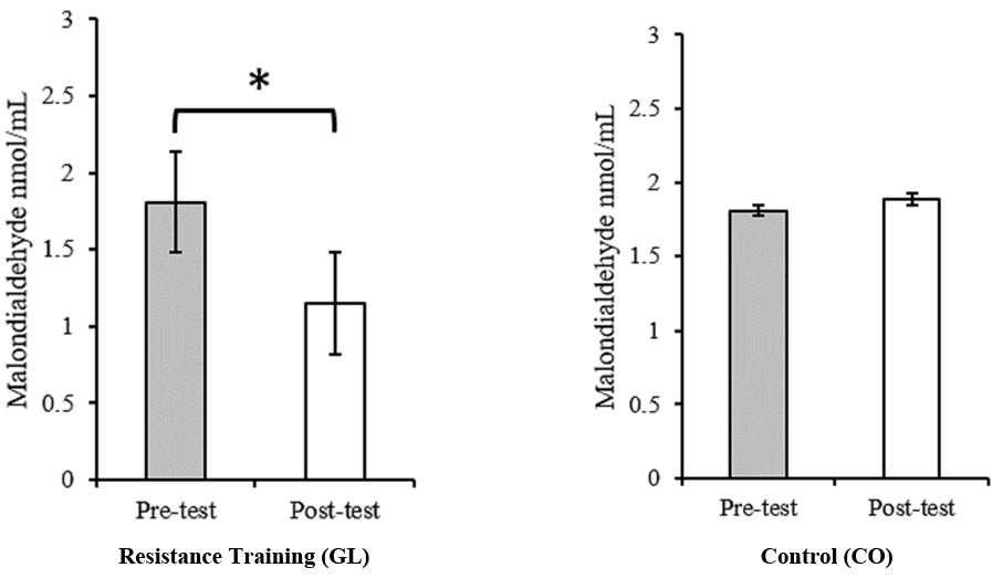
Data Group Paired-Samples t-test mean±SD p-value Malondialdehyde Resistance Training (Pre-test) 1.81±0.58 *0.001 Resistance Training (Post-test) 1.15±0.43 Control (Pre-test) 1.81±0.71 0.224 Control (Post-test) 1.89±1.23 Notes. There was a significant difference in the (GL) group with resistance training intervention (*p<0.05) and there was no significant difference in the (CO) group (p>0.05).
Resistance Training and Oxidative Stress
Exercise causes the body's ROS levels to rise, but it also raises endogenous antioxidant levels, which controls the body's defensive mechanism [21]. An imbalance between the body's greater amounts of free radicals and antioxidants causes oxidative stress, which happens during exercise [22]. The body can reach a stable state of health in a variety of methods, including exercise, which can boost cardiac output, improve cardiovascular function, and improve the metabolic system to meet the needs of the musculoskeletal system. Maximal oxygen absorption, mitochondrial ATP synthesis, oxidative capability, and post-exercise muscle exhaustion can all be impacted by aerobic exercise [23].
Regular exercise will also gradually reduce sarcopenia, or the aging-related loss of muscle mass [12]. Muscle endurance is increased by increased skeletal muscle contraction during exercise [24]. Exercise-induced oxidative stress is assessed using malondialdehyde (MDA) as a biomarker [25]. Endogenous antioxidants, such as important enzymes such as catalase (CAT), superoxide dismutase (SOD), and glutathione peroxidase (GPX), which support the body's defensive mechanism against free radicals, also rise during physical activity [25]. Glutathione peroxidase and catalase help SOD, an endogenous antioxidant, transform H2O2 into H2O [26].
Physiological Mechanisms of Resistance Training in Reducing Malondialdehyde Levels
As a result of regular cellular metabolism, living things' cells typically produce reactive oxygen species (ROS), which are essential for preserving cellular homeostasis. Low to moderate levels of ROS are implicated in immunological responses, inflammation, learning, memory, and synaptic plasticity in physiological contexts [27]. Nevertheless, excessive ROS generation can be dangerous, leading to damaging oxidative changes in cellular components, such as mitochondrial structures, which are the initial site of ROS-induced damage [28]. However, the human body has a number of antioxidants, such as the glutathione (GSH) and superoxide dismutase (SOD) systems, that work to counteract the effects of oxidants [29]. The creation of ROS can overwhelm antioxidant defenses when the generation of free radicals and detoxification are out of balance. This can result in oxidative stress, a hazardous condition, and a general deterioration in cellular function. Both aging and other clinical instances involving mitochondrial malfunction exhibit this behavior [30]. The brain is especially susceptible to oxidative stress and injury due to its high oxygen consumption, inadequate antioxidant defenses, and high concentration of oxidation-sensitive polyunsaturated lipids [31].
Another study found that while glutathione levels dropped after six months of soccer training, blood catalase (CAT), superoxide dismutase (SOD), and lipid peroxidation increased [20]. These results suggest a strong correlation between elevated catalase activity and elevated lipid peroxidation index, a sign of oxidative damage. Additionally, it should be mentioned that CAT is regarded as an intracellular enzyme, and that tissue and cellular damage contribute to its rise in serum levels [32].The concept and mechanism of oxidative stress, which refers to the imbalance between oxidants and antioxidants that causes cell damage, was originally presented in 1985 [33]. At first, it was understood that oxygen is primarily utilized by living cells and that it initiates redox reactions [34]. The connection between oxidants and antioxidants in metabolism has been the subject of much research as a result of this theory [34].
Oxidation reactions take place throughout the human respiratory chain, such as when oxygen is taken in and transformed into energy in the mitochondria, which also causes an increase in ROS [12]. Numerous environmental factors, including changes in nutrition, antigens, immunity, stress, and oxygen concentrations, all have an impact on cellular physiology and cause constant cellular adaptation. Given its extensive distribution in human tissues, SOD may play a significant role in shielding the organism from oxidative stress brought on by free radicals [35]. It has been demonstrated that after exercise, SOD, glutathione, and catalase levels rise [36]. Therefore, body's enhanced antioxidant levels, which can neutralize free radicals, are the reason for the drop in MDA levels that happens following resistance training. Free radicals are transformed into H2O2 (hydrogen peroxide) by the enzyme SOD [35].
A transition metal is necessary for all three of the SOD enzyme's isoforms to activate the enzyme's active site [37]. One of the main antioxidant enzymes that is present outside of cells is plasma SOD, also known as EC SOD [38]. The body, on the other hand, achieves a dynamic equilibrium between the antioxidant and oxidation systems by reducing or eliminating oxidative byproducts through a sequence of metabolic events [36]. High-intensity exercise can change antioxidant levels, which can affect redox-related physiological processes, according to a preliminary 2019 study [39]. In short, the decrease in MDA levels after resistance training is due to a physiological increase in SOD, an antioxidant, in the body. This neutralizes MDA levels to prevent oxidative stress and further cell damage triggered by free radicals. This is a physiological response to exercise, which is part of the oxidation reaction process to maintain homeostasis.
Strength and Limitations
The most reliable scientific approach, a randomized controlled trial, eliminates the risk of unclear cause-and-effect relationships, making it the strength of our study. We also discuss the limitations of our study, such as its small sample size. We acknowledge that a larger sample size would provide a better understanding of the results. This is indeed a limitation of our study. Furthermore, our intervention focused solely on resistance training. Therefore, it is unclear which type of exercise is effective in lowering MDA levels. Therefore, further research is recommended to examine this, for example, by comparing resistance training and aerobic exercise in lowering MDA levels. This is important to determine which type of exercise is most effective in lowering MDA levels. The impact of resistance training on other biomarkers, such as creatine kinase, interleukin 6, and protein carbonyl levels, needs further exploration. This is crucial for understanding the inflammatory response to exercise.
CONCLUSIONS
- The study results showed that resistance training can significantly reduce malondialdehyde levels, a marker of oxidative stress.
- The decrease in malondialdehyde levels after resistance training occurs due to the physiological response to exercise, which increases antioxidant levels in the body, which in turn neutralizes free radicals such as malondialdehyde.
- Therefore, resistance training is highly recommended to reduce the adverse effects of oxidative stress.
Acknowledgement. The Republic of Indonesia's Ministry of Higher Education, Science, and Technology provided financial assistance for this study under contract number 128/C3/DT.05.00/PL/2025.
Contributors:
Wibawa J.C. – conceptualization, writing – original draft;
Ayubi N. – conceptualization, writing – original draft;
Herawati L., Putro B.N.,Sceisarriya V.M., Pratama H.G., Febrianto N. – data curation, visualization;
Fudin M.S., Kurnaz M. – methodology, formal analysis.
Funding. This research received no external funding.
Conflict of interests. The authors declare no conflict of interest.
REFERENCES
- Park JH, Moon JH, Kim HJ, Kong MH, Oh YH. Sedentary Lifestyle: Overview of Updated Evidence of Potential Health Risks. Korean J Fam Med. 2020;41(6):365-73. doi: https://doi.org/10.4082/kjfm.20.0165
- Maidartati Hayati S, Anggraeni DE, Irawan E, Damayanti A, Silviani DAR. Gambaran Sedentary Lifestyle Pada Remaja Di SMA Kota Bandung. J Keperawatan BSI [Internet]. 2022 [cited 2026 Mar 02];10(2):250-65. Available from: https://ejurnal.ars.ac.id/index.php/keperawatan/index
- Afzal D, Tariq Z, Batool SA, Jabeen J, Sajjad S, Rehman S. Association of Hypertension with Sedentary Lifestyle Among Elderly in Urban Population. Heal J Physiother Rehabil Sci. 2023;3(5):532-9. doi: https://doi.org/10.55735/hjprs.v3i5.138
- Sánchez-Rodríguez MA, Zacarías-Flores M, Correa-Muñoz E, Arronte-Rosales A, Mendoza-Núñez VM. Oxidative Stress Risk Is Increased with a Sedentary Lifestyle during Aging in Mexican Women. Oxid Med Cell Longev. 2021 Oct 25;2021:9971765. doi: https://doi.org/10.1155/2021/9971765
- Kaldur T, Unt E, Ööpik V, Zilmer M, Eha J, Paapstel K, et al. The acute effects of passive heat exposure on arterial stiffness, oxidative stress, and inflammation. Medicina (Kaunas). 2016;52(4):211-6. doi: http://doi.org/10.1016/j.medici.2016.06.001
- Ceci R, Beltran Valls MR, Duranti G, Dimauro I, Quaranta F, Pittaluga M, et al. Oxidative stress responses to a graded maximal exercise test in older adults following explosive-type resistance training. Redox Biol. 2014;2(1):65-72. doi: http://doi.org/10.1016/j.redox.2013.12.004
- Yıldız M, Alp HH, Gül P, Bakan N, Özcan M. Lipid peroxidation and DNA oxidation caused by dental filling materials. J Dent Sci. 2017;12(3):233-40. doi: https://doi.org/10.1016/j.jds.2017.02.002
- Campbell JP, Turner JE. Debunking the myth of exercise-induced immune suppression: Redefining the impact of exercise on immunological health across the lifespan. Front Immunol. 2018;9:648. doi: https://doi.org/10.3389/fimmu.2018.00648
- Huang Y, Chen H, Liu Q, Hu J, Hu D, Huang Z, et al. Obesity difference on association blood malondialdehyde level and diastolic hypertension in the elderly population: a cross-sectional analysis. Eur J Med Res. 2023;28(1):44. doi: https://doi.org/10.1186/s40001-022-00983-7
- Zhao F, Su W, Sun Y, Wang J, Lu B, Yun H. Optimal resistance training parameters for improving bone mineral density in postmenopausal women : a systematic review and meta-analysis. J Orthop Surg Res. 2025;20(1):523. doi: https://doi.org/10.1186/s13018-025-05890-1
- Baraibar MA, Ladouce R, Friguet B. Proteomic quantification and identification of carbonylated proteins upon oxidative stress and during cellular aging. J Proteomics. 2013;92:63-70. doi: https://doi.org/10.1016/j.jprot.2013.05.008
- Ayubi N, Wibawa JC, Sceisarriya VM, Hariyadi K, Fudin MS, Febrianto N, et al. Mechanism of physical exercise increases malondialdehyde levels as a marker of oxidative stress: A systematic review. Fizjoterapia Pol. 2024;24(3):236-41. doi: https://doi.org/10.56984/8ZG020A43V
- Gibala MJ, Little JP, Macdonald MJ, Hawley JA. Physiological adaptations to low-volume, high-intensity interval training in health and disease. J Physiol. 2012;590(5):1077-84. doi: https://doi.org/10.1113/jphysiol.2011.224725
- Marcos-Pardo PJ, Vaquero-Cristóbal R, Huber G. The Power of Resistance Training: Evidence-based Recommendations for Middle-aged and Older Women’s Health. Retos. 2024;51:319-31. doi: https://doi.org/10.47197/retos.v51.99638
- Fragala MS, Cadore EL, Dorgo S, Izquierdo M, Kraemer WJ, Peterson MD, et al. Resistance training for older adults: Position statement from the national strength and conditioning association. J Strength Cond Res. 2019;33(8):2019-52. doi: https://doi.org/10.1519/JSC.0000000000003230
- Lopes M, Marques MC, Fonseca N, Marques DL, Nunes C, Marinho DA, et al. Effects of Eight-Week Single-Set Resistance Training on Muscle Health, Metabolic Profile and Oxidative Stress in Individuals with Cognitive Impairment. Appl Sci. 2025;15(13):7091. doi: https://doi.org/10.3390/app15137091
- Amanat S, Sinaei E, Panji M, Mohammadpor Hodki R, Bagheri-Hosseinabadi Z, Asadimehr H, et al. A Randomized Controlled Trial on the Effects of 12 Weeks of Aerobic, Resistance, and Combined Exercises Training on the Serum Levels of Nesfatin-1, Irisin-1 and HOMA-IR. Front Physiol. 2020;11:1-14. doi: https://doi.org/10.3389/fphys.2020.562895
- Brzycki M. Strength Testing – Predicting a One-Rep Max from Reps-to-Fatigue. J Phys Educ Recreat Danc. 1993;64(1):88-90. doi: https://doi.org/10.1080/07303084.1993.10606684
- Ayubi N, Rusdiawan A, Hartoto S, Wibawa J, Rizki A, Afandi A, et al. Limited Impact of Roselle Flower Extract on Protein Carbonyl Levels After Physical Exercise in Healthy Men. Medicni Perspektivi. 2025;30(1):117-26. doi: https://doi.org/10.26641/2307-0404.2025.1.325371
- Niknam A, Gaeini AA, Hamidvand A, Jahromi MK, Oviedo GR, Kordi M, et al. High-intensity functional training modulates oxidative stress and improves physical performance in adolescent male soccer players: a randomized controlled trial. BMC Sports Sci Med Rehabil. 2025 Mar 4;17(1):38.
doi: https://doi.org/10.1186/s13102-024-01037-7 - Lu Z, Xu Y, Song Y, Bíró I, Gu Y. A Mixed Comparisons of Different Intensities and Types of Physical Exercise in Patients With Diseases Related to Oxidative Stress: A Systematic Review and Network Meta-Analysis. Front Physiol. 2021;12:700055. doi: https://doi.org/10.3389/fphys.2021.700055
- Kawamura T, Higashida K, Muraoka I. Application of Molecular Hydrogen as a Novel Antioxidant in Sports Science. Oxid Med Cell Longev. 2020 Jan 14;2020:2328768. doi: https://doi.org/10.1155/2020/2328768
- van der Zwaard S, Brocherie F, Jaspers RT. Under the Hood: Skeletal Muscle Determinants of Endurance Performance. Front Sport Act Living. 2021 Aug 4;3:719434. doi: https://doi.org/10.3389/fspor.2021.719434
- Mcleod JC, Currier BS, Lowisz CV, Phillips SM. The influence of resistance exercise training prescription variables on skeletal muscle mass, strength, and physical function in healthy adults: An umbrella review. J Sport Heal Sci. 2024;13(1):47-60. doi: https://doi.org/10.1016/j.jshs.2023.06.005
- Cordiano R, Di Gioacchino M, Mangifesta R, Panzera C, Gangemi S, Minciullo PL. Malondialdehyde as a Potential Oxidative Stress Marker for Allergy-Oriented Diseases: An Update. Molecules. 2023;28(16):5979. doi: https://doi.org/10.3390/molecules28165979
- Cho SY, Chung YS, Yoon HK, Roh HT. Impact of Exercise Intensity on Systemic Oxidative Stress, Inflammatory Responses, and Sirtuin Levels in Healthy Male Volunteers. Int J Environ Res Public Health. 2022;19(18):11292. doi: https://doi.org/10.3390/ijerph191811292
- Massaad CA, Klann E. Reactive oxygen species in the regulation of synaptic plasticity and memory. AntioxidantsRedox 2011;14(10):2013-54. doi: https://doi.org/10.1089/ars.2010.3208
- Cenini G, Lloret A, Cascella R. Oxidative stress in neurodegenerative diseases: From a mitochondrial point of view. Oxid Med Cell Longev. 2019 May 9:2019:2105607. doi: https://doi.org/10.1155/2019/2105607
- Indo HP, Yen HC, Nakanishi I, Matsumoto KI, Tamura M, Nagano Y, et al. A mitochondrial superoxide theory for oxidative stress diseases and aging. J Clin Biochem2015;56(1):1-7. doi: https://doi.org/10.3164/jcbn.14-42
- Grimm A, Friedland K, Eckert A. Mitochondrial dysfunction: the missing link between aging and sporadic Alzheimer’s disease. Biogerontology. 2016;17(2):281-96. doi: https://doi.org/10.1007/s10522-015-9618-4
- Cobley JN, Fiorello ML, Bailey DM. 13 Reasons Why the Brain Is Susceptible To Oxidative Stress. Redox Biol.2018;15:490-503. doi: https://doi.org/10.1016/j.redox.2018.01.008
- Pal S, Chaki B, Chattopadhyay S, Bandyopadhyay A. High-intensity exercise induced oxidative stress and skeletal muscle damage in postpubertal boys and girls: A comparative study. J Strength Cond Res. 2018;32(4):1045-52. doi: https://doi.org/10.1519/JSC.0000000000002167
- Sies H. Oxidative stress: A concept in redox biology and medicine. Redox Biol. 2015;4:180-3. doi: https://doi.org/10.1016/j.redox.2015.01.002
- Lorenzen I, Eble JA, Hanschmann EM. Thiol switches in membrane proteins – Extracellular redox regulation in cell biology. Biol Chem. 2021;402(3):253-69. doi: https://doi.org/10.1515/hsz-2020-0266
- Zhao H, Zhang R, Yan X, Fan K. Superoxide dismutase nanozymes: an emerging star for anti-oxidation. J MaterChem 2021;9(35):6939-57. doi: https://doi.org/10.1039/D1TB00720C
- Powers SK, Deminice R, Ozdemir M, Yoshihara T, Bomkamp MP, Hyatt H. Exercise-induced oxidative stress: Friend or foe? J Sport Heal Sci. 2020;9(5):415-25. doi: https://doi.org/10.1016/j.jshs.2020.04.001
- Fetherolf MM, Boyd SD, Taylor AB, Kim HJ, Wohlschlegel JA, et Copper-zinc superoxide dismutase is activated through a sulfenic acid intermediate at a copper ion entry site. J Biol Chem. 2017;292(29):12025-40. doi: https://doi.org/10.1074/jbc.M117.775981
- Xu Y, Liang M, Ugbolue UC, Fekete G, Gu Y. Effect of Physical Exercise Under Different Intensity and Antioxidative Supplementation for Plasma Superoxide Dismutase in Healthy Adults: Systematic Review and Network Meta-Analysis. Front Physiol. 2022;13:707176. doi: https://doi.org/10.3389/fphys.2022.707176
- Souza AV, Giolo JS, Teixeira RR, Vilela DD, Peixoto LG, Justino AB, et al. Salivary and Plasmatic Antioxidant Profile following Continuous, Resistance, and High-Intensity Interval Exercise: Preliminary Study. Oxid Med Cell Longev. 2019 Nov 26:2019:5425021. doi: https://doi.org/10.1155/2019/5425021