

 UK
 EN



Вернігора Д.Г.1, 2
 Дігтяр В.А.1
Бойко М.В.2
Обертинський А.В.2
Островська О.А.2
Гладкий О.П.2
Ключові слова: сечокам’яна хвороба, діти, уретероскопія, ретроградна інтраренальна хірургія, мініперкутанна нефролітотомія, інфрачервона спектроскопія з перетворенням Фур’є, ретроспективні дослідження
Key words: urolithiasis, child, ureteroscopy, retrograde intrarenal surgery, nephrolithotomy, percutaneous, Fourier transform infrared spectroscopy, retrospective studies
Реферат
Мета дослідження – описати характеристики конкрементів і ранні результати ендоскопічного лікування сечокам’яної хвороби в дітей з роздільним поданням уретероскопії (URS), ретроградної інтраренальної хірургії (RIRS), мініперкутанної нефролітотомії (mini-PCNL) та цистолітотрипсії. Проведено ретроспективне одноцентрове обсерваційне дослідження 72 дітей віком до 18 років, яких лікували з 01.01.2025 до 12.03.2026. Оцінювали локалізацію, розмір, комп’ютерно-томографічну щільність і склад каменів, вид втручання, залишкові фрагменти та ранні ускладнення за Clavien-Dindo. Вибір методики був клінічним і нерандомізованим. Усім 72 дітям виконували післяопераційне ультразвукове дослідження та контрольну низькодозову нативну КТ без внутрішньовенного контрастування через один місяць для уніфікованого оцінювання залишкових фрагментів. Критерієм результату була відсутність залишкових фрагментів >4 мм; фрагменти ≤4 мм допускалися. Камені верхніх сечових шляхів виявлено в 68 (94%) дітей: ниркові – у 47 (65%), сечовідні – у 21 (29%); камені сечового міхура – у 4 (6%). Розмір конкрементів становив 4-18 мм, середнє значення – 8,8 мм, медіана – 8 (6; 11) мм. Щільність становила 390-1410 HU, середнє значення – 895 HU, медіана – 1015 (928; 1132,5) HU. Кальцій-оксалатні камені виявлено в 39 (54%) дітей. URS виконано у 20 (28%) дітей, RIRS – у 36 (50%), mini-PCNL – у 12 (17%), цистолітотрипсію – у 4 (6%). Через один місяць прийнятого критерію досягнуто в 71 із 72 дітей (99%): після URS – у 20/20 (100%), RIRS – у 36/36 (100%), mini-PCNL – у 11/12 (92%), цистолітотрипсії – у 4/4 (100%). Зареєстровано три ускладнення I ступеня (4%): дві післяопераційні кровотечі після mini-PCNL та один випадок вираженого больового синдрому після RIRS. Отримані дані характеризують ранні результати лікування в одному центрі за визначеним критерієм і строком контролю. Вони не встановлюють переваги окремої методики. Досягнення прийнятого критерію не тотожне повній відсутності каменів. Аналіз складу каменів методом інфрачервоної спектроскопії з перетворенням Фур’є (FTIR) доповнює хірургічне лікування та допомагає планувати метаболічне обстеження й профілактику. Віддалені рецидиви не оцінювали.
Abstract
Modern endoscopic treatment of pediatric urolithiasis: early outcomes of a single-center retrospective study. Vernihora D.H., Dihtiar V.A., Boyko M.V., Obertinskyi A.V., Ostrovska O.O., Hladkyi O.P. The aim was to describe stone characteristics and early outcomes of endoscopic treatment for pediatric urolithiasis, reporting ureteroscopy (URS), retrograde intrarenal surgery (RIRS), mini-percutaneous nephrolithotomy (mini-PCNL), and cystolithotripsy separately. This retrospective single-center observational study included 72 children younger than 18 years treated between 01 January 2025 and 12 March 2026. Stone location, size, computed tomography (CT) attenuation, and composition, as well as procedure type, residual fragments, and early complications according to the Clavien-Dindo classification, were assessed. Treatment selection was clinical and nonrandomized. All 72 children underwent postoperative ultrasonography and follow-up low-dose non-contrast CT in one month for standardized residual-fragment assessment. The outcome criterion was the absence of residual fragments >4 mm; fragments ≤4 mm were allowed. Upper urinary tract stones were present in 68 children (94%): renal stones in 47 (65%) and ureteral stones in 21 (29%); bladder stones were present in 4 (6%). Stone size ranged from 4 to 18 mm, with a mean of 8.8 mm and a median of 8 (6; 11) mm. Attenuation ranged from 390 to 1410 HU, with a mean of 895 HU and a median of 1015 (928; 1132.5) HU. Calcium oxalate stones were found in 39 children (54%). URS was performed in 20 children (28%), RIRS in 36 (50%), mini-PCNL in 12 (17%), and cystolithotripsy in 4 (6%). At one month, 71/72 children (99%) met the specified criterion: 20/20 (100%) after URS, 36/36 (100%) after RIRS, 11/12 (92%) after mini-PCNL, and 4/4 (100%) after cystolithotripsy. Three grade I complications (4%) were recorded: two cases of postoperative bleeding after mini-PCNL and one case of pronounced pain after RIRS. These findings describe early institutional outcomes under an explicit criterion and follow-up interval. They do not establish the superiority of any procedure. Meeting the outcome criterion is not equivalent to complete stone clearance. Fourier-transform infrared spectroscopy (FTIR) complements surgery by informing metabolic evaluation and prevention. Long-term recurrence was not assessed.
Urolithiasis in children remains a complex clinical problem that combines urgent manifestations, risk of obstructive and infectious complications, the need for metabolic investigation, and high requirements for selecting the optimal surgical strategy. Current recommendations emphasize an individualized approach to diagnosis, treatment and follow-up in children with urinary stones [1, 2, 3].
Modern pediatric endourology allows treatment to be individualized according to stone location, size, density, and morphology. Ureteroscopy, retrograde intrarenal surgery, mini-percutaneous nephrolithotomy, and cystolithotripsy are used in clinical practice. Selection should consider the child’s age and anatomy, stone number and aggregate size, residual-fragment risk, and the availability of small-caliber instruments [1, 2, 3, 4, 5].
Stone composition analysis complements surgical management by informing metabolic evaluation and individualized prevention [1, 7].
Single-center observational data can describe stone characteristics, procedures actually used, and early outcomes. Separate reporting by the procedure performed provides a description of clinical experience. Establishing comparative effectiveness requires clinically comparable groups and individual patient data.
Aim – to describe stone characteristics and early outcomes of endoscopic treatment for pediatric urolithiasis, reporting URS, RIRS, mini-PCNL, and cystolithotripsy separately.
MATERIALS AND METHODS OF RESEARCH
Study design. A retrospective single-center observational cohort study was conducted including children with urolithiasis treated at the Regional Medical Center of Family Health from 01 January 2025 to 12 March 2026. The manuscript structure was prepared taking into account the STROBE recommendations for reporting observational studies [6].
The analysis included 72 children younger than 18 years with confirmed urinary tract stones. Inclusion criteria were: confirmed urolithiasis on ultrasonography and/or computed tomography; available data on stone location, size, and density; endoscopic treatment; and documented residual-fragment assessment in one month. Exclusion criteria were: age 18 years or older, incomplete clinical or imaging data, and absence of post-treatment outcome assessment.
Children were divided into four groups according to the procedure performed: URS for ureteral stones, n=20; RIRS, n=36 (35 children with renal stones and one with a ureteral stone); mini-PCNL for renal stones, n=12; and cystolithotripsy for bladder stones, n=4. URS and RIRS were treated as distinct procedures. Treatment allocation was nonrandomized and depended on stone location and characteristics, urinary tract anatomy, obstruction, infection, and access feasibility [1, 2, 3, 4, 5].
Data source and analytical population. The descriptive analysis used de-identified aggregate clinical measures for a cohort of 72 children and procedure-specific absolute counts supplied by the authors. Individual records from the primary clinical dataset were not used to verify group-specific Hounsfield units (HU) or structural anomalies; these fields were omitted from the group table. The unit of observation was a child. Outcomes are described for the entire cohort; group characteristics are reported separately for 36 children after RIRS and 12 after mini-PCNL. These groups reflect actual clinical selection. Eligibility of each patient for both procedures was not established; factors determining procedure selection were therefore not analysed.
For each case, stone location and size in millimeters, composition, computed tomography attenuation in Hounsfield units, treatment modality, and residual fragments were assessed. HU values characterize stone attenuation and are considered alongside other clinical and imaging features [1, 7].
Computed tomography protocol. Low-dose non-contrast spiral CT (LD-NCCT) was performed without intravenous contrast administration. Children were scanned in the supine position using a single non-contrast acquisition, with coverage limited to the upper renal poles through the urinary bladder. Pediatric acquisition settings were adapted to the child’s anthropometric characteristics according to the ALARA principle (As Low As Reasonably Achievable). Tube voltage was 70-100 kVp according to body size; automatic tube-current modulation was used, with a rotation time of 0.3-0.5 s and a pitch of 0.8-1.2 according to scanner capabilities. Iterative reconstruction with overlapping 1-2 mm slices was used. Residual-fragment number, location, and maximum diameter were assessed. Individual CTDIvol (mGy) and DLP (mGy·cm) values are not reported in this analysis.
Operative techniques. Contact lithotripsy was performed using a Dornier Medilas H Solvo 35 holmium laser (Dornier MedTech, Ho:YAG; maximum rated output 35 W). Dusting and fragmentation modes and fibers of 200 μm diameter were used. Accessible fragments were retrieved with endoscopic loops or baskets. Irrigation during endoscopic procedures was delivered using an irrigation pump. The baseline pump pressure setting was 15 mmHg and was increased up to 20 mmHg when required.
Ureteroscopy (URS). Ureteral stones were treated through a retrograde approach using KARL STORZ 27002L / 27002KK ureterorenoscopes of nominal caliber 9.5 Fr, working length 34-43 cm, and direction of view 6°. The working channel was 6 Fr, accommodating instruments up to 5 Fr. Following visualization, laser lithotripsy and retrieval of accessible fragments were performed. Drainage used a 5 Fr ureteral catheter, removed the following day, or a ureteral stent of 16 or 26 cm according to the child’s age. A Foley catheter was left for 2-3 days, with its caliber selected according to age. Ureteral stents were removed after 3 weeks.
Retrograde intrarenal surgery (RIRS). The collecting system was inspected and renal stones underwent contact lithotripsy using a single-use flexible video ureterorenoscope, Innovex US27F-12-EU (Shanghai AnQing Medical Instrument Co., Ltd.): nominal shaft diameter 7.5 Fr, distal tip 8.1 Fr, and working channel 3.6 Fr. A 9.5 Fr ureteral access sheath (UAS) was used to facilitate repeated access to the renal collecting system when atraumatic placement was feasible. Laser lithotripsy during RIRS used pulse energies of 0.5-0.8 J and frequencies of 10-20 Hz. Clinically narrow ureters underwent preoperative stenting, usually for approximately one month. Stent length, 16 or 26 cm, was selected according to the child’s age. A ureteral stent was placed after RIRS and removed after 3 weeks.
Mini-percutaneous nephrolithotomy (mini-PCNL). Renal stones were removed via a percutaneous approach using a SUODE SNJ-I-005430 endoscope, 8/9.8 Fr. Percutaneous access to the renal collecting system was established using a 16 Fr working sheath. Renal puncture was performed under ultrasonographic and fluoroscopic guidance using an image intensifier. After access formation, laser lithotripsy used pulse energies of 0.8-2.0 J and frequencies of 5-15 Hz, followed by stone fragment extraction. A nephrostomy tube was placed in all mini-PCNL cases and removed on postoperative day 3.
Cystolithotripsy. Access to the bladder was transurethral during cystoureteroscopy. Bladder stones were fragmented and removed endoscopically using a SUODE SNJ-I-005430 endoscope, 8/9.8 Fr. The bladder was drained with a catheter, with its type and caliber selected according to the patient’s age. The catheter was removed on postoperative day 3.
Outcome assessment. All 72 children underwent postoperative ultrasonography and follow-up low-dose non-contrast CT one month after surgery. CT was used as the uniform follow-up imaging method for the entire cohort to assess residual-fragment number, location, and size. This provided a consistent imaging method for outcome assessment across all procedure groups. The criterion was the absence of detected residual fragments >4 mm on CT, including patients with no stones and those with fragments ≤4 mm. This operational definition allows small residual fragments and is not equivalent to complete clearance. Definitions vary in the literature, and a 0 mm criterion should be reported separately [1, 9, 10, 11].
Complications. Early postoperative complications were graded using the Clavien–Dindo classification [12]. Residual fragments and complications were considered separate outcomes. Long-term recurrence assessment, including evaluation in one year, was outside the scope of this analysis.
Retrieved stones or fragments were sent for composition analysis using Fourier-transform infrared spectroscopy (FTIR). Results informed subsequent metabolic assessment and individualized recurrence-prevention recommendations [1, 7].
This retrospective observational study analysed de-identified clinical data. The study protocol was approved by the Commission on Medical Ethics at the Communal Non-Profit Society “Regional Medical Center of Family Health” of the Dnipropetrovsk Regional Council, protocol No. 8.1 dated 18 August 2026. The Commission issued a positive decision on conducting the retrospective single-center observational study of pediatric urolithiasis. The institution hosting the committee is located at 13, Kosmichna street, Dnipro, 49100, Ukraine. The study was conducted in accordance with the Declaration of Helsinki of the World Medical Association. For all 72 children, written informed consent was obtained from parents or legal representatives before inclusion of data in the analysis, covering treatment and the scientific use of de-identified clinical data and examination results.
The analysis was descriptive. Quantitative clinical measures are reported as means, ranges, medians, and lower and upper quartiles, Me (Q1; Q3); categorical variables as counts and percentages. Fluoroscopy time and the reported fluoroscopic dose metric are expressed as mean ± standard deviation (SD). Operative durations are reported as approximate intervals for URS/RIRS and mini-PCNL; separate procedure-specific means are not reported. Statistical processing used STATISTICA v. 6.1 (StatSoft Inc., USA). Percentages were rounded to whole numbers, with corresponding counts stated in the text and tables. Within-procedure proportions used the number of children in that procedure group as the denominator. Independently rounded percentages may not sum to 100%. Treatment-selection models and outcome-prediction models were not used. Residual-fragment outcomes were described without modeling because only one child did not meet the specified outcome criterion.
RESULTS AND DISCUSSION
The analysis included 72 children with urolithiasis. Upper urinary tract stones predominated, being detected in 68 children (94%). Renal or collecting-system stones were present in 47 (65%), ureteral stones in 21 (29%), and bladder stones in 4 (6%).
Stone size ranged from 4 to 18 mm, with a mean of 8.8 mm and a median of 8 (6; 11) mm. Density ranged from 390 to 1410 HU, with a mean of 895 HU and a median of 1015 (928; 1132.5) HU. General stone characteristics are presented in Table 1.
Parameter n/value %/range Total number of children 72 100 Kidney / pyelocaliceal system stones 47 65 Ureteral stones 21 29 Bladder stones 4 6 Stone size, mm, mean 8.8 4-18 Stone size, mm, Me (Q1; Q3) 8 (6; 11) - Stone density, HU, mean 895 390-1410 Stone density, HU, Me (Q1; Q3) 1015 (928; 1132.5) -
Calcium oxalate stones predominated in the structure of stone composition. Calcium phosphate, struvite/infectious, uric acid, cystine and mixed stones accounted for a smaller proportion; however, these groups are clinically important because they may be associated with specific metabolic or infectious mechanisms of stone formation (Table 2).
URS was performed in 20 children (28%), RIRS in 36 (50%), mini-PCNL in 12 (17%), and cystolithotripsy in 4 (6%). These proportions use the entire cohort, n=72, as the denominator. Available clinical characteristics of the RIRS and mini-PCNL groups are presented in Table 3. Outcomes by each of the four procedures are shown in Table 4.
Structure / composition of stone n % Calcium oxalate 39 54 Calcium phosphate 15 21 Struvite / infectious 8 11 Uric acid 5 7 Cystine 3 4 Mixed 2 3
Characteristic RIRS, n=36 mini-PCNL, n=12 Number of children, n 36 12 Age, years, mean 9 11 Sex, male / female, n (%) 22 (61%) / 14 (39%) 8 (67%) / 4 (33%) Renal / ureteral location, n (%) 35 (97%) / 1 (3%) 12 (100%) / 0 (0%) Stone number: single / multiple, n (%) 13 (36%) / 23 (64%) 5 (42%) / 7 (58%) Obstruction, n (%) 8 (22%) 3 (25%) Preoperative infection, n (%); assessment 2 (6%) 4 (33%) Preoperative stenting, n (%) 15 (42%) 1 (8%) Treatment stages, n (%) 1 stage — 32 (89%); 1 stage — 9 (75%); Repeat or auxiliary procedures, n (%) 1 (3%) 4 (33%) Notes: Percentages: 36 children in the RIRS group and 12 in the mini-PCNL group as denominators. Stone attenuation in HU in Table 1 describes the entire cohort; group-specific HU, structural anomalies, and urinary tract dilatation were not analysed in Table 3.
by urinalysis and urine culture
2 stages — 4 (11%)
2 stages — 2 (17%);
3 stages — 1 (8%)
Treatment modality n No fragments >4 mm detected, n Fragments >4 mm, n Criterion met, % URS 20 20 0 100 RIRS 36 36 0 100 mini-PCNL 12 11 1 92 Cystolithotripsy 4 4 0 100 Total 72 71 1 99 Notes: URS, ureteroscopy; RIRS, retrograde intrarenal surgery; mini-PCNL, mini-percutaneous nephrolithotomy. The outcome criterion was the absence of residual fragments >4 mm on follow-up low-dose non-contrast CT one month after surgery; fragments ≤4 mm were allowed. This outcome is not equivalent to complete fragment clearance.
On one-month follow-up CT, 71/72 children (99%) had no residual fragments >4 mm. The criterion was met by 20/20 children (100%) after URS, 36/36 (100%) after RIRS, 11/12 (92%) after mini-PCNL, and 4/4 (100%) after cystolithotripsy. The single case of residual fragments >4 mm after mini-PCNL is reported descriptively.
Approximate operative duration was 25-40 min for URS/RIRS and 90-126 min for mini-PCNL. Hospital stay after mini-PCNL was 3-4 days. After URS/RIRS, hospital stay was generally one day, with a two-day stay in three cases.
In the mini-PCNL group, using combined ultrasonographic and fluoroscopic guidance, mean fluoroscopy time was 32.55±18.00 s and the recorded fluoroscopic dose metric was 6.00±2.60 mGy (mean ± SD).
Three early Clavien-Dindo grade I complications were recorded (3/72; 4%): two cases of postoperative bleeding after mini-PCNL (2/12; 17%) and one case of pronounced pain after RIRS (1/36; 3%). No early complications after URS or cystolithotripsy were recorded in the reported clinical data.
Stone composition analysis
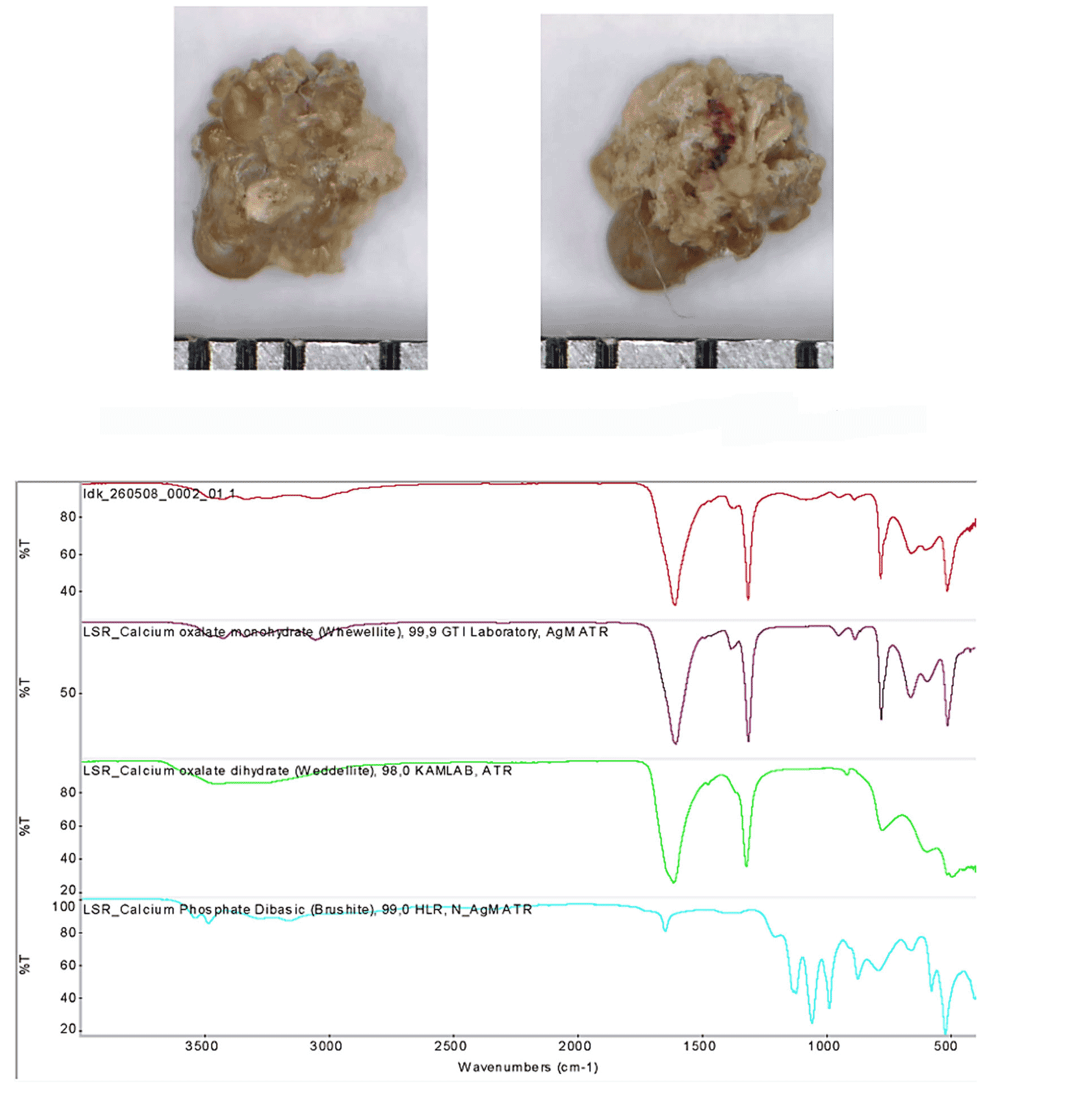
Fourier-transform infrared spectroscopy identifies stone composition and defines the direction of metabolic evaluation. Clinically, this supports individualized fluid and dietary recommendations, nephrology involvement, and infection control [1, 7]. The effect of these preventive measures on long-term recurrence was not evaluated in this study (Figure).
Upper urinary tract stones predominated in this cohort. This distribution highlights the need for access to different pediatric endourological approaches. Current guidelines recommend consideration of age, anatomy, location, number and aggregate size of stones, and the risk of recurrence [1, 2, 3].
Treatment selection reflected clinical decisions. Renal stones predominated in the RIRS group; one case had a ureteral location. Mini-PCNL was used for renal stones, URS for ureteral stones, and cystolithotripsy for bladder stones. Comparison of the two renal procedures should account for age, anatomy, collecting-system location, stone number and aggregate size, attenuation, infection, obstruction, and preoperative stenting. Assessment of the independent contribution of these characteristics requires individual data and a clinically comparable analytical population.
Calcium oxalate and calcium phosphate stones accounted for most observations. Their composition informs assessment of metabolic abnormalities, diet, and fluid intake. Struvite, cystine, uric acid, and mixed stones warrant evaluation for the relevant infectious or metabolic mechanisms. Composition analysis complements surgery, but does not itself demonstrate the effectiveness of subsequent prevention [1, 7].
The high proportion meeting the specified criterion at one month characterizes early outcomes at our center. Differences in patient selection prevent conclusions about superiority of either procedure. Comparative pediatric studies indicate that evaluation of RIRS and mini-PCNL should include complications, treatment duration, and auxiliary procedures in addition to residual fragments [4, 5, 8, 9, 13].
Computed tomography attenuation is one of the characteristics considered during treatment planning. A recent randomized study of high-density pediatric renal stones highlights the need to assess multiple outcomes and patient characteristics [13]. In this study, HU was reported only as a descriptive stone characteristic; its independent association with treatment selection or outcome was not determined. Imaging follow-up in children should account for radiation exposure: EAU guidelines recommend ultrasonography and plain abdominal radiography as the primary follow-up methods, with low-dose non-contrast CT used for diagnostically uncertain or complex cases [2]. In the randomized study by Salama et al., the combined ultrasound and fluoroscopy guidance group (n=25) had a fluoroscopy time of 29.44±17.01 s and a reported radiation dose of 6.07±3.57 mGy [14]. These published values are not results from our cohort.
During endourological procedures, the pressure set on an irrigation pump should be distinguished from intrarenal pressure (IRP). Physiological IRP in an unobstructed kidney is approximately 0-15 mmHg (0-20 cm H₂O). Sustained IRP near or above 30 mmHg is associated with pyelovenous backflow and infectious risk; maintaining IRP below this level is considered a risk-reduction target [1]. A universal safe threshold has not been established; IRP depends on the balance between irrigation inflow and outflow [15]. These published targets are not measured values from our cohort.
Published U.S. diagnostic reference levels (DRLs) for non-contrast abdominal and pelvic CT in age categories spanning 1-18 years range from 2.6 to 11.0 mGy for CTDIvol and 95 to 510 mGy cm for DLP [16]. These reference levels support imaging optimization and are not measured dose values from our cohort.
Limitations include the retrospective single-center design, the small mini-PCNL group, and clinical selection. Group-specific HU, structural anomalies, urinary tract dilatation, and aggregate stone size were not included in the between-group description. Aggregate clinical measures do not establish clinical comparability of RIRS and mini-PCNL or distinguish the outcome of one procedure from that of completed staged treatment. The outcome criterion allows residual fragments ≤4 mm, so meeting it is not equivalent to complete stone clearance. Even small residual fragments in children require attention during follow-up [1, 2, 10]. Further analysis requires separate recording of 0 mm, ≤4 mm, and >4 mm residual-fragment outcomes and the precise timing of assessment relative to treatment stages and auxiliary procedures. All 72 children underwent follow-up low-dose non-contrast CT in one month for uniform outcome assessment; individual CTDIvol and DLP values are not reported in this analysis, limiting assessment of radiation exposure. Operative durations are reported as approximate intervals, without separate means for URS and RIRS. The specific fluoroscopic dose metric was not identified, limiting comparison of radiation exposure with other series. One-year outcomes and preventive effectiveness were outside the scope of this analysis. Direct IRP measurements are not reported, limiting assessment of the actual collecting-system pressure.
The study describes the use of four endoscopic procedures in children and their early outcomes assessed at a defined interval using the described imaging methods. The findings support separate reporting of URS and RIRS, an explicit residual-fragment criterion, and completion of outcome reporting with treatment stages, repeat interventions, and long-term follow-up [11].
CONCLUSIONS
1. Upper urinary tract stones predominated in this cohort, and calcium oxalate was the most frequent stone composition.
2. Endoscopic treatment was associated with a high proportion of patients without residual fragments larger than 4 mm at one month. This outcome should not be equated with complete fragment clearance.
3. Recorded early complications were classified as Clavien-Dindo grade I. Small and clinically heterogeneous groups limit conclusions about comparative safety and effectiveness.
4. RIRS and mini-PCNL were used for renal stones. Nonrandomized selection and limited aggregate data do not establish the superiority of either approach or independent factors determining treatment selection.
5. Stone composition analysis using Fourier-transform infrared spectroscopy helps plan metabolic evaluation and individualized prevention; its long-term effectiveness requires separate evaluation.
Contributors:
Vernihora D.H. – concept, study design, data collection and analysis, manuscript writing;
Dihtiar V.A. – scientific supervision and manuscript editing;
Boyko M.V., Obertinskyi A.V., Ostrovska O.O. –clinical data collection, interpretation of results, critical review of the manuscript;
Hladkyi O.P. – organizational support, critical revision of the manuscript.
Funding. This research received no external funding.
Conflict of interests. The authors declare no conflict of interest.
REFERENCES
1. European Association of Urology. EAU Guidelines on Urolithiasis [Internet]. Arnhem: EAU Guidelines Office; 2026 [cited 2026 Jun 12]. Available from: https://uroweb.org/guidelines/urolithiasis
2. European Association of Urology. EAU Guidelines on Paediatric Urology [Internet]. Arnhem: EAU Guidelines Office; 2026 [cited 2026 Jun 12]. Available from: https://uroweb.org/guidelines/paediatric-urology
3. Güven S, Tokas T, Tozsin A, Haid B, Lendvay TS, Silay S, et al. Consensus statement addressing controversies and guidelines on pediatric urolithiasis. World J Urol. 2024;42:473. doi: https://doi.org/10.1007/s00345-024-05161-4
4. Aydamirov M, Karkin K. Comparison of two methods for treatment of 1-2 cm kidney stones in pediatric patients: mini PCNL and RIRS. Langenbecks Arch Surg. 2024;409:207. https://doi.org/10.1007/s00423-024-03402-z
5. Yuan Y, Liang YN, Li KF, Ho YR, Wu QL, Zhao Z. A meta-analysis: retrograde intrarenal surgery vs. percutaneous nephrolithotomy in children. Front Pediatr. 2023;11:1086345. doi: https://doi.org/10.3389/fped.2023.1086345
6. von Elm E, Altman DG, Egger M, Pocock SJ, Gøtzsche PC, Vandenbroucke JP, et al. The Strengthening the Reporting of Observational Studies in Epidemiology (STROBE) statement: guidelines for reporting observational studies. Lancet. 2007;370(9596):1453-7.
doi: https://doi.org/10.1016/S0140-6736(07)61602-X
7. Cao B, Daniel R, McGregor R, Tasian GE. Pediatric nephrolithiasis. Healthcare (Basel). 2023;11(4):552. doi: https://doi.org/10.3390/healthcare11040552
8. Mahmood SN, Said SHA, Mohammed RO, Jaafar MS. Safety and efficacy of mini-percutaneous nephrolithotomy in management of renal stones in pediatric age group. BMC Nephrol. 2025;26:111.
doi: https://doi.org/10.1186/s12882-025-04031-4
9. Mahmoud MA, Shawki AS, Abdallah HM, Mostafa D, Elawady H, Samir M. Use of retrograde intrarenal surgery (RIRS) compared with mini-percutaneous nephrolithotomy (mini-PCNL) in pediatric kidney stones. World J Urol. 2022;40(12):3083-9. doi: https://doi.org/10.1007/s00345-022-04186-x
10. Lombardo R, Tzelves L, Geraghty R, Davis NF, Neisius A, Petřík A, et al. Follow-up of urolithiasis patients after treatment: an algorithm from the EAU Urolithiasis Panel. World J Urol. 2024;42:202. doi: https://doi.org/10.1007/s00345-024-04872-y
11. Juliebø-Jones P, Ulvik Ø, Beisland C, Somani BK. Paediatric percutaneous nephrolithotomy (P-PCNL) reporting checklist. Scand J Urol. 2023;58:115-9. doi: https://doi.org/10.2340/sju.v58.13392
12. Clavien PA, Barkun J, de Oliveira ML, Vauthey JN, Dindo D, Schulick RD, et al. The Clavien-Dindo classification of surgical complications: five-year experience. Ann Surg. 2009;250(2):187-96. doi: https://doi.org/10.1097/SLA.0b013e3181b13ca2
13. Mahmoud O, Abdallah M, Abdellatif A, Mostafa AH, Albudairat A, Elmarakbi AA, et al. Comparative evaluation of flexible ureterorenoscopy and mini-percutaneous nephrolithotomy for the management of renal pelvic stones less than 2 cm with high attenuation value in pediatric patients: a prospective randomized study. Urolithiasis. 2026;54:48. doi: https://doi.org/10.1007/s00240-026-01938-x
14. Salama AK, ElSabbagh MA, Fahmy AM, Youssif M, Badawy H, Ghozlan A, et al. Can Ultrasound Guided Fluoroscopy Efficiently Reduce Radiation Time and Dose in Mini-Percutaneous Nephrolithotomy in Children, a Prospective Randomized Trial. Urology. 2025;201:95-100. doi: https://doi.org/10.1016/j.urology.2025.03.012
15. Somani B, Davis N, Emiliani E, Göcke MI, Jung H, Keller EX, et al. Intrarenal Pressure Monitoring During Ureteroscopy: A Delphi Panel Consensus. Eur Urol Open Sci. 2025;73:43-50. doi: https://doi.org/10.1016/j.euros.2025.01.005
16. Kanal KM, Butler PF, Chatfield MB, Wells J, Samei E, Simanowith M, et al. U.S. Diagnostic Reference Levels and Achievable Doses for 10 Pediatric CT Examinations. Radiology. 2022;302(1):164-74. doi: https://doi.org/10.1148/radiol.2021211241