

 UK
UK  EN
EN 



Taulant Gishto *1
 Edlira Rruci1
Naltin Shuka1
Silvia Methoxha1
Gentjan Vyshka2
Edlira Rruci1
Naltin Shuka1
Silvia Methoxha1
Gentjan Vyshka2
Key words: acute coronary syndrome, carotid intima-media thickness, epicardial adipose tissue, uric acid
Ключові слова: гострий коронарний синдром, товщина комплексу інтима-медіа сонних артерій, епікардіальна жирова тканина, сечова кислота
Abstract
Acute coronary syndrome (ACS) can occur even when the burden of traditional cardiovascular risk factors is limited, highlighting residual risk and the potential value of non-traditional markers. The purpose of this study was to assess carotid intima-media thickness and echocardiographic epicardial/paracardial adipose tissue thickness, together with selected biomarkers, in acute coronary syndrome compared with coronary angiography or coronary computed tomography-negative controls. Carotid intima-media thickness (CIMT) and epicardial adipose tissue (EAT) capture structural and metabolic alterations that may cluster with systemic inflammatory and metabolic markers in patients with acute coronary syndrome.In this hospital-based case–control study, we enrolled 150 patients with ACS and 150 control subjects in whom obstructive coronary artery disease was ruled out by invasive coronary angiography or coronary CT angiography. Demographic and clinical data, carotid ultrasound for CIMT, transthoracic echocardiography for EAT and pericardial adipose tissue (PAT), and biochemical markers (C-reactive protein, fibrinogen, white blood cell and neutrophil counts, serum uric acid) were collected. Group comparisons, correlation analyses and receiver operating characteristic (ROC) curves were used to assess associations and discriminative performance. Compared with controls, ACS patients had significantly higher mean CIMT (1.02 vs 0.65 mm), thicker EAT (6.5 vs 3.7 mm) and PAT (5.8 vs 3.1 mm) and higher levels of all non-traditional biochemical markers (all p<0.001). CIMT, EAT and PAT correlated positively with fibrinogen, C-reactive protein, leukocyte and neutrophil counts, serum uric acid and body mass index, with positive correlations observed in both groups. CIMT demonstrated excellent discrimination for ACS (area under the curve [AUC] >0.90; sensitivity >93%; specificity >85%), with an optimal cut-off around 0.72 mm. EAT and PAT also showed strong discrimination (AUC 0.902 and 0.875, respectively), while serum uric acid achieved an AUC of 0.860 with high specificity.Patients with ACS exhibit a distinct non-traditional risk profile characterised by increased CIMT, epicardial/pericardial fat and heightened inflammatory–metabolic activation. CIMT and EAT are structural expressions of this adverse milieu and, when combined with selected non-traditional biochemical markers, demonstrate strong discrimination in this cohort. Prospective studies are needed to validate structural-inflammatory phenotypes and define their role in personalised risk stratification and prevention.
Реферат
Товщина комплексу інтима-медіа сонних артерій, епікардіальний/перикардіальний жир та нетрадиційні біомаркери при гострому коронарному синдромі: дослідження типу «випадок-контроль». Ґішто Т., Рручі Е., Шука Н., Метходжа С., Вишка Дж. Гострий коронарний синдром може виникати навіть за відносно невеликого тягаря традиційних серцево-судинних факторів ризику, що підкреслює наявність резидуального ризику та потенційну цінність нетрадиційних маркерів. Метою цього дослідження було оцінити товщину комплексу інтима-медіа сонних артерій (carotid intima-media thickness, CIMT) та ехокардіографічно визначену товщину епікардіальної/паракардіальної (перикардіальної) жирової тканини (epicardial adipose tissue/pericardial adipose tissue, EAT/PAT), а також вибрані біомаркери в пацієнтів з гострим коронарним синдромом порівняно з контрольною групою, у якій обструктивну ішемічну хворобу серця було виключено за допомогою інвазивної коронарографії або коронарної КТ-ангіографії. CIMT та епікардіальна жирова тканина відображають структурні й метаболічні зміни, які можуть поєднуватися із системними запальними та метаболічними маркерами у хворих з гострим коронарним синдромом. У цьому госпітальному дослідженні типу «випадок-контроль» було залучено 150 пацієнтів з гострим коронарним синдромом та 150 осіб контрольної групи. Збирали демографічні й клінічні дані, виконували ультразвукове дослідження сонних артерій для визначення CIMT, трансторакальну ехокардіографію для оцінки EAT і PAT, а також лабораторні показники (С-реактивний білок, фібриноген, кількість лейкоцитів і нейтрофілів, сечова кислота сироватки). Для аналізу застосовували порівняння груп, кореляційний аналіз і ROC-криві для оцінювання асоціацій та дискримінаційної здатності. Порівняно з контролем, у пацієнтів з гострим коронарним синдромом були достовірно вищими середні значення CIMT (1,02 проти 0,65 мм), товщина EAT (6,5 проти 3,7 мм) і PAT (5,8 проти 3,1 мм), а також рівні всіх досліджених нетрадиційних біохімічних маркерів (усі p<0,001). CIMT, EAT і PAT позитивно корелювали з фібриногеном, С-реактивним білком, кількістю лейкоцитів і нейтрофілів, сечовою кислотою сироватки та індексом маси тіла; позитивні кореляції спостерігалися в обох групах. CIMT продемонструвала відмінну здатність розрізняти гострий коронарний синдром (площа під кривою, AUC>0,90; чутливість >93%; специфічність >85%) з оптимальним пороговим значенням близько 0,72 мм. EAT і PAT також мали високу дискримінаційну здатність (AUC 0,902 і 0,875 відповідно), тоді як сечова кислота сироватки досягла AUC 0,860 з високою специфічністю. Пацієнти з гострим коронарним синдромом характеризуються вираженим профілем нетрадиційного ризику, що включає збільшення CIMT, епікардіальної/перикардіальної жирової тканини та посилену запально-метаболічну активацію. CIMT та EAT є структурними проявами цього несприятливого середовища і, у поєднанні з вибраними нетрадиційними біохімічними маркерами, демонструють сильну дискримінаційну здатність у цій когорті. Потрібні проспективні дослідження для валідації структурно-запальних фенотипів і визначення їх ролі в персоналізованій стратифікації ризику та профілактиці.
Acute coronary syndrome (ACS) refers to a continuum of myocardial ischemia comprising unstable angina and acute myocardial infarction [1].
According to the Fourth Universal Definition, acute myocardial infarction is myocardial necrosis secondary to prolonged ischemia [2].
Acute myocardial infarction may occur even in individuals without traditional cardiovascular risk factors and may represent distinct underlying mechanisms [3].
Atherosclerosis is the dominant substrate for most acute coronary events, and contemporary evidence highlights inflammation-driven pathways as major therapeutic targets [4].
Epicardial adipose tissue (EAT) and pericardial adipose tissue (PAT) represent metabolically active visceral fat depots that surround the coronary arteries and myocardium. EAT is in direct contact with the vascular adventitia and lacks a fascial barrier, allowing paracrine interactions through adipokines, cytokines and free fatty acids that can promote local inflammation, endothelial dysfunction and plaque vulnerability. Several studies and meta-analyses have shown that increased epicardial fat thickness or volume is associated with the presence and severity of coronary artery disease (CAD) and improves prediction of obstructive CAD and coronary events beyond traditional risk factors and coronary calcium score. Systematic reviews focused on ACS suggest that EAT thickness correlates with angiographic severity scores and established ACS risk scores, but heterogeneity between studies remains substantial [9,10].
Despite this evidence, the interplay between carotid intima-media thickness (CIMT), EAT/PAT and non-traditional circulating risk markers in the specific context of ACS has not been fully clarified. Most studies have examined these markers in isolation, in stable CAD populations or in general community cohorts, and only a minority have evaluated how structural vascular changes and epicardial fat relate simultaneously to systemic inflammatory and metabolic risk markers in patients with acute coronary events. Furthermore, data from South-Eastern Europe and other middle-income settings remain scarce, although these regions face a growing burden of premature cardiovascular disease.
In this case-control study, we investigated patients with confirmed ACS and individuals without ACS in whom obstructive coronary disease had been ruled out by coronary angiography or coronary CT angiography. Within this population, we measured CIMT and echocardiographic indices of EAT and PAT alongside a panel of non-traditional risk markers, including inflammatory and metabolic parameters. This design allowed us to explore whether structural vascular markers and epicardial fat depots are closely linked to non-traditional biochemical risk factors and whether their combined assessment provides discrimination between ACS and non-ACS individuals.
Primary aim – to compare CIMT, EAT and PAT between ACS cases and angiography/CT-negative controls.
Secondary aims – to evaluate the relationships between CIMT and EAT/PAT and a panel of non-traditional risk markers (e.g. inflammatory and metabolic parameters such as fibrinogen, leukocyte and neutrophil counts, C-reactive protein and serum uric acid); to evaluate the discriminative performance of CIMT, EAT/PAT and selected non-traditional biochemical markers for discriminating ACS from non-ACS individuals, using receiver operating characteristic (ROC) analysis.
MATERIALS AND METHODS OF RESEARCH
This was a hospital-based case-control study designed to explore the relationship between non-traditional risk markers and structural vascular phenotypes – carotid intima-media thickness (CIMT) and epicardial/pericardial fat – in patients with acute coronary syndrome (ACS). The study was conducted at the Cardiology Service of the University Hospital Center “Mother Teresa” in Tirana, Albania, between January 2024 and May 2025.
Study population and groups
A total of 300 participants were enrolled and divided into two groups: 150 consecutive patients hospitalized with confirmed ACS (ST-elevation myocardial infarction [STEMI], non-ST-elevation myocardial infarction [NSTEMI] or unstable angina), and 150 controls without ACS, selected from the same hospital population. ACS was diagnosed according to contemporary ESC guidelines, based on compatible symptoms, electrocardiographic changes and elevated cardiac troponin I.
Controls had no clinical/ECG/troponin evidence of ACS, and obstructive coronary artery disease was excluded by invasive coronary angiography or coronary CT angiography. Controls underwent angiography/CT as part of routine clinical evaluation for suspected coronary artery disease (e.g., chest pain) and were recruited from the same hospital service.
Inclusion criteria: age ≥18 years and, for the case group, a confirmed diagnosis of ACS; for the control group, no current or previous ACS (no clinical/ECG/troponin evidence of ACS).
Exclusion criteria: active inflammatory or malignant disease, end-stage renal failure and other severe systemic illnesses such as hepatic cirrhosis or systemic lupus erythematosus [1,2].
Clinical and demographic data
Demographic and basic clinical variables collected for all participants included age, sex, body mass index (BMI) and place of residence (urban vs rural). BMI was calculated as weight (kg) divided by height squared (m²) and classified into four categories: underweight (<18.5 kg/m²), normal weight (18.5-24.9 kg/m²), overweight (25.0-29.9 kg/m²) and obesity (≥30 kg/m²).
Laboratory measurements and non-traditional biochemical markers
Venous blood samples were obtained on admission after standard clinical stabilization. Routine hematological and biochemical analyses were performed according to hospital laboratory protocols. For the purposes of this analysis, the main non-traditional biochemical markers of interest were:
- Serum uric acid (mg/dL);
- C-reactive protein (CRP, mg/L);
- Fibrinogen (mg/dL);
- Total white blood cell (WBC) count (×10⁹/L);
- Absolute neutrophil count (×10⁹/L).
Markers were pre-specified based on biological plausibility and routine availability in our setting. Cardiac troponin I (TPI) was measured for diagnostic confirmation of ACS but was not treated as a “non-traditional” marker in the main models.
Ultrasonographic assessment of carotid intima-media thickness
Carotid ultrasound was performed in all participants using high-resolution B-mode ultrasonography by trained operators, blinded to clinical and laboratory information (including angiography/CT results) whenever feasible. CIMT measurements were obtained bilaterally from the common carotid arteries (right and left), following a standardized protocol based on the Mannheim consensus. For each artery, CIMT was measured along predefined segments of the distal common carotid, at the far wall, over a plaque-free segment of at least 10 mm. Multiple measurements were taken in anterior and posterior projections and averaged to derive segment-specific values as well as mean CIMT for each side. Summary vascular parameters such as mean right CIMT and mean left CIMT were then calculated and used in the statistical analyses as continuous variables [7, 8].
Echocardiographic assessment of epicardial and pericardial fat
Epicardial adipose tissue (EAT) and pericardial adipose tissue (PAT) were assessed via transthoracic echocardiography. Standard parasternal long-axis and short-axis views were acquired in left lateral decubitus position. Epicardial fat thickness was defined as the echo-free space between the outer wall of the myocardium and the visceral layer of the pericardium, measured perpendicularly on the free wall of the right ventricle at end-systole over three consecutive cardiac cycles; the average value was recorded as EAT thickness. Pericardial fat (PAT) was measured as the hypoechoic space external to the parietal pericardium in the same projections, again averaged over three cycles [9,10].
For ROC reporting, EAT and PAT were analysed as continuous variables; where dichotomisation was presented, cut-offs were derived using the Youden index.
Definition of non-traditional risk marker set for this analysis
For this first paper, “non-traditional risk markers” were operationalized as the combination of:
1. Structural vascular markers: mean CIMT of the right and left common carotid arteries.
2. Cardiac adiposity markers: echocardiographic EAT and PAT thickness.
3. Inflammatory and metabolic markers: fibrinogen, WBC and neutrophil counts, serum uric acid and CRP.
Traditional cardiovascular risk factors were not analysed in detail in this paper and are outside the scope of the present report. Detailed analysis of standard modifiable risk factors is planned for a separate report.
Ethical approval was obtained from the responsible institutional ethics committee of Faculty of Medicine, Tirana Medical University (Protocol no 337/2, date 21.12.2023). The study was conducted in accordance with the Declaration of Helsinki and applicable national regulations. All participants provided written informed consent prior to enrolment. Clinical and imaging data were analysed in anonymised form to protect confidentiality.
All analyses were conducted using MedCalc® Statistical Software version 23.3.7 (MedCalc Software Ltd, Ostend, Belgium; https://www.medcalc.org; 2025). Continuous variables are presented as mean ± standard deviation (SD) or, when non-normally distributed, as median (interquartile range [IQR]); categorical variables are presented as counts and percentages. Normality of distributions was assessed with the Kolmogorov-Smirnov test.
Comparisons between ACS cases and controls were performed using the independent-samples Student’s t-test for continuous variables (or the Mann-Whitney U test when normality assumptions were not met) and the χ² test or Fisher’s exact test for categorical variables, as appropriate. Correlations between structural markers (CIMT, EAT and PAT) and biochemical non-traditional markers were explored with Spearman correlation coefficients (ρ). Binary logistic regression was used with ACS status (ACS vs controls) as the dependent variable to assess whether CIMT, EAT and PAT were independently associated with ACS. Each imaging marker (CIMT, EAT and PAT) was entered in a separate model and adjusted for age, sex and BMI. To improve interpretability, CIMT was rescaled and odds ratios were reported per 0.1-mm increase (EAT and PAT per 1-mm increase).
The discriminative ability of CIMT, EAT, PAT and key inflammatory/metabolic markers for ACS was assessed using receiver operating characteristic (ROC) curve analysis according to standard methodology [11]. For each marker, the area under the curve (AUC) was calculated, and optimal cut-off values were derived from the Youden index, with corresponding sensitivity, specificity, positive predictive value (PPV) and negative predictive value (NPV). A two-sided p-value < 0.05 was considered statistically significant for all analyses. PPV and NPV were calculated within the study sample and are interpreted cautiously given the case–control design and prevalence dependence. Given the number of correlations and ROC analyses, findings are interpreted as exploratory and no formal adjustment for multiplicity was applied.
RESULTS AND DISCUSSION
A total of 300 individuals were included: 150 patients with acute coronary syndrome (ACS) and 150 controls without ACS, in whom obstructive coronary artery disease had been ruled out by invasive coronary angiography or coronary CT angiography. Demographic characteristics are presented in Table. ACS patients were older and more frequently male than controls.
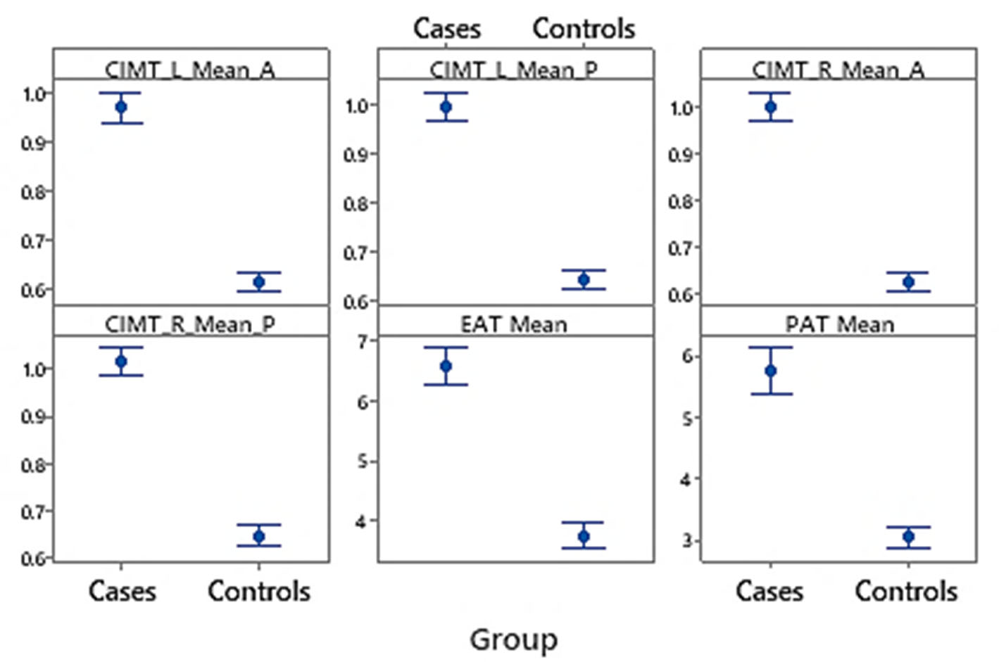
Cardiac troponin I was higher in the ACS group (p<0.001). CRP, fibrinogen, WBC, absolute neutrophil count and uric acid were also higher in ACS (all p<0.001) (Fig. 1).
When CIMT was summarized as a global mean value, the ACS group showed a mean CIMT of 1.02 mm versus 0.65 mm in controls (p<0.001). This represents a substantial absolute difference in carotid wall thickness between individuals with and without ACS. Cardiac adipose depots followed the same direction. Mean epicardial adipose tissue (EAT) thickness was 6.5 mm in ACS patients and 3.7 mm in controls (p<0.001), while mean pericardial adipose tissue (PAT) thickness was 5.8 mm versus 3.1 mm, respectively (p<0.001). All mean EAT and PAT measurements across the three cardiac cycles were significantly higher in the case group (p<0.001 for every comparison).
In multivariable binary logistic regression models adjusted for age, sex and BMI, all three imaging markers remained independently associated with ACS. CIMT showed a strong association with ACS (adjusted OR 3.52 per 0.1-mm increase, 95% CI 2.46-5.04, p<0.001). Similarly, higher epicardial adipose tissue thickness was independently associated with ACS (adjusted OR 2.17 per 1-mm increase, 95% CI 1.69-2.79, p<0.001), and pericardial adipose tissue thickness also remained independently associated (adjusted OR 2.77 per 1-mm increase, 95% CI 2.00-3.83, p<0.001). There was no evidence of problematic multicollinearity among predictors (all VIFs <2).
Variables Controls (n=150) Cases (n=150) Sex, n (%) Female 51 (34.0) 38 (25.3) Male 99 (66.0) 112 (74.7) Age, mean (SD), years 57.4±10.9 66.1±10.7 BMI, mean (SD), kg/m² 27.7±3.5 27.4±3.9 BMI category, n (%) Underweight 0 (0.0) 2 (1.3) Normal weight 27 (18.0) 33 (22.0) Overweight 84 (56.0) 81 (54.0) Obesity 39 (26.0) 34 (22.7) Place of residence, n (%) Rural 31 (20.7) 42 (28.0) Urban 119 (79.3) 108 (72.0)
In correlation analyses, both carotid thickness and cardiac fat were strongly linked to non-traditional risk markers. BMI showed a positive relationship with carotid wall thickness and with EAT and PAT. Across the cohort, ACS cases exhibited higher CIMT and EAT/PAT values than controls, suggesting that ACS is associated with a heavier structural and adipose burden beyond adiposity alone. All inflammatory and metabolic markers – serum uric acid, CRP, fibrinogen, WBC and neutrophils – demonstrated positive associations with carotid thickness across all four CIMT indicators, in both groups. Similarly, these markers were positively correlated with both EAT and PAT, in both groups, indicating that accumulation of epicardial and pericardial adipose tissue is closely linked to systemic inflammation, particularly among individuals with elevated cardiovascular risk (Fig. 2).
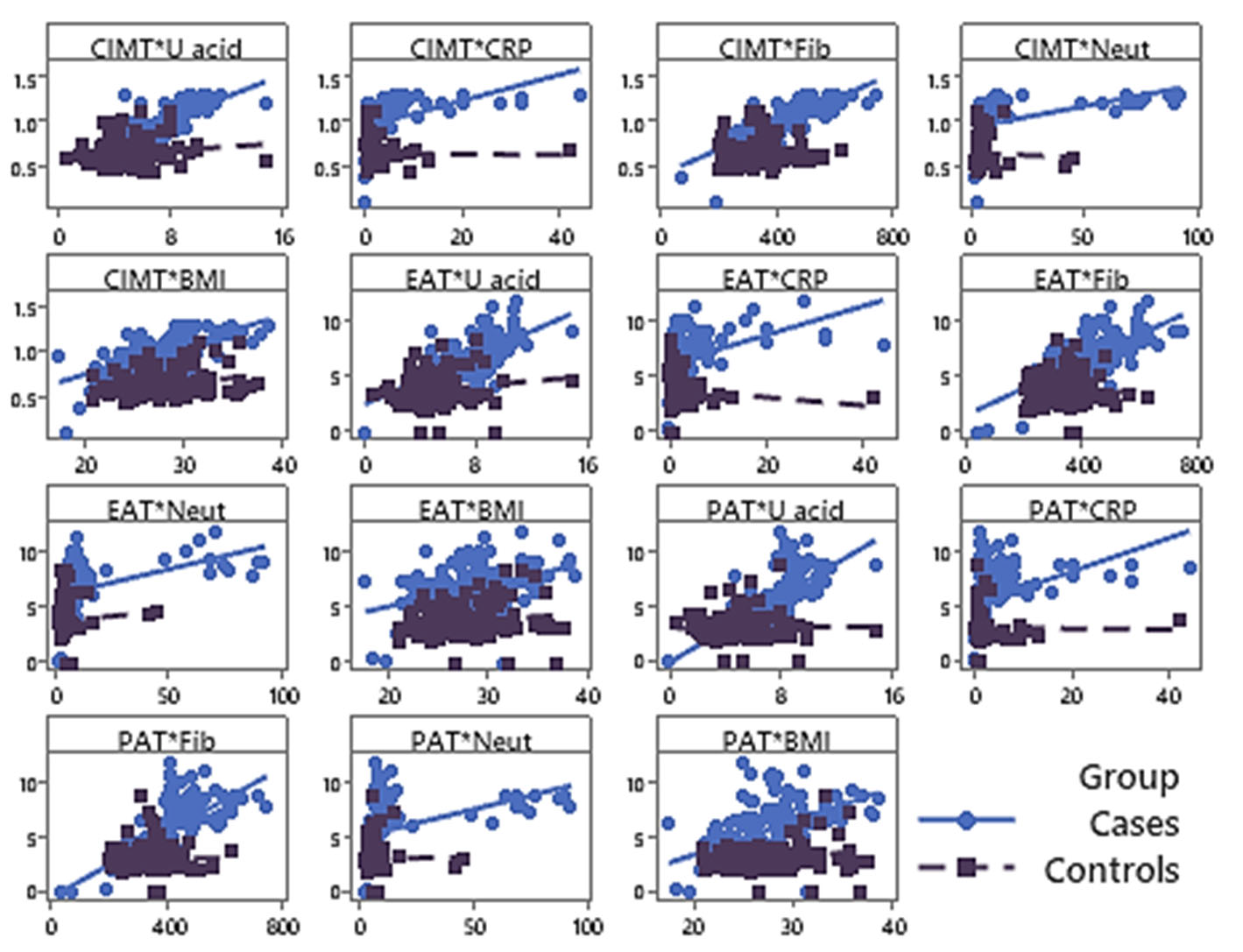
In clinical terms, these patterns indicate that non-traditional biochemical markers and structural indices (CIMT, EAT, PAT) cluster together: higher inflammatory and metabolic burden is paralleled by thicker carotid walls and greater epicardial/pericardial fat.
Discriminative performance for differentiating ACS from controls
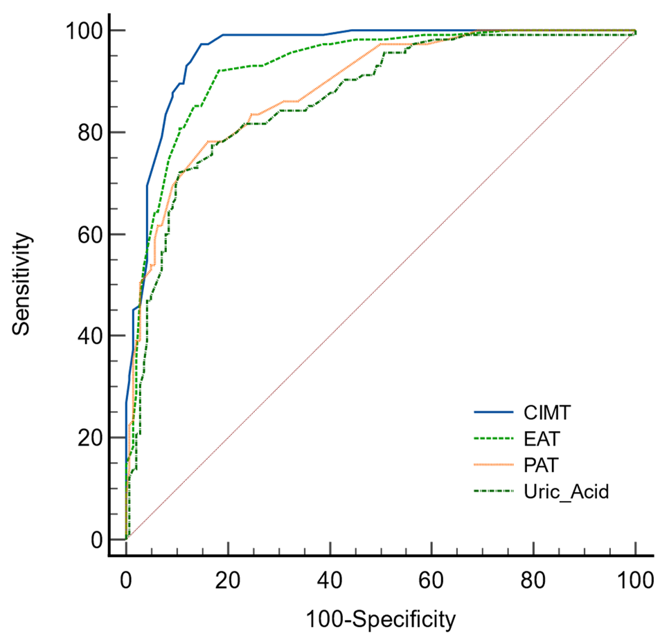
Receiver operating characteristic (ROC) analysis showed that structural and biochemical non-traditional markers provided strong discrimination between ACS patients and controls. Mean CIMT indicators demonstrated AUC values above 0.90, with sensitivity >93% and specificity >85%, and positive likelihood ratios up to 9.93, indicating outstanding discriminative performance within this cohort. A global CIMT cut-off around 0.72 mm emerged as a useful threshold for differentiating ACS from non-ACS individuals in this cohort; this cut-off should be externally validated before clinical use. Mean EAT and PAT thickness also showed strong discrimination, with AUC 0.902 and 0.875, respectively (both p<0.001). An EAT cut-off of approximately 4.8 mm showed high sensitivity in this cohort; this cut-off should be externally validated before clinical use (Fig. 3).
Among biochemical non-traditional markers, serum uric acid yielded an AUC of 0.860, with high specificity (around 88.9%) and a positive likelihood ratio of 6.46, although sensitivity (around 71.8%) was lower than that of the morphological parameters. Overall, the combination of increased CIMT, elevated EAT/PAT and higher levels of inflammatory/metabolic markers clearly distinguished ACS patients from controls. These markers showed good within-sample discrimination between cases and controls; cut-offs require external validation.
This case-control study demonstrates that patients with acute coronary syndrome exhibit a characteristic non-traditional risk profile that integrates structural vascular changes, cardiac adiposity and systemic inflammatory/metabolic activation. Compared with angiographically negative controls, ACS patients had strikingly higher CIMT, thicker epicardial and pericardial fat layers, and consistently elevated inflammatory and metabolic biomarkers. The discriminative performance of these markers – particularly CIMT and EAT/PAT was excellent, with CIMT AUC values above 0.90 and EAT AUC around 0.90 for discriminating ACS from non-ACS individuals. Taken together, the data support the concept of an “unconventional” structural – inflammatory phenotype that complements classical risk factors in ACS [12].
CIMT as a structural expression of cumulative risk
Our findings reinforce the role of carotid intima-media thickness as a robust marker of generalized atherosclerotic burden. The difference in mean CIMT between ACS patients and controls (1.02 mm vs 0.65 mm) is clinically meaningful and aligns with large meta-analyses showing that higher CIMT predicts myocardial infarction, stroke and cardiovascular death. The fact that all four CIMT indicators (left-right, anterior-posterior) were uniformly elevated among ACS cases suggests a diffuse pattern of arterial remodeling rather than focal changes limited to a single segmen [13].
From a risk-prediction standpoint, our ROC analyses confirm that CIMT carries substantial discriminative information for distinguishing ACS from non-ACS populations, with AUC values above 0.90 and very high likelihood ratios. This is consistent with previous work indicating that CIMT improves risk stratification when added to traditional risk scores, particularly in intermediate-risk individuals. In settings where advanced coronary imaging is not routinely available, standardised CIMT assessment – when measured using a standardized far-wall protocol – could therefore serve as a practical tool to capture cumulative vascular damage and identify patients with a heavier atherosclerotic profile.
Epicardial/pericardial fat and the inflammatory–metabolic milieu
The markedly higher EAT and PAT thickness in ACS patients, together with their strong correlation with inflammatory markers, highlight the importance of cardiac visceral adipose tissue as a metabolically active organ rather than a passive fat depot. Epicardial fat shares the coronary blood supply and is in direct anatomical continuity with the myocardium and coronary arteries. Experimental and clinical data show that EAT secretes pro-inflammatory cytokines and adverse adipokines and has reduced adiponectin production in obesity and metabolic syndrome, shifting its profile towards a pro-atherogenic, pro-inflammatory phenotype. These mechanisms provide a plausible biological link between increased EAT thickness and coronary plaque development, vulnerability and thrombosis [14,15].
Our results fit well with meta-analyses and cohort studies reporting that higher EAT thickness or volume is independently associated with coronary artery disease, plaque vulnerability and major adverse cardiac events. In the present study, mean EAT achieved an AUC of 0.902 for ACS discrimination, and values below approximately 4.8 mm were associated with a lower probability of ACS within this dataset. However, predictive values are prevalence-dependent and should not be extrapolated to clinical rule-out decisions without external validation in representative cohorts. PAT showed slightly lower but still very good discriminative performance, indicating that pericardial fat also captures adverse cardiometabolic burden, even though its direct paracrine impact on coronary arteries is less pronounced than that of EAT [16,17].
Biochemical non-traditional markers: uric acid and inflammation
The biochemical profile of ACS patients in this study is consistent with the structural findings. Fibrinogen, CRP, WBC and neutrophil counts were substantially higher in cases, and all these markers correlated positively with both CIMT and EAT/PAT. This reinforces the idea that low-grade systemic inflammation and structural atherosclerotic remodeling are tightly linked processes rather than independent domains [18, 19, 20].
Serum uric acid, in particular, emerged as a useful non-traditional marker, with an AUC of 0.860, high specificity and a positive likelihood ratio around 6.5 for ACS discrimination. Meta-analyses have reported that elevated uric acid is associated with higher risk of ACS and poorer short-term outcomes, even after adjustment for classical risk factors, supporting its role as a marker of oxidative stress and endothelial dysfunction. In our cohort, the positive correlations between uric acid, CIMT and EAT/PAT suggest that hyperuricaemia may be one of the biochemical signatures of the structural-inflammatory phenotype we describe [21].
Interplay between structural and non-traditional markers
A key message of this work is the interplay between structural measures (CIMT, EAT, PAT) and non-traditional biochemical markers, rather than their isolated performance. Higher BMI, inflammatory markers and uric acid levels clustered with thicker carotid walls and increased epicardial/pericardial fat, especially among ACS cases. This clustering pattern supports the view that what we often treat as separate domains – obesity, inflammation, structural atherosclerosis – are in practice different faces of the same underlying pathophysiology [22].
For clinical practice, this means that CIMT and EAT/PAT should not necessarily be seen merely as “add-on imaging tests” but as structural readouts of a broader inflammatory and metabolic environment. In patients with intermediate risk or with few classical risk factors, the presence of thickened CIMT and high EAT/PAT, together with elevated fibrinogen and uric acid, could justify more aggressive prevention or earlier imaging, even when traditional scores appear only moderately elevated [23].
Strengths and limitations
The study has several strengths and a clear novelty: it combines CIMT, echocardiographic EAT/PAT, and a targeted inflammatory – metabolic panel within the same ACS cohort, using angiography/CT-negative controls to minimise disease misclassification. This integrated design enables the description of a structural – inflammatory phenotype relevant for hypothesis generation and future risk stratification in a South-Eastern European, middle-income setting.
However, some limitations must be acknowledged. First, inter- and intra-observer reproducibility for CIMT and EAT/PAT measurements was not formally quantified, which may affect measurement precision. This is a single-centre, hospital-based case–control study with a moderate sample size, which may limit generalisability. The analyses are mainly unadjusted; therefore, residual confounding (including differences in age and sex) cannot be excluded, and the incremental value of each marker beyond conventional risk factors cannot be fully quantified. Biomarkers measured during the acute presentation may partly reflect the inflammatory response to the index event rather than baseline risk. Finally, ROC-derived cut-offs are cohort-specific and require external validation in independent cohorts. Positive and negative predictive values are prevalence-dependent and may not be directly generalisable due to the case–control design.
Clinical implications and future directions
Despite these limitations, the present findings have clear clinical implications. In resource-constrained environments where advanced coronary imaging is not universally available, simple non-invasive measures such as CIMT and EAT/PAT may enhance risk stratification, particularly when interpreted alongside inflammatory and metabolic markers such as fibrinogen, neutrophils and uric acid. Our findings support incorporating structural–inflammatory markers into risk stratification and phenotyping frameworks for patients with suspected ACS or those at high cardiovascular risk, particularly when conventional risk scores and routine tests leave residual uncertainty. In this context, CIMT and EAT/PAT may help identify residual risk and prioritise patients for intensified preventive strategies and/or further diagnostic testing, rather than serving as acute stand-alone diagnostic tests. These may complement existing clinical assessment and biomarker-based pathways, but should not be considered stand-alone diagnostic tools [8,12,13].
Future work should validate these findings in larger, prospective cohorts, evaluate the incremental prognostic value of combined CIMT/EAT – inflammation phenotypes for long-term outcomes and formally test phenotype-based approaches (structural, inflammatory, combined) for guiding personalised therapy. In parallel, implementation studies will be needed to define standardised protocols, training requirements and cost-effectiveness of incorporating CIMT and EAT/PAT into routine cardiovascular risk assessment.
CONCLUSIONS
- Patients with acute coronary syndrome demonstrate a distinct non-traditional cardiovascular risk profile characterized by increased carotid intima–media thickness, increased epicardial and pericardial adipose tissue thickness, and enhanced inflammatory and metabolic activity.
- Carotid intima-media thickness and cardiac adipose tissue thickness reflect structural and metabolic alterations that are closely associated with systemic inflammatory markers, including C-reactive protein, fibrinogen, leukocyte and neutrophil counts, serum uric acid levels, and body mass index.
- Carotid intima-media thickness shows excellent discriminative ability for the identification of acute coronary syndrome, while epicardial and pericardial adipose tissue thickness also demonstrate strong diagnostic performance.
- The combination of vascular imaging markers, cardiac adipose tissue assessment, and selected non-traditional biochemical markers may improve risk stratification beyond traditional cardiovascular risk factors in patients presenting with acute coronary syndrome.
- Further prospective studies are required to validate these structural and inflammatory phenotypes and to clarify their role in personalized cardiovascular risk assessment and prevention strategies.
Contributors:
Gishto T. – conceptualization, methodology, formal analysis, writing – original draft;
Rruci E. – resources, data curation;
Shuka N. – software, formal analysis;
Methoxha S. – investigation;
Vyshka G. – writing – review & editing.
Funding.Thisresearchreceivednoexternalfunding.
Conflict of interests. The authors declare no conflict of interest.
REFERENCES
- Byrne RA, Rossello X, Coughlan JJ, Barbato E, Berry C, Chieffo A, et al. 2023 ESC Guidelines for the management of acute coronary syndromes. Eur Heart J. 2023;44(38):3720-826. doi: https://doi.org/10.1093/eurheartj/ehad191
- Thygesen K, Alpert JS, Jaffe AS, et al. Fourth universal definition of myocardial infarction. Circulation. 2018;138(20):e618-e651. doi: https://doi.org/10.1161/CIR.0000000000000617
- Saito Y, Tsujita K, Kobayashi Y. Acute myocardial infarction without standard modifiable cardiovascular risk factors: prevalence, mechanisms and prognosis. Cardiovasc Interv Ther. 2024;39(4):403-11. doi: https://doi.org/10.1007/s12928-024-01022-4
- Libby P. The changing landscape of atherosclerosis. Nature. 2021;592:524-33. doi: https://doi.org/10.1038/s41586-021-03392-8
- Ling Y, Wan Y, Barinas-Mitchell E, et al. Varying definitions of carotid intima-media thickness and future cardiovascular disease: a systematic review and meta-analysis. J Am Heart Assoc. 2023;12:e031217. doi: https://doi.org/10.1161/JAHA.123.031217
- Mitra S, Biswas RK, Jung M, et al. Carotid intima-media thickness, cardiovascular disease, and risk factors in 29,000 UK Biobank adults. Am J Prev Cardiol. 2025;22:101011. doi: https://doi.org/10.1016/j.ajpc.2025.101011
- Naylor R, Rantner B, Ancetti S, de Borst GJ, De Carlo M, et al. Editor’s choice – european society for vascular surgery (ESVS). 2023 Clinical practice guidelines on the management of atherosclerotic carotid and vertebral artery disease. Eur J Vasc Endovasc Surg. 2023;65(1):7-111. doi: https://doi.org/10.1016/j.ejvs.2022.04.011
- Albricker ACL, Biagioni LC, dos Santos SN, Freire CMV, et al. Recommendation update for vascular ultrasound evaluation of carotid and vertebral artery disease: DIC, CBR and SBACV – 2023. Arq Bras Cardiol. 2023;120(10):e20230695. doi: https://doi.org/10.36660/abc.20230695
- Demmert M, Klambauer K, Moser LJ, Mergen V, Eberhard M, Alkadhi H. Epicardial and pericardial adipose tissue: anatomy, physiology, imaging, segmentation, and treatment effects. Br J Radiol. 2025 Sep 13:tqaf223. doi: https://doi.org/10.1093/bjr/tqaf223
- Hanley JA, McNeil BJ. The meaning and use of the area under a receiver operating characteristic (ROC) curve. Radiology. 1982;143(1):29-36. doi: https://doi.org/0.1148/radiology.143.1.7063747
- Iacobellis G, Malavazos AE. Epicardial adipose tissue in contemporary cardiology. Nat Rev Cardiol. 2022;19:593-606.
doi: https://doi.org/10.1038/s41569-022-00679-9 - Li C, Liu X, Adhikari BK, et al. The role of epicardial adipose tissue dysfunction in cardiovascular diseases: an overview of pathophysiology, evaluation, and management. Front Endocrinol (Lausanne). 2023;14:1167952. doi: https://doi.org/10.3389/fendo.2023.1167952
- Abbassi M, Besbes B, Elkadri N, et al. Characterization of epicardial adipose tissue thickness and structure by ultrasound radiomics in acute and chronic coronary patients. Int J Cardiovasc Imaging. 2025;41:477-88. doi: https://doi.org/10.1007/s10554-025-03329-6
- Chong B, Jayawardana R, Wong SM, et al. Epicardial adipose tissue and coronary artery disease: a systematic review and meta-analysis. Circ Cardiovasc Imaging. 2023;16:e015159. doi: https://doi.org/10.1161/CIRCIMAGING.122.015159
- Napoli G, Pergola V, Basile P, De Feo D, Bertrandino F, Baggiano A, et al. Epicardial and pericoronary adipose tissue, coronary inflammation, and acute coronary syndromes. J Clin Med. 2023;12(23):7212. doi: https://doi.org/10.3390/jcm12237212
- Jehn M, Thiele H, Abdelhady M, et al. Epicardial adipose tissue and obstructive coronary artery disease in acute chest pain: the EPIC-ACS study. Eur Heart J Open. 2023;3(3):oead041. doi: https://doi.org/10.1093/ehjopen/oead041
- Rong J, Fang C, Chen X, Hong C, Huang L. Association of serum uric acid with prognosis in patients with myocardial infarction: an update systematic review and meta-analysis. BMC Cardiovasc Disord. 2023;23(1):512. doi: https://doi.org/10.1186/s12872-023-03523-1
- Wang JJ, Yi JK, Zhou LR, Chen J, Zhang BQ, Huang HM, et al. A systematic review of the connection between serum uric acid levels and the risk of cardiovascular disease. Front Cardiovasc Med. 2025;12:1577952. doi: https://doi.org/10.3389/fcvm.2025.1577952
- Liu Y, Li Z, Xu Y, Mao H, Huang N. Uric acid and atherosclerosis in patients with chronic kidney disease: recent progress, mechanisms, and prospect. Kidney Dis. 2025;11(1):112-27. doi: https://doi.org/10.1159/000543781
- Islam F, Nukala SK, Shrestha P, et al. Air pollution and cardiovascular disease: a systematic review of the effects of air pollution, including bushfire smoke, on cardiovascular disease. Am Heart J Plus. 2025;54:100546. doi: https://doi.org/10.1016/j.ahjo.2025.100546
- Powell-Wiley TM, Poirier P, Burke LE, et al. Social determinants of cardiovascular disease. Circ Res. 2022;130(5):782-99. doi: https://doi.org/10.1161/CIRCRESAHA.121.319811
- Yeghiazarians Y, Jneid H, Tietjens JR, et al. Obstructive sleep apnea and cardiovascular disease: a scientific statement from the American Heart Association. Circulation. 2021;144(3):e56-e67. doi: https://doi.org/10.1161/CIR.0000000000000988
- Lopez-Jimenez F, Almahmeed W, Bays H, Cuevas A, Di Angelantonio E, le Roux CW, et al. Obesity and cardiovascular disease: mechanistic insights and management strategies. A joint position paper by the World Heart Federation and World Obesity Federation. Eur J Prev Cardiol. 2022;29(17):2218-37. doi: https://doi.org/10.1093/eurjpc/zwac187