

 UK
 EN


Mamai O.V.
 Bilash S.M.
Pronina O.M.
Bieliaieva O.M.
Pirog-Zakaznikova A.V.
Koptev M.M.
Bilash V.P.
Bilanov O.S.
Oliinichenko Ya.O.
Donchenko S.V.
Oleksiienko V.V.
Kolomiiets S.V.
Mazur B.R.
Key words: spleen, food additives, oxidative stress, morphology, cell proliferation, B lymphocytes, experiment, rats
Ключові слова: селезінка, харчові добавки, оксидативний стрес, морфологія, проліферація клітин,
В-лімфоцити, експеримент, щури
Abstract
Morphofunctional characteristics of remodeling processes in the structural components of the rat spleen during prolonged exposure to a mixture of food additives. Mamai O.V., Bilash S.M., Pronina O.M., Bieliaieva O.M., Pirog-Zakaznikova A.V., Koptev M.M., Bilash V.P., Bilanov O.S., Oliinichenko Ya.O., Donchenko S.V., Oleksiienko V.V., Kolomiiets S.V., Mazur B.R. The aim of the study was to determine the morphofunctional characteristics of spleen remodeling in rats during prolonged oral administration of a mixture of food additives at subtoxic doses. The study included 70 Wistar rats, with equal numbers of males and females, divided into seven groups of 10 animals each: one week-0 group and six experimental groups. The animals received monosodium glutamate at a dose of 20 mg/kg, Ponceau 4R at a dose of 5 mg/kg, and sodium nitrite at a dose of 0.6 mg/kg and were euthanized after 1, 4, 8, 12, 16, or 20 weeks by sodium thiopental overdose under ether anesthesia. Superoxide dismutase and catalase activities and malondialdehyde and nitrite concentrations were measured spectrophotometrically in a 10% spleen homogenate; Ki-67-positive and CD20-positive cells were evaluated immunohistochemically. Data were presented as mean±standard deviation and analyzed using the Shapiro-Wilk test, Welch analysis of variance, the Kruskal-Wallis test, Holm-adjusted pairwise comparisons, quadratic regression, and Spearman rank correlation. At week 12, superoxide dismutase and catalase activities were 58.5% and 57.8% lower than in the week-0 group, respectively, whereas malondialdehyde concentration increased 7.23-fold. Nitrite concentration peaked at week 8 and was 83.3% above the level in the week-0 group. The number of Ki-67-positive cells was lowest at week 16, (by 90.7% below that in the week-0 group); the number of CD20-positive cells increased by 56.6% at week 8 and decreased by 28.1% at week 20, with Holm-adjusted p<0.05 for all reported comparisons. Thus, prolonged exposure to the mixture of food additives was accompanied by oxidative-nitrosative stress and exposure duration-dependent changes in proliferative activity and the B-cell component of the spleen.
Реферат
Морфофункціональні особливості процесів ремоделювання структурних компонентів селезінки щурів при тривалому впливі комплексу харчових добавок. Мамай О.В., Білаш С.М., Проніна О.М., Бєляєва О.М., Пирог-Заказнікова А.В., Коптев М.М., Білаш В.П., Біланов О.С., Олійніченко Я.О., Донченко С.В., Олексієнко В.В., Коломієць С.В., Мазур Б.Р. Метою дослідження було встановити морфофункціональні особливості ремоделювання селезінки щурів за умов тривалого перорального введення комплексу харчових добавок у субтоксичних дозах. Дослідження виконано на 70 щурах лінії Вістар обох статей порівну, розподілених на сім груп по 10 тварин: групу 0-го тижня та шість експериментальних. Тварини отримували глутамат натрію в дозі 20 мг/кг, Понсо 4R у дозі 5 мг/кг та нітрит натрію в дозі 0,6 мг/кг; їх евтаназували після 1, 4, 8, 12, 16 або 20 тижнів шляхом передозування тіопенталу натрію під ефірним наркозом. У 10% гомогенаті селезінки спектрофотометрично визначали активність супероксиддисмутази й каталази, концентрацію малонового діальдегіду та нітритів; кількість Ki-67- і CD20-позитивних клітин оцінювали імуногістохімічно. Дані наведено як середнє±стандартне відхилення; застосовано критерії Шапіро–Вілка, Уелча та Краскела–Уолліса, попарні порівняння з поправкою Холма, квадратичну регресію і рангову кореляцію Спірмена. На 12-му тижні активність супероксиддисмутази й каталази була нижчою за показник групи нульового тижня відповідно на 58,5% і 57,8%, а концентрація малонового діальдегіду зросла в 7,23 раза. Найвищу концентрацію нітритів зареєстровано на 8-му тижні (на 83,3% вище рівня групи нульового тижня). Кількість Ki-67-позитивних клітин була найнижчою на 16-му тижні (на 90,7% нижче за групу нульового тижня); кількість CD20-позитивних клітин на 8-му тижні зросла на 56,6%, а на 20-му тижні зменшилася на 28,1% (для всіх наведених порівнянь скориговане за Холмом p<0,05). Отже, тривалий вплив комплексу харчових добавок супроводжувався оксидативно-нітрозативним стресом і залежними від тривалості експозиції змінами проліферативної активності та В-клітинного компонента селезінки.
Natural and synthetic food additives are widely used in modern food production to improve the taste, color, consistency, shelf life, and commercial appearance of products [1, 2]. In a usual diet, a person consumes several foods at the same time, each of which may contain its own set of technological ingredients. Consequently, the body is exposed not only to individual compounds, but also to their combinations.
The safety of most food additives is assessed primarily when they are used separately in regulated amounts. At the same time, the toxicological effects of mixtures may differ from those of the individual components and depend on composition, dose, and duration of intake [3, 4]. Current reviews and experimental studies also indicate that some food additives may affect the intestinal barrier, microbiota, redox status, and immune response [23, 24]. Prolonged combined exposure to low doses of additives with different functional purposes requires particular attention because gradual metabolic and cellular changes may develop without signs of acute intoxication.
The spleen is the largest peripheral organ of the immune system and combines filtration, circulatory, and immunoregulatory functions. Prolonged metabolic or pro-oxidant load may alter the state of its lymphoid tissue, cellular proliferative activity, and tissue redox balance. Oxidative-nitrosative stress is regarded as one of the universal mechanisms of cellular injury and impaired immune response [5, 6].
Previous studies using this experimental model separately established impaired antioxidant defense in the splenic parenchyma and changes in its macrophage component [7, 8]. However, it remains insufficiently understood how biochemical manifestations of oxidative-nitrosative stress are combined with changes in proliferative activity and the B-cell component of lymphoid tissue at different durations of food additive mixture administration.
Therefore, a comprehensive study of biochemical and immunohistochemical changes in the spleen during prolonged combined exposure to food additives is relevant for clarifying the characteristics of remodeling in peripheral organs of the immune system.
The study parameters were selected to permit simultaneous assessment of the pro-oxidant-antioxidant balance and cellular changes in the spleen. Superoxide dismutase and catalase activities characterize the enzymatic component of antioxidant defense, malondialdehyde concentration reflects the intensity of lipid peroxidation, and nitrite concentration is an integrated indicator of changes in nitric oxide metabolism [5]. Ki-67 was used to assess the proliferative activity of cells in the splenic red pulp, and CD20 to characterize the B-cell component of lymphoid tissue in the splenic red pulp [20, 21]. Combined measurement of these parameters made it possible to compare redox changes with the dynamics of proliferative activity and the B-cell component of the splenic red pulp.
The aim of this study was to determine the morphofunctional characteristics of remodeling in the structural components of the rat spleen during prolonged exposure to a mixture of food additives at subtoxic doses, based on biochemical parameters and immunohistochemical expression of Ki-67 and CD20.
MATERIALS AND METHODS OF RESEARCH
The study was conducted on 70 white laboratory Wistar rats, with equal numbers of both sexes, aged 6 months at the beginning of the experiment and weighing (190±1.34) g. The animals were kept under standard conditions in the vivarium of Poltava State Medical University. The rats were housed four per standard cage measuring 90×60×120 cm on corn bedding. Room temperature was maintained at (22±2)°C, relative humidity at (50±10)%, and the light cycle at 12 h light/12 h dark. The study was conducted from March to September 2024.
The animals were divided into seven groups of 10 animals each: one week-0 group and six experimental groups. Every morning before the main feeding, animals in the experimental groups received 0.5 mL of a freshly prepared solution of the food additive mixture orally through a soft gavage tube for 1-20 weeks. The animals had free access to drinking water and the standard vivarium diet. The solution contained monosodium glutamate (E621) at a dose of 20 mg/kg (Meihua Holdings Group Co., Ltd., China), Ponceau 4R (E124) at a dose of 5 mg/kg (Ajanta Chemical Industries, India), and sodium nitrite (E250) at a dose of 0.6 mg/kg (BASF, Germany). These substances were selected because they belong to different functional classes: monosodium glutamate is a flavor enhancer, Ponceau 4R is a synthetic colorant, and sodium nitrite is a preservative. The regulatory basis for selecting the initial levels comprised Order No. 45 of the Ministry of Health of Ukraine dated 08.01.2024 [13] and the Requirements for Food Additives approved by this order [14], which define the general conditions of use, functional classes, and maximum levels of food additives in foods. The experimental doses were 50% of the corresponding initial levels adopted in the approved study model. Interspecies conversion to rat body weight was performed using biological activity coefficients according to the Rybolovlev formula: D_rat = D_human ×3.62/0.57=D_human × 6.35 [15]. The animals were euthanized after 0, 1, 4, 8, 12, 16, or 20 weeks by sodium thiopental overdose under ether anesthesia. Taking the duration of the experiment into account, age at euthanasia ranged from 6 months in the week-0 group to 10.5 months in the week-20 group. No separate concurrent age-matched control groups or gavage control groups receiving water or saline at the corresponding time points were formed. After euthanasia, the spleen was removed, rinsed with saline, and used for biochemical and immunohistochemical examination.
The experimental studies were conducted in accordance with the General Ethical Principles for Experiments on Animals (Kyiv, 2001), Law of Ukraine No. 3447-IV dated 21.02.2006, On the Protection of Animals from Cruelty, as amended, and the Procedure for Conducting Experiments on Animals by Scientific Institutions, approved by Order No. 249 of the Ministry of Education and Science, Youth and Sports of Ukraine dated 01.03.2012. The study design was approved by the Biomedical Ethics Commission of Poltava State Medical University, protocol No. 219 dated 21.09.2023.
The following were determined in a 10% spleen homogenate: superoxide dismutase activity, based on the ability of the sample to inhibit epinephrine auto-oxidation in an alkaline medium at 26°C, with the result expressed in arbitrary units [16]; catalase activity, based on the amount of hydrogen peroxide decomposed in the presence of the sample after reaction with ammonium molybdate at a wavelength of 405 nm, with the result expressed in μkat/g [17]; malondialdehyde concentration, determined spectrophotometrically by reaction with 1-methyl-2-phenylindole at 586 nm, with the result expressed in μmol/g tissue [18]; nitrite concentration, determined by the Griess-Ilosvay reaction with photometry at 530 nm, with the result expressed in nmol/g tissue [19].
Immunohistochemical examination was performed on 3-4 μm formalin-fixed, paraffin-embedded spleen sections using a standard indirect immunoperoxidase method and antibodies validated by the manufacturer for immunohistochemistry of paraffin-embedded rat tissues [20, 21]. After standard preparation of the sections, recombinant rabbit Anti-CD20 antibody, clone SP32 (ab64088, Abcam, Cambridge, UK) [20], and Anti-Ki-67 antibody, clone SP6 (ab16667, Abcam, Cambridge, UK) [21], were applied. Visualization was performed using the Mouse/Rabbit PolyVue™ HRP/DAB polymer system (Diagnostic BioSystems, USA), and the sections were counterstained with Mayer's hematoxylin (Biognost, Croatia). Microscopy was performed using an Olympus C 3040-ADU light microscope with Olympus DP-Soft software. Ki-67-positive and CD20-positive cells were counted in 10 non-overlapping fields of the red pulp using a ×40 objective. For each animal, the mean number of positive cells in the red pulp was calculated in10 fields of view.
Statistical analysis was performed at the level of the individual rat as the experimental unit using IBM SPSS Statistics 26.0 (IBM Corp., Armonk, NY, USA, 2020; subscription identifier 515892337). Distribution normality was assessed using the Shapiro-Wilk test. Data are presented as mean±standard deviation (M±SD). Differences between groups were assessed using Welch analysis of variance for seven independent groups, and the Kruskal-Wallis test was used under nonparametric conditions. Pairwise comparisons of each experimental group with the week-0 group were performed using Welch's t-test for two independent samples, with Holm correction of p-values [22]. To assess the pattern of temporal dynamics, exposure duration was analyzed as an ordered quantitative variable (0, 1, 4, 8, 12, 16, and 20 weeks) in models that included linear and quadratic terms. Exploratory associations between parameters were assessed using Spearman's rank correlation coefficient [22]. All statistical tests were two-sided, and differences were considered statistically significant at p<0.05.
RESULTS AND DISCUSSION
For all study groups, mean biochemical values and mean numbers of positive cells in 10 non-overlapping fields of view in the splenic red pulp were calculated (Table 1).
Outcome Week 0 Week 1 Week 4 Week 8 Week 12 Week 16 Week 20 SOD, a.u. 10.42±0.76 8.55±0.71* 6.79±0.45* 7.50±0.51* 4.32±0.24* 5.77±0.35* 5.10±0.90* Catalase, μkat/g 0.500±0.002 0.487±0.002* 0.453±0.004* 0.474±0.002* 0.211±0.004* 0.297±0.002* 0.248±0.004* MDA, μmol/g 0.43±0.14 0.81±0.14* 1.66±0.12* 1.38±0.16* 3.11±0.33* 1.72±0.16* 2.27±0.17* Nitrites, nmol/g 0.042±0.004 0.069±0.013* 0.054±0.009* 0.077±0.018* 0.071±0.004* 0.050±0.005* 0.061±0.011* Ki-67+cells in 10 fields of view 77.1±22.1 38.2±11.8* 30.5±6.5* 18.5±6.9* 46.5±11.0* 7.2±2.4* 46.6±10.6* CD20+cells in 10 fields of view 24.2±3.5 32.3±4.2* 27.3±4.5 37.9±2.8* 30.4±1.7* 21.3±1.9 17.4±2.7* Notes: data are presented as M±SD, where M is the mean and SD is the standard deviation; n=10 in each group; SOD – superoxide dismutase; MDA – malondialdehyde; Ki-67+ - Ki-67-positive cells; CD20+ - CD20-positive cells; * – p<0.05 versus the week-0 group after Holm correction for multiple comparisons.
The overall between-group effect was statistically significant for all study parameters according to Welch analysis of variance and was confirmed by the Kruskal-Wallis test (Table 2).
Outcome Welch Welch Kruskal- Kruskal- R² β p β p SOD, a.u. 160.28 <0.001 64.03 <0.001 0.752 -0.5724 <0.001 0.0178 <0.001 Catalase, μkat/g 15590.61 <0.001 67.63 <0.001 0.757 -0.0188 <0.001 0.0002 0.180 MDA, μmol/g 190.75 <0.001 65.49 <0.001 0.653 0.2489 <0.001 -0.0085 <0.001 Nitrites, nmol/g 50.34 <0.001 44.48 <0.001 0.159 0.0032 <0.001 -0.0002 <0.001 Ki-67+ cells 66.36 <0.001 55.25 <0.001 0.354 -6.9477 <0.001 0.3027 <0.001 CD20+ cells 62.95 <0.001 56.37 <0.001 0.585 1.6925 <0.001 -0.1124 <0.001 Notes: F – Welch analysis of variance statistic; H – Kruskal-Wallis statistic; p – significance level; R² – coefficient of determination; β– regression coefficient; week² – quadratic term of the model; p<0.05 – statistically significant result.
F
p
Wallis H
Wallis p
trend
week
week
week²
week²
All experimental groups showed lower superoxide dismutase and catalase activities and higher malondialdehyde and nitrite concentrations than the week-0 group. The most pronounced changes in the antioxidant system were found at week 12 – superoxide dismutase and catalase activities were by 58.5% and 57.8% lower than in the week-0 group, respectively, and malondialdehyde concentration increased 7.23-fold. The highest nitrite concentration was recorded at week 8, 83.3% above the level in the week-0 group. At weeks 16 and 20, the biochemical parameters remained significantly altered, indicating persistent oxidative-nitrosative disturbances.
Immunohistochemical examination revealed divergent dynamics of the two markers. The number of Ki-67-positive cells was lower than in the week-0 group at all observation times, and the lowest value was recorded at week 16 – 90.7% below the value in the week-0 group.
The number of CD20-positive cells changed in a phase-dependent manner. It increased at the early time points, reaching a maximum at week 8 (+56.6% compared with week 0), and subsequently decreased. At week 20, the value was 28.1% lower than at week 0.
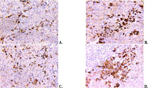
The results of light microscopic analysis of sections stained for Ki-67 and CD20 were consistent with the quantitative data. In the series of CD20 micrographs, denser clusters of positive cells were observed in the red pulp at the early time points (Fig. 1A, B), whereas their number decreased at the later time points (Fig. 1C, D).
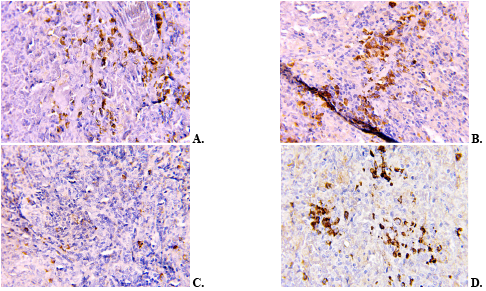
The series of Ki-67 micrographs showed an overall decrease in the number of positive nuclei (Fig. 2A, B), most pronounced at week 16 (Fig. 2C). At week 20, the number of positive cells was higher than at week 16, but remained lower than in the week-0 group (Fig. 2D).
Quadratic models of temporal dynamics showed a statistically significant quadratic component for superoxide dismutase activity, malondialdehyde and nitrite concentrations, and the numbers of Ki-67-positive and CD20-positive cells in the red pulp of the rat spleen. This confirmed the nonlinear pattern of changes during the experiment. The heatmap of standardized mean values reflected the sequential change in the relationships among the study parameters (Fig. 3).
SOD – superoxide dismutase; MDA – malondialdehyde; Ki-67+ - Ki-67-positive cells; CD20+ - CD20-positive cells; Z-score - standardized deviation of the group mean from the overall mean, expressed in standard deviation units. Positive values indicate a parameter level above the overall mean, and negative values indicate a level below it. 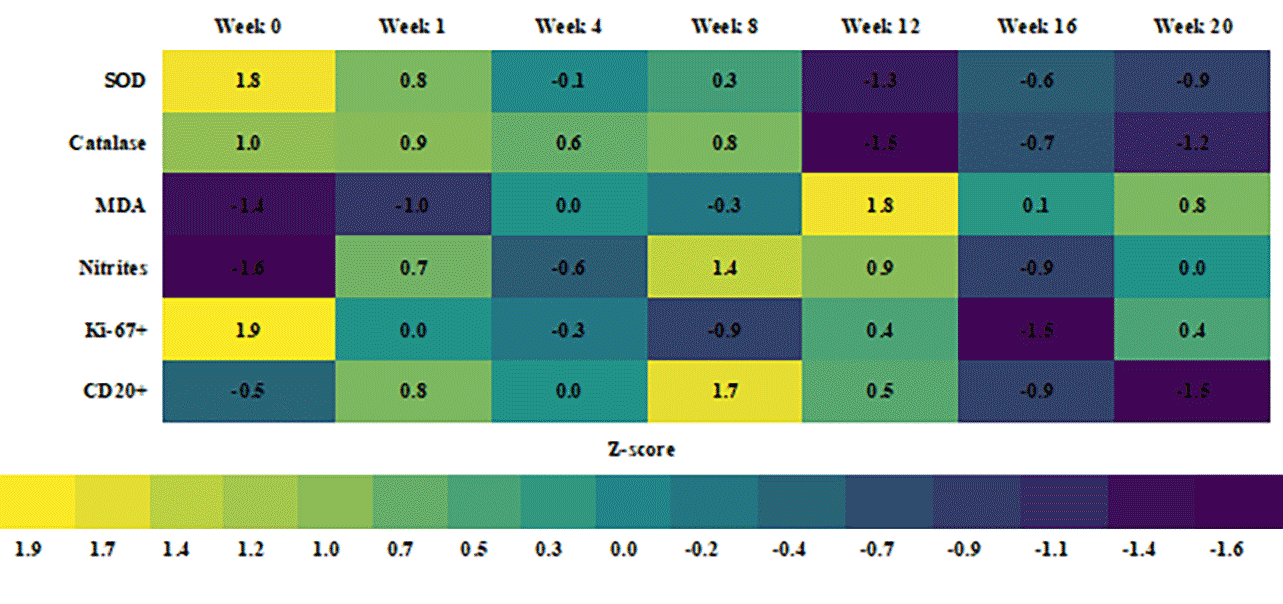
Exploratory analysis using Spearman's rank correlation coefficient at the level of individual animals revealed direct relationships between the measures of enzymatic antioxidant defense and inverse associations between antioxidant enzyme activities, concentrations of lipid peroxidation products, and immunohistochemical parameters. Given the nonlinear temporal dynamics, these correlations were regarded as an additional description of the relationships rather than independent evidence of causality.
A synthesis of the data made it possible to provisionally distinguish three consecutive periods of change. In the early period (weeks 1-8), the development of oxidative-nitrosative disturbances was accompanied by an increase in the number of CD20-positive cells. The most pronounced biochemical changes were observed at week 12. In the late period (weeks 16-20), antioxidant defense remained reduced, the number of CD20-positive cells decreased, and proliferative activity was most markedly suppressed.
The observed decrease in superoxide dismutase and catalase activities against a background of increased malondialdehyde concentration is consistent with reports of redox-dependent injury to lymphoid organs after exposure to monosodium glutamate. Das et al. showed that four weeks of monosodium glutamate administration was accompanied by oxidative changes and signs of immune dysfunction in the rat thymus and spleen [9]. The similar direction of the changes supports the assumption that impairment of enzymatic antioxidant defense is one of the factors involved in remodeling of the cellular composition of the spleen.
Recent studies of Ponceau 4R also describe biological effects associated with impaired cell proliferation and redox balance. Kara et al. found a decrease in the mitotic index with increasing Ponceau 4R concentration, whereas signs of genotoxicity were detected mainly at high concentrations [10]. In an experimental model using Artemia franciscana, E124 altered the activity of mitochondrial respiratory chain components and the redox state [11]. Differences between the models preclude direct extrapolation of these results to the rat spleen, although they are consistent with the combination of redox disturbances and a lower number of Ki-67-positive cells observed in our study.
Sodium nitrite can enhance the formation of reactive nitrogen species and secondary lipid peroxidation. In the study by Ahmed et al., its administration to rats was accompanied by an increase in malondialdehyde concentration and a decrease in antioxidant enzyme activities in the tissues examined [12]. In our study, the peak nitrite concentration at week 8 preceded the maximum increase in malondialdehyde at week 12, which may reflect a temporal sequence of nitrosative and lipid peroxidation changes.
The time difference between the maximum manifestations of oxidative-nitrosative stress at week 12 and the lowest number of Ki-67-positive cells at week 16 may reflect delayed suppression of proliferation due to accumulated redox-dependent injury. The increase in this parameter at week 20 was probably associated with compensatory and adaptive renewal of cell populations [9].
The effect of the mixture should not be regarded as a simple sum of the effects of its components. Studies of food additives in combination show that the cellular response to a mixture may differ from the response to individual substances [3], and combined intake of common additives in an animal model was accompanied by simultaneous changes in oxidative status, the inflammatory response, and immune cells [4]. Reviews of the effects of food additives on intestinal microecology and experimental data on interactions between the intestinal barrier and the immune system confirm that the effects of some technological ingredients may extend beyond the organ of initial contact [23, 24]. The results of this study complement previously established changes in antioxidant defense [7] and the macrophage component of the spleen [8] in the same experimental model by linking them to the dynamics of Ki-67-positive and CD20-positive cells.
The combination of reduced proliferative activity and a late decrease in the number of CD20-positive cells may indicate functional suppression and remodeling of the B-cell component of the spleen. These dynamics may be associated with disruption of the stromal-follicular microenvironment that supports the organization of B-cell follicles [25]. At the same time, lower CD20 expression may reflect not only a decrease in the number of mature B cells, but also altered marker expression or their differentiation into CD20-negative plasma cells [26].
A separate limitation of this study is the absence of daily gavage control groups. Because animals of the same initial age were euthanized at different time points, their age at euthanasia differed. All study time points fell within the adult period of the rats' lifespan; however, in the absence of concurrent age-matched control groups, the contribution of age to the observed biochemical and immunohistochemical changes cannot be fully excluded.
Repeated restraint and gavage administration of the solution may potentially induce a short-term stress response, the magnitude of which depends on the technical performance of the procedure and the volume and properties of the solution. In a separate study in male Sprague-Dawley rats acclimated to handling, daily orogastric administration of water at a volume of 5 mL/kg for 28 days, when the procedure was performed properly, was not accompanied by signs of chronic stress [27]. In our study, the initial dosing volume was 2.6 mL/kg; however, exposure lasted up to 20 weeks, and specific markers of the stress response were not measured. Therefore, the contribution of repeated gavage and associated handling stress cannot be qualitatively or quantitatively separated from other factors.
CONCLUSIONS
- Prolonged oral administration of a mixture of food additives (monosodium glutamate, Ponceau 4R, and sodium nitrite) at subtoxic doses was accompanied by the development of persistent oxidative-nitrosative stress in rat spleen tissue, manifested by lower superoxide dismutase and catalase activities and higher malondialdehyde and nitrite concentrations.
- The most pronounced changes in free radical oxidation and lipid peroxidation were observed at week 12 of administration of the food additive mixture, whereas the highest nitrite concentration was recorded at week 8.
- The number of cells representing the B-cell component changed in a phase-dependent manner: the increase at the early time points was followed by a subsequent decrease, most pronounced at week 20, which may indicate functional suppression or reduced marker expression during differentiation into plasma cells.
- The number of proliferatively active cells was lower than the control value at all time points of the experiment; the most pronounced decrease in proliferative activity was observed at week 16 as a result of delayed redox-dependent injury, with partial compensatory and adaptive recovery at week 20.
- Splenic remodeling during prolonged exposure to the food additive mixture followed a nonlinear pattern dependent on exposure duration and combined early reactive changes with a subsequent decrease in proliferative activity and the B-cell component at the later time points.
Contributors:
Mamai O.V. – conceptualization, investigation, writing – original draft;
Bilash S.M. – conceptualization, project supervision, methodology;
Pronina O.M. – project administration;
Bieliaieva O.M. – writing, review and editing;
Pirog-Zakaznikova A.V. – methodology;
Koptev M.M. – data curation;
Bilash V.P., Donchenko S.V. – investigation;
Bilanov O.S., Mazur B.R. – visualization;
Oliinichenko Ya.O. – resources;
Oleksiienko V.V. – validation;
Kolomiiets S.V. – formal analysis;
Funding.The study was conducted as part of the investigator-initiated research project of the Department of Anatomy with Clinical Anatomy and Operative Surgery, Poltava State Medical University, entitled “Pathogenetic mechanisms of post-stress disorders under exogenous and wartime influences and the search for methods of their correction” (state registration No. 0124U003313).
Conflict of interests. The authors declare no conflict of interest.
REFERENCES
- Amchova P, Siska F, Ruda-Kucerova J. Food safety and health concerns of synthetic food colors: an update. Toxics. 2024;12(7):466. doi: https://doi.org/10.3390/toxics12070466
- de Oliveira ZB, Silva da Costa DV, da Silva dos Santos AC, da Silva Júnior AQ, de Lima Silva A, de Santana RCF, et al. Synthetic colors in food: a warning for children’s health. Int J Environ Res Public Health. 2024;21(6):682. doi: https://doi.org/10.3390/ijerph21060682
- Recoules C, Touvier M, Pierre F, Audebert M. Evaluation of the toxic effects of food additives, alone or in mixture, in four human cell models. Food Chem Toxicol. 2025;196:115198. doi: https://doi.org/10.1016/j.fct.2024.115198
- Gomaa S, Mohamed N, Nassef M. Frequently utilized food additives contribute to immune system dysfunction, chronic inflammation and oxidative stress. Drug Chem Toxicol. 2026 Jun 8:1-14. Online ahead of print. doi: https://doi.org/10.1080/01480545.2026.2681174
- Shannon OM, Easton C, Shepherd AI, Siervo M, Bailey SJ, Clifford T. Dietary nitrate and population health: a narrative review of the translational potential of existing laboratory studies. BMC Sports Sci Med Rehabil. 2021;13(1):65. doi: https://doi.org/10.1186/s13102-021-00292-2
- Pant A, Dasgupta D, Tripathi A, Pyaram K. Beyond antioxidation: Keap1-Nrf2 in the development and effector functions of adaptive immune cells. Immunohorizons. 2023;7(4):288-98. doi: https://doi.org/10.4049/immunohorizons.2200061
- Bilash SM, Mamai OV, Pronina OM, Koptev MM, Pirog-Zakaznikova AV, Oliinichenko YaO, et al. State of antioxidant defence of the spleen parenchyma of rats under the influence of a complex of chemical food additives. World Med Biol. 2025;21(92):155-9. doi: https://doi.org/10.26724/2079-8334-2025-2-92-155-159
- Mamai OV, Bilash SM, Pronina OM, Koptev MM, Pirog-Zakaznikova AV, Kolomiiets SV, et al. Morphological and functional state of the macrophagal link of the spleen under conditions of oxidative-nitrosative stress caused by prolonged administration of a complex of chemical food additives. World Med Biol. 2026;22(95):199-202. doi: https://doi.org/10.26724/2079-8334-2026-1-95-199-202
- Das D, Banerjee A, Manna K, Sarkar D, Shil A, Sikdar M, et al. Quercetin counteracts monosodium glutamate to mitigate immunosuppression in the thymus and spleen via redox-guided cellular signaling. Phytomedicine. 2024;126:155226. doi: https://doi.org/10.1016/j.phymed.2023.155226
- Kara SG, Yuzbasioglu D, Avuloglu-Yilmaz E, Unal F. Do the azo food colorings carmoisine and Ponceau 4R have a genotoxic potential? Toxicol Res (Camb). 2025;14(2):tfaf033. doi: https://doi.org/10.1093/toxres/tfaf033
- Fasciolo G, Napolitano G, Muscari Tomajoli MT, Geremia E, Petito A, Gravato C, et al. Synthetic and natural red food dyes affect oxidative metabolism and the redox state in the nauplii of brine shrimp Artemia franciscana. Antioxidants (Basel). 2025;14(6):634. doi: https://doi.org/10.3390/antiox14060634
- Ahmed EH, Abdeen MA, Soliman S. Impact of chlorogenic acid on submandibular salivary gland and liver of albino rats exposed to sodium nitrite. BMC Oral Health. 2024;24:911.
doi: https://doi.org/10.1186/s12903-024-04661-4 - [On approval of the requirements for food flavorings, food additives and food enzymes. Order Ministry of Health of Ukraine No. 45 of 2024 Jan 8]. [Internet]. 2024 [cited 2026 Jul 14]. Ukrainian. Available from: https://zakon.rada.gov.ua/go/z0119-24
- [Requirements for food additives. Order of the Ministry of Health of Ukraine dated 2024 Jan 8 No. 45]. [Internet]. 2024 [cited 2026 Jul 14]. Ukrainian. Available from: https://zakon.rada.gov.ua/go/z0120-24
- Rybolovlev YuR, Rybolovlev RS. [Dosage of substances for mammals according to biological activity constants]. Dokl Akad Nauk SSSR. 1979;247(6):1513-6. Russian.
- Berkalo LV, Bobovych OV, Bobrova NO, Heyko OO, Kaydashev IP, Kutsenko LO, et al. [Methods of clinical and experimental research in medicine]. Poltava: Polimet; 2003. p. 96-99. Ukrainian.
- Korolyuk MA, Ivanova LI, Mayorova IG, Tokarev VE. [Method for determining catalase activity]. Laboratornoe delo. 1988;1:16-19. Russian.
- Gérard-Monnier D, Erdelmeier I, Régnard K, Moze-Henry N, Yadan JC, Chaudière J. Reactions of 1-methyl-2-phenylindole with malondialdehyde and 4-hydroxyalkenals: analytical applications to a colorimetric assay of lipid peroxidation. Chem Res Toxicol. 1998;11(10):1176-83. doi: https://doi.org/10.1021/tx9701790
- Akimov OYe, Kostenko VO. Functioning of nitric oxide cycle in gastric mucosa of rats under excessive combined intake of sodium nitrate and fluoride. Ukr Biochem2016;88(6):70-75. doi: https://doi.org/10.15407/ubj88.06.070
- Anti-CD20 antibody [SP32] (ab64088) [Internet]. Cambridge: Abcam; [cited 2026 Jul 14]. Available from: https://www.abcam.com/en-us/products/primary-antibodies/cd20-antibody-sp32-ab64088
- Anti-Ki67 antibody [SP6] (ab16667) [Internet]. Cambridge: Abcam; [cited 2026 Jul 14]. Available from: https://www.abcam.com/en-us/products/primary-antibodies/ki67-antibody-sp6-ab16667
- Field A. Discovering statistics using IBM SPSS Statistics. 6th ed. London: SAGE Publications; 2024. 1144 p.
- Wang H, Bai J, Miao P, Wei Y, Chen X, Lan H, et al. The key to intestinal health: a review and perspective on food additives. Front Nutr. 2024;11:1420358. doi: https://doi.org/10.3389/fnut.2024.1420358
- Lamas B, Evariste L, Houdeau E. Dysregulation along the gut microbiota-immune system axis after oral exposure to titanium dioxide nanoparticles: a possible environmental factor promoting obesity-related metabolic disorders. Environ Pollut. 2023;330:121795. doi: https://doi.org/10.1016/j.envpol.2023.121795
- Heesters BA, van Megesen K, Tomris I, de Vries RP, Magri G, Spits H. Characterization of human FDCs reveals regulation of T cells and antigen presentation to B cells. J Exp Med. 2021;218(10):e20210790. doi: https://doi.org/10.1084/jem.20210790
- Kläsener K, Jellusova J, Andrieux G, Salzer U, Böhler C, Steiner SN, et al. CD20 as a gatekeeper of the resting state of human B cells. Proc Natl Acad Sci U S A. 2021;118(7):e2021342118. doi: https://doi.org/10.1073/pnas.2021342118
- Turner PV, Vaughn E, Sunohara-Neilson J, Ovari J, Leri F. Oral gavage in rats: animal welfare evaluation. J Am Assoc Lab Anim Sci. 2012 Jan;51(1):25-30. PMID: 22330864; PMCID: PMC3276962.