


When citing articles published in our journal,
please use the official title
Medicni Perspektivi
and include the article’s DOI
Follow us on
Open Journal Systems


Key words: spleen, food additives, oxidative stress, morphology, cell proliferation, B lymphocytes, experiment, rats
Ключові слова: селезінка, харчові добавки, оксидативний стрес, морфологія, проліферація клітин,
В-лімфоцити, експеримент, щури
Abstract
Morphofunctional characteristics of remodeling processes in the structural components of the rat spleen during prolonged exposure to a mixture of food additives. Mamai O.V., Bilash S.M., Pronina O.M., Bieliaieva O.M., Pirog-Zakaznikova A.V., Koptev M.M., Bilash V.P., Bilanov O.S., Oliinichenko Ya.O., Donchenko S.V., Oleksiienko V.V., Kolomiiets S.V., Mazur B.R. The aim of the study was to determine the morphofunctional characteristics of spleen remodeling in rats during prolonged oral administration of a mixture of food additives at subtoxic doses. The study included 70 Wistar rats, with equal numbers of males and females, divided into seven groups of 10 animals each: one week-0 group and six experimental groups. The animals received monosodium glutamate at a dose of 20 mg/kg, Ponceau 4R at a dose of 5 mg/kg, and sodium nitrite at a dose of 0.6 mg/kg and were euthanized after 1, 4, 8, 12, 16, or 20 weeks by sodium thiopental overdose under ether anesthesia. Superoxide dismutase and catalase activities and malondialdehyde and nitrite concentrations were measured spectrophotometrically in a 10% spleen homogenate; Ki-67-positive and CD20-positive cells were evaluated immunohistochemically. Data were presented as mean±standard deviation and analyzed using the Shapiro-Wilk test, Welch analysis of variance, the Kruskal-Wallis test, Holm-adjusted pairwise comparisons, quadratic regression, and Spearman rank correlation. At week 12, superoxide dismutase and catalase activities were 58.5% and 57.8% lower than in the week-0 group, respectively, whereas malondialdehyde concentration increased 7.23-fold. Nitrite concentration peaked at week 8 and was 83.3% above the level in the week-0 group. The number of Ki-67-positive cells was lowest at week 16, (by 90.7% below that in the week-0 group); the number of CD20-positive cells increased by 56.6% at week 8 and decreased by 28.1% at week 20, with Holm-adjusted p<0.05 for all reported comparisons. Thus, prolonged exposure to the mixture of food additives was accompanied by oxidative-nitrosative stress and exposure duration-dependent changes in proliferative activity and the B-cell component of the spleen.
Реферат
Морфофункціональні особливості процесів ремоделювання структурних компонентів селезінки щурів при тривалому впливі комплексу харчових добавок. Мамай О.В., Білаш С.М., Проніна О.М., Бєляєва О.М., Пирог-Заказнікова А.В., Коптев М.М., Білаш В.П., Біланов О.С., Олійніченко Я.О., Донченко С.В., Олексієнко В.В., Коломієць С.В., Мазур Б.Р. Метою дослідження було встановити морфофункціональні особливості ремоделювання селезінки щурів за умов тривалого перорального введення комплексу харчових добавок у субтоксичних дозах. Дослідження виконано на 70 щурах лінії Вістар обох статей порівну, розподілених на сім груп по 10 тварин: групу 0-го тижня та шість експериментальних. Тварини отримували глутамат натрію в дозі 20 мг/кг, Понсо 4R у дозі 5 мг/кг та нітрит натрію в дозі 0,6 мг/кг; їх евтаназували після 1, 4, 8, 12, 16 або 20 тижнів шляхом передозування тіопенталу натрію під ефірним наркозом. У 10% гомогенаті селезінки спектрофотометрично визначали активність супероксиддисмутази й каталази, концентрацію малонового діальдегіду та нітритів; кількість Ki-67- і CD20-позитивних клітин оцінювали імуногістохімічно. Дані наведено як середнє±стандартне відхилення; застосовано критерії Шапіро–Вілка, Уелча та Краскела–Уолліса, попарні порівняння з поправкою Холма, квадратичну регресію і рангову кореляцію Спірмена. На 12-му тижні активність супероксиддисмутази й каталази була нижчою за показник групи нульового тижня відповідно на 58,5% і 57,8%, а концентрація малонового діальдегіду зросла в 7,23 раза. Найвищу концентрацію нітритів зареєстровано на 8-му тижні (на 83,3% вище рівня групи нульового тижня). Кількість Ki-67-позитивних клітин була найнижчою на 16-му тижні (на 90,7% нижче за групу нульового тижня); кількість CD20-позитивних клітин на 8-му тижні зросла на 56,6%, а на 20-му тижні зменшилася на 28,1% (для всіх наведених порівнянь скориговане за Холмом p<0,05). Отже, тривалий вплив комплексу харчових добавок супроводжувався оксидативно-нітрозативним стресом і залежними від тривалості експозиції змінами проліферативної активності та В-клітинного компонента селезінки.
Natural and synthetic food additives are widely used in modern food production to improve the taste, color, consistency, shelf life, and commercial appearance of products [1, 2]. In a usual diet, a person consumes several foods at the same time, each of which may contain its own set of technological ingredients. Consequently, the body is exposed not only to individual compounds, but also to their combinations.
The safety of most food additives is assessed primarily when they are used separately in regulated amounts. At the same time, the toxicological effects of mixtures may differ from those of the individual components and depend on composition, dose, and duration of intake [3, 4]. Current reviews and experimental studies also indicate that some food additives may affect the intestinal barrier, microbiota, redox status, and immune response [23, 24]. Prolonged combined exposure to low doses of additives with different functional purposes requires particular attention because gradual metabolic and cellular changes may develop without signs of acute intoxication.
The spleen is the largest peripheral organ of the immune system and combines filtration, circulatory, and immunoregulatory functions. Prolonged metabolic or pro-oxidant load may alter the state of its lymphoid tissue, cellular proliferative activity, and tissue redox balance. Oxidative-nitrosative stress is regarded as one of the universal mechanisms of cellular injury and impaired immune response [5, 6].
Previous studies using this experimental model separately established impaired antioxidant defense in the splenic parenchyma and changes in its macrophage component [7, 8]. However, it remains insufficiently understood how biochemical manifestations of oxidative-nitrosative stress are combined with changes in proliferative activity and the B-cell component of lymphoid tissue at different durations of food additive mixture administration.
Therefore, a comprehensive study of biochemical and immunohistochemical changes in the spleen during prolonged combined exposure to food additives is relevant for clarifying the characteristics of remodeling in peripheral organs of the immune system.
The study parameters were selected to permit simultaneous assessment of the pro-oxidant-antioxidant balance and cellular changes in the spleen. Superoxide dismutase and catalase activities characterize the enzymatic component of antioxidant defense, malondialdehyde concentration reflects the intensity of lipid peroxidation, and nitrite concentration is an integrated indicator of changes in nitric oxide metabolism [5]. Ki-67 was used to assess the proliferative activity of cells in the splenic red pulp, and CD20 to characterize the B-cell component of lymphoid tissue in the splenic red pulp [20, 21]. Combined measurement of these parameters made it possible to compare redox changes with the dynamics of proliferative activity and the B-cell component of the splenic red pulp.
The aim of this study was to determine the morphofunctional characteristics of remodeling in the structural components of the rat spleen during prolonged exposure to a mixture of food additives at subtoxic doses, based on biochemical parameters and immunohistochemical expression of Ki-67 and CD20.
MATERIALS AND METHODS OF RESEARCH
The study was conducted on 70 white laboratory Wistar rats, with equal numbers of both sexes, aged 6 months at the beginning of the experiment and weighing (190±1.34) g. The animals were kept under standard conditions in the vivarium of Poltava State Medical University. The rats were housed four per standard cage measuring 90×60×120 cm on corn bedding. Room temperature was maintained at (22±2)°C, relative humidity at (50±10)%, and the light cycle at 12 h light/12 h dark. The study was conducted from March to September 2024.
The animals were divided into seven groups of 10 animals each: one week-0 group and six experimental groups. Every morning before the main feeding, animals in the experimental groups received 0.5 mL of a freshly prepared solution of the food additive mixture orally through a soft gavage tube for 1-20 weeks. The animals had free access to drinking water and the standard vivarium diet. The solution contained monosodium glutamate (E621) at a dose of 20 mg/kg (Meihua Holdings Group Co., Ltd., China), Ponceau 4R (E124) at a dose of 5 mg/kg (Ajanta Chemical Industries, India), and sodium nitrite (E250) at a dose of 0.6 mg/kg (BASF, Germany). These substances were selected because they belong to different functional classes: monosodium glutamate is a flavor enhancer, Ponceau 4R is a synthetic colorant, and sodium nitrite is a preservative. The regulatory basis for selecting the initial levels comprised Order No. 45 of the Ministry of Health of Ukraine dated 08.01.2024 [13] and the Requirements for Food Additives approved by this order [14], which define the general conditions of use, functional classes, and maximum levels of food additives in foods. The experimental doses were 50% of the corresponding initial levels adopted in the approved study model. Interspecies conversion to rat body weight was performed using biological activity coefficients according to the Rybolovlev formula: D_rat = D_human ×3.62/0.57=D_human × 6.35 [15]. The animals were euthanized after 0, 1, 4, 8, 12, 16, or 20 weeks by sodium thiopental overdose under ether anesthesia. Taking the duration of the experiment into account, age at euthanasia ranged from 6 months in the week-0 group to 10.5 months in the week-20 group. No separate concurrent age-matched control groups or gavage control groups receiving water or saline at the corresponding time points were formed. After euthanasia, the spleen was removed, rinsed with saline, and used for biochemical and immunohistochemical examination.
The experimental studies were conducted in accordance with the General Ethical Principles for Experiments on Animals (Kyiv, 2001), Law of Ukraine No. 3447-IV dated 21.02.2006, On the Protection of Animals from Cruelty, as amended, and the Procedure for Conducting Experiments on Animals by Scientific Institutions, approved by Order No. 249 of the Ministry of Education and Science, Youth and Sports of Ukraine dated 01.03.2012. The study design was approved by the Biomedical Ethics Commission of Poltava State Medical University, protocol No. 219 dated 21.09.2023.
The following were determined in a 10% spleen homogenate: superoxide dismutase activity, based on the ability of the sample to inhibit epinephrine auto-oxidation in an alkaline medium at 26°C, with the result expressed in arbitrary units [16]; catalase activity, based on the amount of hydrogen peroxide decomposed in the presence of the sample after reaction with ammonium molybdate at a wavelength of 405 nm, with the result expressed in μkat/g [17]; malondialdehyde concentration, determined spectrophotometrically by reaction with 1-methyl-2-phenylindole at 586 nm, with the result expressed in μmol/g tissue [18]; nitrite concentration, determined by the Griess-Ilosvay reaction with photometry at 530 nm, with the result expressed in nmol/g tissue [19].
Immunohistochemical examination was performed on 3-4 μm formalin-fixed, paraffin-embedded spleen sections using a standard indirect immunoperoxidase method and antibodies validated by the manufacturer for immunohistochemistry of paraffin-embedded rat tissues [20, 21]. After standard preparation of the sections, recombinant rabbit Anti-CD20 antibody, clone SP32 (ab64088, Abcam, Cambridge, UK) [20], and Anti-Ki-67 antibody, clone SP6 (ab16667, Abcam, Cambridge, UK) [21], were applied. Visualization was performed using the Mouse/Rabbit PolyVue™ HRP/DAB polymer system (Diagnostic BioSystems, USA), and the sections were counterstained with Mayer's hematoxylin (Biognost, Croatia). Microscopy was performed using an Olympus C 3040-ADU light microscope with Olympus DP-Soft software. Ki-67-positive and CD20-positive cells were counted in 10 non-overlapping fields of the red pulp using a ×40 objective. For each animal, the mean number of positive cells in the red pulp was calculated in10 fields of view.
Statistical analysis was performed at the level of the individual rat as the experimental unit using IBM SPSS Statistics 26.0 (IBM Corp., Armonk, NY, USA, 2020; subscription identifier 515892337). Distribution normality was assessed using the Shapiro-Wilk test. Data are presented as mean±standard deviation (M±SD). Differences between groups were assessed using Welch analysis of variance for seven independent groups, and the Kruskal-Wallis test was used under nonparametric conditions. Pairwise comparisons of each experimental group with the week-0 group were performed using Welch's t-test for two independent samples, with Holm correction of p-values [22]. To assess the pattern of temporal dynamics, exposure duration was analyzed as an ordered quantitative variable (0, 1, 4, 8, 12, 16, and 20 weeks) in models that included linear and quadratic terms. Exploratory associations between parameters were assessed using Spearman's rank correlation coefficient [22]. All statistical tests were two-sided, and differences were considered statistically significant at p<0.05.
RESULTS AND DISCUSSION
For all study groups, mean biochemical values and mean numbers of positive cells in 10 non-overlapping fields of view in the splenic red pulp were calculated (Table 1).
Outcome Week 0 Week 1 Week 4 Week 8 Week 12 Week 16 Week 20 SOD, a.u. 10.42±0.76 8.55±0.71* 6.79±0.45* 7.50±0.51* 4.32±0.24* 5.77±0.35* 5.10±0.90* Catalase, μkat/g 0.500±0.002 0.487±0.002* 0.453±0.004* 0.474±0.002* 0.211±0.004* 0.297±0.002* 0.248±0.004* MDA, μmol/g 0.43±0.14 0.81±0.14* 1.66±0.12* 1.38±0.16* 3.11±0.33* 1.72±0.16* 2.27±0.17* Nitrites, nmol/g 0.042±0.004 0.069±0.013* 0.054±0.009* 0.077±0.018* 0.071±0.004* 0.050±0.005* 0.061±0.011* Ki-67+cells in 10 fields of view 77.1±22.1 38.2±11.8* 30.5±6.5* 18.5±6.9* 46.5±11.0* 7.2±2.4* 46.6±10.6* CD20+cells in 10 fields of view 24.2±3.5 32.3±4.2* 27.3±4.5 37.9±2.8* 30.4±1.7* 21.3±1.9 17.4±2.7* Notes: data are presented as M±SD, where M is the mean and SD is the standard deviation; n=10 in each group; SOD – superoxide dismutase; MDA – malondialdehyde; Ki-67+ - Ki-67-positive cells; CD20+ - CD20-positive cells; * – p<0.05 versus the week-0 group after Holm correction for multiple comparisons. Table 1. Changes in biochemical and immunohistochemical parameters over the course of the experiment (* – p<0.05 after Holm correction) ↓
The overall between-group effect was statistically significant for all study parameters according to Welch analysis of variance and was confirmed by the Kruskal-Wallis test (Table 2).
Outcome Welch Welch Kruskal- Kruskal- R² β p β p SOD, a.u. 160.28 <0.001 64.03 <0.001 0.752 -0.5724 <0.001 0.0178 <0.001 Catalase, μkat/g 15590.61 <0.001 67.63 <0.001 0.757 -0.0188 <0.001 0.0002 0.180 MDA, μmol/g 190.75 <0.001 65.49 <0.001 0.653 0.2489 <0.001 -0.0085 <0.001 Nitrites, nmol/g 50.34 <0.001 44.48 <0.001 0.159 0.0032 <0.001 -0.0002 <0.001 Ki-67+ cells 66.36 <0.001 55.25 <0.001 0.354 -6.9477 <0.001 0.3027 <0.001 CD20+ cells 62.95 <0.001 56.37 <0.001 0.585 1.6925 <0.001 -0.1124 <0.001 Notes: F – Welch analysis of variance statistic; H – Kruskal-Wallis statistic; p – significance level; R² – coefficient of determination; β– regression coefficient; week² – quadratic term of the model; p<0.05 – statistically significant result. Table 2. Overall between-group tests and models of temporal dynamics ↓
F
p
Wallis H
Wallis p
trend
week
week
week²
week²
All experimental groups showed lower superoxide dismutase and catalase activities and higher malondialdehyde and nitrite concentrations than the week-0 group. The most pronounced changes in the antioxidant system were found at week 12 – superoxide dismutase and catalase activities were by 58.5% and 57.8% lower than in the week-0 group, respectively, and malondialdehyde concentration increased 7.23-fold. The highest nitrite concentration was recorded at week 8, 83.3% above the level in the week-0 group. At weeks 16 and 20, the biochemical parameters remained significantly altered, indicating persistent oxidative-nitrosative disturbances.
Immunohistochemical examination revealed divergent dynamics of the two markers. The number of Ki-67-positive cells was lower than in the week-0 group at all observation times, and the lowest value was recorded at week 16 – 90.7% below the value in the week-0 group.
The number of CD20-positive cells changed in a phase-dependent manner. It increased at the early time points, reaching a maximum at week 8 (+56.6% compared with week 0), and subsequently decreased. At week 20, the value was 28.1% lower than at week 0.
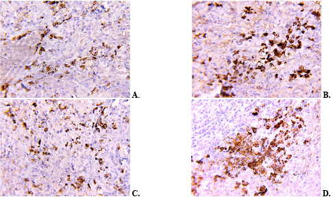
The results of light microscopic analysis of sections stained for Ki-67 and CD20 were consistent with the quantitative data. In the series of CD20 micrographs, denser clusters of positive cells were observed in the red pulp at the early time points (Fig. 1A, B), whereas their number decreased at the later time points (Fig. 1C, D).
Fig. 1. Representative micrographs of CD20-positive cells in the red pulp of the rat spleen: A – week 4; B – week 8; C – week 16; D – week 20. Magnification: ×10 eyepiece, ×40 objective ↓
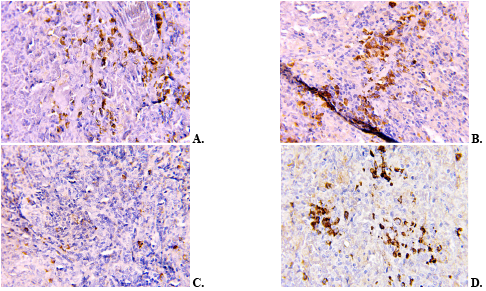
The series of Ki-67 micrographs showed an overall decrease in the number of positive nuclei (Fig. 2A, B), most pronounced at week 16 (Fig. 2C). At week 20, the number of positive cells was higher than at week 16, but remained lower than in the week-0 group (Fig. 2D).
Fig. 2. Representative micrographs of Ki-67-positive cells in the red pulp of the rat spleen: A – week 4; B – week 8; C – week 16; D – week 20. Magnification: ×10 eyepiece, ×40 objective ↓
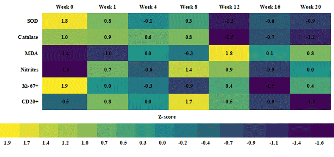
Quadratic models of temporal dynamics showed a statistically significant quadratic component for superoxide dismutase activity, malondialdehyde and nitrite concentrations, and the numbers of Ki-67-positive and CD20-positive cells in the red pulp of the rat spleen. This confirmed the nonlinear pattern of changes during the experiment. The heatmap of standardized mean values reflected the sequential change in the relationships among the study parameters (Fig. 3).
SOD – superoxide dismutase; MDA – malondialdehyde; Ki-67+ - Ki-67-positive cells; CD20+ - CD20-positive cells; Z-score - standardized deviation of the group mean from the overall mean, expressed in standard deviation units. Positive values indicate a parameter level above the overall mean, and negative values indicate a level below it. Fig. 3. Heatmap of standardized group means ↓
Exploratory analysis using Spearman's rank correlation coefficient at the level of individual animals revealed direct relationships between the measures of enzymatic antioxidant defense and inverse associations between antioxidant enzyme activities, concentrations of lipid peroxidation products, and immunohistochemical parameters. Given the nonlinear temporal dynamics, these correlations were regarded as an additional description of the relationships rather than independent evidence of causality.
A synthesis of the data made it possible to provisionally distinguish three consecutive periods of change. In the early period (weeks 1-8), the development of oxidative-nitrosative disturbances was accompanied by an increase in the number of CD20-positive cells. The most pronounced biochemical changes were observed at week 12. In the late period (weeks 16-20), antioxidant defense remained reduced, the number of CD20-positive cells decreased, and proliferative activity was most markedly suppressed.
The observed decrease in superoxide dismutase and catalase activities against a background of increased malondialdehyde concentration is consistent with reports of redox-dependent injury to lymphoid organs after exposure to monosodium glutamate. Das et al. showed that four weeks of monosodium glutamate administration was accompanied by oxidative changes and signs of immune dysfunction in the rat thymus and spleen [9]. The similar direction of the changes supports the assumption that impairment of enzymatic antioxidant defense is one of the factors involved in remodeling of the cellular composition of the spleen.
Recent studies of Ponceau 4R also describe biological effects associated with impaired cell proliferation and redox balance. Kara et al. found a decrease in the mitotic index with increasing Ponceau 4R concentration, whereas signs of genotoxicity were detected mainly at high concentrations [10]. In an experimental model using Artemia franciscana, E124 altered the activity of mitochondrial respiratory chain components and the redox state [11]. Differences between the models preclude direct extrapolation of these results to the rat spleen, although they are consistent with the combination of redox disturbances and a lower number of Ki-67-positive cells observed in our study.
Sodium nitrite can enhance the formation of reactive nitrogen species and secondary lipid peroxidation. In the study by Ahmed et al., its administration to rats was accompanied by an increase in malondialdehyde concentration and a decrease in antioxidant enzyme activities in the tissues examined [12]. In our study, the peak nitrite concentration at week 8 preceded the maximum increase in malondialdehyde at week 12, which may reflect a temporal sequence of nitrosative and lipid peroxidation changes.
The time difference between the maximum manifestations of oxidative-nitrosative stress at week 12 and the lowest number of Ki-67-positive cells at week 16 may reflect delayed suppression of proliferation due to accumulated redox-dependent injury. The increase in this parameter at week 20 was probably associated with compensatory and adaptive renewal of cell populations [9].
The effect of the mixture should not be regarded as a simple sum of the effects of its components. Studies of food additives in combination show that the cellular response to a mixture may differ from the response to individual substances [3], and combined intake of common additives in an animal model was accompanied by simultaneous changes in oxidative status, the inflammatory response, and immune cells [4]. Reviews of the effects of food additives on intestinal microecology and experimental data on interactions between the intestinal barrier and the immune system confirm that the effects of some technological ingredients may extend beyond the organ of initial contact [23, 24]. The results of this study complement previously established changes in antioxidant defense [7] and the macrophage component of the spleen [8] in the same experimental model by linking them to the dynamics of Ki-67-positive and CD20-positive cells.
The combination of reduced proliferative activity and a late decrease in the number of CD20-positive cells may indicate functional suppression and remodeling of the B-cell component of the spleen. These dynamics may be associated with disruption of the stromal-follicular microenvironment that supports the organization of B-cell follicles [25]. At the same time, lower CD20 expression may reflect not only a decrease in the number of mature B cells, but also altered marker expression or their differentiation into CD20-negative plasma cells [26].
A separate limitation of this study is the absence of daily gavage control groups. Because animals of the same initial age were euthanized at different time points, their age at euthanasia differed. All study time points fell within the adult period of the rats' lifespan; however, in the absence of concurrent age-matched control groups, the contribution of age to the observed biochemical and immunohistochemical changes cannot be fully excluded.
Repeated restraint and gavage administration of the solution may potentially induce a short-term stress response, the magnitude of which depends on the technical performance of the procedure and the volume and properties of the solution. In a separate study in male Sprague-Dawley rats acclimated to handling, daily orogastric administration of water at a volume of 5 mL/kg for 28 days, when the procedure was performed properly, was not accompanied by signs of chronic stress [27]. In our study, the initial dosing volume was 2.6 mL/kg; however, exposure lasted up to 20 weeks, and specific markers of the stress response were not measured. Therefore, the contribution of repeated gavage and associated handling stress cannot be qualitatively or quantitatively separated from other factors.
CONCLUSIONS
Contributors:
Mamai O.V. – conceptualization, investigation, writing – original draft;
Bilash S.M. – conceptualization, project supervision, methodology;
Pronina O.M. – project administration;
Bieliaieva O.M. – writing, review and editing;
Pirog-Zakaznikova A.V. – methodology;
Koptev M.M. – data curation;
Bilash V.P., Donchenko S.V. – investigation;
Bilanov O.S., Mazur B.R. – visualization;
Oliinichenko Ya.O. – resources;
Oleksiienko V.V. – validation;
Kolomiiets S.V. – formal analysis;
Funding.The study was conducted as part of the investigator-initiated research project of the Department of Anatomy with Clinical Anatomy and Operative Surgery, Poltava State Medical University, entitled “Pathogenetic mechanisms of post-stress disorders under exogenous and wartime influences and the search for methods of their correction” (state registration No. 0124U003313).
Conflict of interests. The authors declare no conflict of interest.
REFERENCES
Key words: experiment, rats, heavy metals, cadmium, liver, accumulation, zinc succinate, iron succinate, copper succinate
Ключові слова: експеримент, щури, важкі метали, кадмій, печінка, накопичення, сукцинат цинку, сукцинат заліза, сукцинат міді
Abstract
The study aimed to determine the dynamics of changes in the accumulation of cadmium and biogenic metals in the liver of experimental pregnant female rats under chronic isolated administration and when combined with biometal succinates. Based on the results of polyelement analysis, the degree of dyselementosis in the liver microelement composition and the bioprotective properties of zinc, iron, and copper succinates were evaluated. The effect of cadmium chloride on the accumulation levels of zinc, iron, and copper in the rat liver was assessed during their simultaneous entry into the body throughout pregnancy. An exceptionally high capacity for cadmium accumulation was established, with a critical excess over the control group by 67-fold on day 13 and by 87-fold at the end of the experiment. Iron, zinc, and copper succinates exhibit a pronounced protective effect, significantly reducing cadmium accumulation in the liver of pregnant rats on days 13 and 19 of the study. In terms of efficacy in limiting toxicant accumulation, the compounds form the following descending order: iron succinate > zinc succinate > copper succinate. Chronic cadmium intoxication was found to disrupt zinc metabolism in the liver of pregnant rats, abnormally increasing its level by 1.9-fold (day 13) and 1.4-fold (day 19) as a protective response for metallothionein biosynthesis. The administration of biogenic metal succinates effectively modulates this process; however, their normalizing effect depends on the gestational age. For the correction of zinc metabolism disorders, copper succinate is priority in the first half of pregnancy, while iron succinate is preferred in the second half. It was established that cadmium chloride intoxication profoundly disrupts iron metabolism and blocks its transport mechanisms, causing a rapid and significant (p<0.05) accumulation of iron in the liver parenchyma by more than 2-fold. This indicates a pronounced toxic effect of the xenobiotic on the iron-storing function of the liver at both gestational stages. The most pronounced protective effect was demonstrated by zinc succinate, which maintained the iron concentration at a level closest to the control values. This confirms the high efficiency of zinc succinate in supporting microelement homeostasis at both periods of the study. The effect of cadmium chloride on copper homeostasis disruption in the liver parenchyma of pregnant females was evaluated. It caused a rapid and significant increase in copper concentration throughout the entire observation period, indicating profound cadmium-induced alterations in the microelement balance and the retention of essential metals in the maternal depot.
Реферат
Динаміка змін мікроелементного балансу печінки щурів за умов підгострого впливу хлоридом кадмію в комбінації із сукцинатами біометалів. Шаторна В.Ф., Захаров С.В., Закут І.М. Метою дослідження було визначення динаміки змін накопичення кадмію та біогенних металів в печінці дослідних вагітних самиць щурів при підгострому ізольованому та комбінованому із сукцинатами біометалів введенні. За результатами поліелементного аналізу визначали ступінь дизелементозу мікроелементного складу печінки та біопротекторні властивості сукцинатів цинку, заліза та міді. Оцінено вплив хлориду кадмію на рівень накопичення цинку, заліза та міді печінкою щурів при їх одночасному надходженні в організм упродовж вагітності. Встановлено надзвичайно високу здатність накопичення кадмію з критичним перевищенням контролю в 67 разів на 13-ту добу та у 87 разів наприкінці експерименту. Сукцинати заліза, цинку та міді чинять виражений захисний ефект, значно знижуючи накопичення кадмію в печінці вагітних щурів на 13-ту та 19-ту добу дослідження. За ефективністю обмеження акумуляції токсиканта сполуки утворюють такий спадний ряд: сукцинат заліза > сукцинат цинку > сукцинат міді. Виявлено, що підгостра інтоксикація кадмієм порушує метаболізм цинку в печінці вагітних щурів, аномально підвищуючи його рівень в 1,9 раза (13-та доба) та в 1,4 раза (19-та доба) як захисну реакцію для біосинтезу металотіонеїнів. Застосування сукцинатів біогенних металів ефективно модулює цей процес, проте їхня нормалізація залежить від терміну гестації. Для корекції порушень обміну цинку пріоритетним у першій половині вагітності є сукцинат міді, а в другій половині – сукцинат заліза. Встановлено, що інтоксикація хлоридом кадмію глибоко порушує метаболізм заліза та блокує його транспортні механізми, викликаючи стрімке й достовірне (p<0,05) накопичення заліза в паренхімі печінки більше ніж у 2 рази, що свідчить про виражений токсичний вплив ксенобіотика на залізодепонуючу функцію печінки на обох термінах гестації. Найбільш виражений протективний ефект продемонстрував сукцинат цинку, де спостерігалося утримання концентрації заліза на рівні, максимально наближеному до контрольних значень, що підтверджує високу ефективність сукцинату цинку для підтримки мікроелементного гомеостазу на обох термінах дослідження. Оцінено вплив хлориду кадмію на порушення гомеостазу міді в паренхімі печінки вагітних самиць, який викликає стрімке й достовірне збільшення її концентрації впродовж усього періоду спостереження, що свідчить про глибокі кадмій-індуковані зміни мікроелементного балансу та затримання есенціальних металів у материнському депо.
Сучасний стан біосфери характеризується зростанням антропогенного навантаження, серед якого особливе місце посідає забруднення важкими металами. Хлорид кадмію є одним з найбільш небезпечних екотоксикантів з вираженою кумулятивною дією. Тривалий період напіввиведення з біологічних систем, який становить від 15 до 30 років, у поєднанні з високою хімічною стійкістю зумовлює персистенцію цього металу в навколишньому середовищі без втрати первинних властивостей [1]. Його тривале надходження до організму призводить до накопичення в організмі та хронічної інтоксикації, що супроводжується деструктивними змінами на клітинному та тканинному рівнях. Одним з ключових механізмів патогенезу кадмієвої токсичності є дезорганізація метаболізму есенціальних елементів.
Функціонування мікроелементів у живих системах ґрунтується на принципах жорсткого синергізму та антагонізму. Біохімічний гомеостаз є настільки взаємозалежним, що відхилення від норми навіть одного компонента неминуче запускає каскадний дисбаланс усієї мікроелементної системи [2, 3]. З огляду на це, об'єктивним критерієм функціонального стану організму є не лише абсолютний вміст конкретних мікроелементів, а, насамперед, їхнє коректне міждіапазонне співвідношення. Розвиток мікроелементозів, викликаний браком життєво необхідних сполук або інтерференцією з боку ксенобіотиків, суттєво модифікує та маскує клінічну картину отруєнь [3, 4]. Це значно ускладнює диференційну діагностику хронічних інтоксикацій.
Після абсорбції в шлунково-кишковому тракті та потрапляння вв системний кровотік мікроелементи розподіляються в органах украй нерівномірно. Найвища концентрація більшості як ендогенних елементів, так і екзогенних токсикантів (зокрема кадмію) реєструється в гепатобіліарній системі [5, 6]. Оскільки печінка виконує роль головного функціонального депо мікроелементів в організмі, вона стає основним органом-мішенню та першою зазнає деструктивного впливу за умов тривалої хімічної агресії [7, 8]. Це дозволяє розглядати печінку як головне функціональне депо мікроелементів, яке першим зазнає системного впливу за умов постійного довготривалого антропогенного навантаження.
Взаємодія мікроелементів в організмі ґрунтується на складних процесах синергізму та антагонізму [4, 5]. Через це дефіцит або надлишок навіть одного компонента неминуче руйнує загальний елементний гомеостаз за принципом ланцюгової реакції. Отже, фізіологічний стан організму визначається не стільки абсолютними значеннями окремих мікроелементів, скільки їхнім збалансованим співвідношенням.
Таким чином, актуальність теми зумовлена широким поширенням кадмієвої інтоксикації в промислових регіонах та її згубним впливом на організм вагітних і плода. Вивчення міжіонних взаємодій кадмію з есенціальними мікроелементами відкриває нові можливості для патогенетичної корекції метаболічних порушень. Застосування біометал-сукцинатів є перспективним напрямком для створення ефективних, безпечних і доступних вітчизняних засобів антенатального метаболічного захисту.
Мета дослідження – визначити динаміку змін накопичення кадмію та біогенних металів у печінці дослідних вагітних самиць щурів при підгострому ізольованому та комбінованому із сукцинатами біометалів введенні. Визначити особливості механізму комбінованого антенатального впливу хлориду кадмію і сукцинатів мікроелементів та ступінь дизелементозу мікроелементного складу печінки.
МАТЕРІАЛИ ТА МЕТОДИ ДОСЛІДЖЕНЬ
Експериментальна частина дослідження була виконана на молодих вагітних самицях щурів лінії Wistar віком 2,5-3 місяці, яких отримували з ліцензованого розплідника «Далі 2001» (м. Київ). Тварин утримували у віварії Дніпровського державного медичного університету в стандартних пластикових клітках (по 3-4 особини) за контрольованих умов: t=20-25°C, вологість ≥50%, регулярна вентиляція, світловий режим «день/ніч» (12/12 год). Раціон був стандартний, з вільним доступом до води.
Дослідження схвалені комісією з питань біомедичної етики Дніпровського державного медичного університету (протокол № 39 від 17.06.2026 р.) та проведені відповідно до вимог Закону України «Про захист тварин від жорстокого поводження» від 21.02.2006 р. № 3447-IV та Наказу Міністерства освіти і науки, молоді та спорту України від 01.03.2012 р. № 249 «Про затвердження Порядку проведення науковими установами дослідів, експериментів на тваринах», а також з урахуванням принципів Директиви 2010/63/ЄС Європейського парламенту і Ради від 22.09.2010 р. про захист тварин, які використовуються в наукових цілях.
Для проведення експерименту у віварію Дніпровського державного медичного університету тварин було розподілено на чотири дослідні групи та контрольну. Введення препаратів здійснювали внутрішньошлунково, щоденно впродовж усього терміну гестації самиць. Група I (контроль, 10 самиць): тварини отримували 0,5 мл ізотонічного 0,9% розчину NaCl для встановлення фонових показників. Група II (модель інтоксикації, 18 самиць): застосовували ізольований вплив хлориду кадмію в дозі 3,0 мг/кг. Застосована доза хлориду кадмію становила 3% від LD₅₀ для щурів при пероральному введенні, що відповідало низькому токсичному рівню. Такий рівень експозиції є достатнім для моделювання гестаційної інтоксикації та порушень гомеостазу металів без розвитку летальних наслідків, що дозволяло проводити тривалий щоденний вплив протягом усієї вагітності. Група III (корекція цинком, 18 самиць): на фоні щоденного введення 3,0 мг/кг хлориду кадмію тварини отримували сукцинат цинку в дозі 5 мг/кг для оцінювання його протективних властивостей. Група IV (корекція залізом, 18 самиць): схема передбачала поєднання кадмію із сукцинатом заліза в дозі 5,0 мг/кг з метою дослідження його коригувальної дії. Група V (корекція купрумом, 18 самиць): схема передбачала поєднання кадмію із сукцинатом міді в дозі 0,1 мг/кг з метою дослідження його коригувальної дії. Дози сукцинатів біметалів вибрані як фізіологічно прийнятні та безпечні для вагітних щурів, проте достатні для впливу на системний пул заліза, цинку для корекції кадмієво-індукованих порушень без розвитку залізо-індукованої токсичності.
Для оцінювання специфіки акумуляції кадмію, цинку та заліза в паренхімі печінки вагітних самиць щурів на 13-ту та 19-ту добу гестації за умов ізольованої інтоксикації хлоридом кадмію та супутньої корекції сукцинатами есенціальних металів застосовували метод атомно-емісійної спектрометрії з електродуговою атомізацією. Цей аналітичний підхід дозволяє здійснювати прецизійний якісний і кількісний поліелементний скринінг біосубстратів різного походження. Елементний аналіз зразків виконували на базі ДП «Український науково-дослідний інститут медицини транспорту МОЗ України» (м. Одеса) в рамках договору про науково-практичне співробітництво. Кількісне визначення мікроелементів у печінці проводили методом атомно-емісійної спектрометрії з дуговою атомізацією на приладі Емас‑200 CCD відповідно до принципів, описаних Кучеруком Д.Д., Головком А.К. [9]. Для порівняння кількісних показників вмісту металів у тканинах печінки щурів на різних термінах вагітності застосовували t-критерій Стьюдента для незалежних вибірок. Статистичний аналіз проводили з метою порівняння кількісних показників вмісту металів у тканинах печінки щурів на різних термінах вагітності. Для оцінювання різниці між незалежними вибірками застосовували t-критерій Стьюдента. Статистична обробка даних проводилась із застосуванням Microsoft Exel R версія 4.3.1 (https//www.r-project.org/) [13]. Аналіз виконували відповідно до сучасних стандартів прозорості та відтворюваності біостатистичних досліджень.
РЕЗУЛЬТАТИ ТА ЇХ ОБГОВОРЕННЯ
Аналіз вмісту кадмію в печінці вагітних самиць щурів за умов щоденної інтоксикації хлоридом кадмію виявив значне накопичення цього металу в органі впродовж усього періоду спостереження (рис. 1). За результатами поліелементного аналізу встановлено, що в печінці самиць контрольної групи вміст кадмію впродовж експерименту залишався на стабільно низькому рівні (близько 0,15-0,18 мкг/г), що відповідало фізіологічній нормі. На 13-ту добу експерименту вміст кадмію в печінках самиць групи ізольованого впливу хлоридом кадмію зріс у десятки разів порівняно з контролем.
Внутрішньошлункове введення хлориду кадмію призвело до стрімкого накопичення металу в печінці, а саме: на 13-ту добу його концентрація зросла до 12,9±1,5 мкг/г, а на 19-ту добу спостерігалося подальше достовірне збільшення показника до 15,9±1,5 мкг/г, що було статистично значущою різницею (p<0,05) та свідчило про високу кумулятивну здатність кадмію в гепатоцитах протягом вагітності. Таким чином, у першій половині вагітності зростання рівня кадмію відбулося порівняно з контролем у 67 разів, а наприкінці експерименту – у 87 разів. Активне накопичення кадмію печінкою порівняно з контролем під час підгострого експерименту зумовлене специфічними фізіологічними, біохімічними та транспортними механізмами печінки щурів. При внутрішньошлунковому введенні печінка першою приймає кров від шлунково-кишкового тракту через ворітну вену, тобто портальну циркуляцію. Орган працює як головний фільтр, затримуючи ксенобіотики для захисту інших систем для детоксикації. Кадмій у печінці індукує масштабний синтез білків металотіонеїнів (МТ) у гепатоцитах, які міцно зв'язують іони Cd, і утворюється тканинне депо. Загальновідомо, що утворені комплекси Cd-MT мають довгий період напіввиведення і затримуються в печінці. Такий рівень кумуляції кадмію ми пояснюємо бар'єрною роллю печінки, яка виступає головним детоксикаційним фільтром організму, першою затримуючи токсикант з портальної циркуляції шлунково-кишкового тракту для захисту інших систем.
Рис. 1. Динаміка накопичення кадмію в паренхімі печінки вагітних щурів на 13-й та 19-й добі гестації (мкг/г) ↓
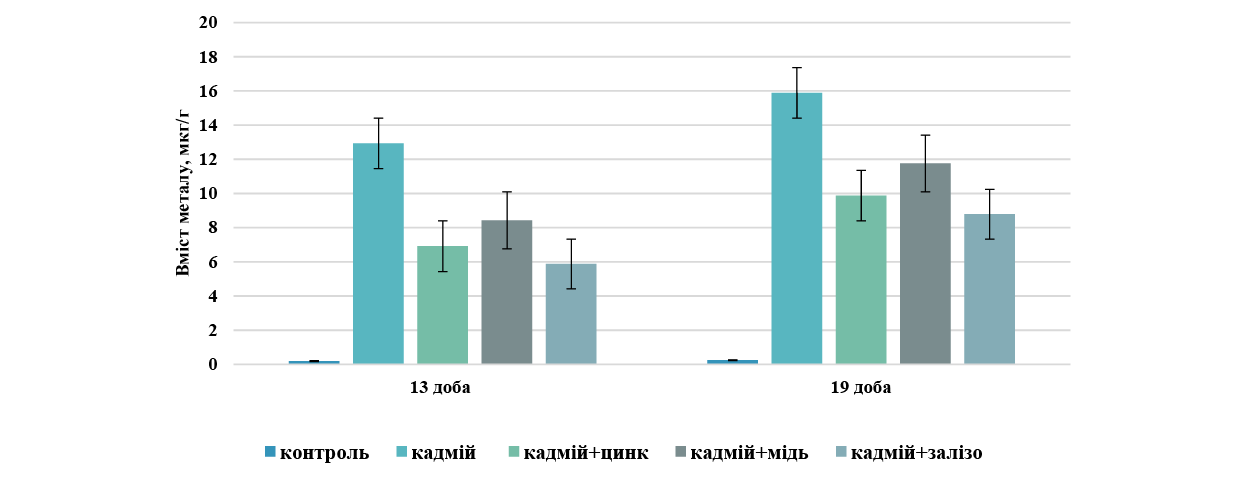
У групі комбінованого введення хлориду кадмію із сукцинатом цинку виявився високий протективний ефект. На 13-ту добу експерименту вміст кадмію в цій групі становив 6,9±1,5 мкг/г, що на 46,5% менше, ніж у групі з ізольованим введенням токсиканта. На 19-ту добу рівень накопичення паренхімою печінки кадмію дещо підвищився до 9,9±1,4 мкг/г, проте він залишався на 37,7% нижчим порівняно з групою інтоксикації. Це підтверджує виражений антагонізм іонів цинку та їх здатність блокувати депонування кадмію.
Проте в групі комбінованого введення хлориду кадмію із сукцинатом заліза ми визначали найбільш виражений протекторний ефект (рис. 1). У цій групі експерименту був найнижчий показник рівня накопичення кадмію, хоча він не завжди мав достовірну різницю з рівнем накопичення групою комбінації хлориду кадмію із сукцинатом цинку.
У групі комбінованого введення хлориду кадмію із сукцинатом міді також визначалось зниження рівня токсиканта, але цей вплив був менш ефективним порівняно з цинком та залізом. На 13-ту добу рівень кадмію становив 8,4±1,6 мкг/г, тобто спостерігалось зниження на 34,9% відносно групи кадмію. На 19-ту добу показник зріс до 11,8±1,7 мкг/г, що на 25,8% менше за показник групи без корекції. Порівнюючи дані про накопичення кадмію печінкою самиць у всіх групах корекції, можна констатувати, що сукцинат цинку та сукцинат заліза ефективніше за сукцинат міді обмежують накопичення кадмію печінкою на обох термінах дослідження в зазначених дозах та способі введення в експерименті на щурах.
Цинк – це мікроелемент, який є життєво важливим для імунної системи, оскільки його дефіцит пригнічує вроджені та набуті імунні механізми, підвищуючи схильність до інфекційних, автоімунних й онкологічних процесів. Крім того, оптимальний рівень цинку відіграє фундаментальну роль в ендокринній системі: він регулює синтез і структуру гормонів щитоподібної залози та забезпечує повноцінну роботу підшлункової залози, беручи безпосередню участь у синтезі та зберіганні інсуліну і глюкагону. Найбільше цинку накопичується в скелетних м'язах та кістковій тканині, проте печінка займає третє місце в ступені накопичення після опорно-рухового апарату та шкіри й волосся.
У нашому експерименті аналіз накопичення цинку паренхімою печінки показав таке. У контрольній групі на 13-ту та 19-ту добу експерименту рівень цинку збільшувався, що ми пояснюємо метаболічними змінами в організмі у зв’язку з перебігом вагітності самиць. На 13-й добі рівень цинку в печінці самиць становив 24,31±3,61 мкг/г, а наприкінці вагітності вже добігав 35,92±4,70 мкг/г, достовірність різниці p<0,05. Ізольоване введення хлориду кадмію змінювало рівень цинку в досліджуваних зразках (рис. 2).
Вже на 13-й добі експерименту в групі ізольованого введення кадмію рівень цинку підвищувався в 1,9 раза, перевищував контрольні дані й становив 45,21±2,40 мкг/г, а на 19-ту добу перевищення відбувалось в 1,4 раза, що становило 48,76±4,31 мкг/г. Порівняння отриманих результатів щодо накопичення цинку не було показовим для групи комбінації кадмію і цинку, тому що рівень цього мікроелемента хоч і був найвищим, але тварини цієї групи його отримували щоденно. Тому основні порівняння нами проводились між іншими групами комбінованого введення.
Неочікуваними виявились кількісні показники цинку в групі комбінованого введення хлориду кадмію із сукцинатом міді. На 13-ту добу експерименту рівень цинку (31,83±4,91 мкг/г) був зіставним з контрольними значеннями, як і в групі комбінації із залізом 33,51± 3,18 мкг/г. Ситуація на 19 -й добі експерименту змінювалась у межах цих груп комбінації. Саме в групі комбінованого введення кадмію із залізом рівень цинку становив 39,87±4,20 мкг/г, що не мало достовірної різниці з контролем, а в групі комбінації із сукцинатом міді показник дорівнював такому в групі кадмієвої інтоксикації. Таким чином, у першій половині вагітності рівень цинку підтримувався введенням сукцинату міді, а в другій половині – сукцинатом заліза.
Нами також розраховувався показник співвідношення кадмію до цинку, який наочно демонструє рівень токсичного навантаження організму важкими металами та ступінь порушення мінерального обміну, оскільки цинк та кадмій є біологічними антагоністами, вони конкурують за одні й ті самі рецептори та транспортні білки в організмі (рис. 2).
Рис. 2. Динаміка індексу співвідношення кадмію до цинку в печінці вагітних щурів на 13-й та 19-й добі гестації ↓
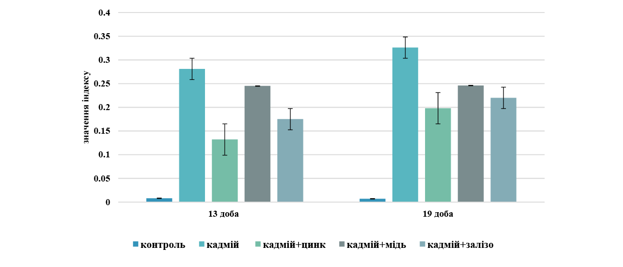
На початковому терміні спостереження показник у контрольній групі залишався мінімальним і становив близько 0,01. Введення кадмію призвело до різкого зростання індексу до 0,28 (p<0,05), що свідчить про гостре накопичення токсичного металу в печінці та порушення балансу мікроелементів.
Застосування біогенних металів у комбінації з тією самою дозою кадмію на 13-ту добу показало різну ефективність досліджуваних сполук. Цинк найбільш ефективно знижував індекс до 0,13, що майже вдвічі менше за групу ізольованого впливу кадмію. Залізо виявляло помірний захисний ефект, знижуючи показник до 0,17, а мідь виявилась найменш ефективною, оскільки індекс зменшувався лише до 0,24.
На 19-й добі експерименту в контрольній групі індекс зберігався на вихідному стабільно низькому рівні (~0,01). Проте в групі з ізольованим введенням кадмію спостерігалося його подальше зростання до пікового значення 0,33, що підтверджувало прогресуючу інтоксикацію. При оцінюванні потенційної корекції сукцинатами біометалів на 19-ту добу виявлено такі зміни. У групі кадмій+цинк індекс зростав порівняно з 13-ю добою до ~0,20, проте цинк і надалі надійно утримував лідерство за рівнем захисту печінки. У групі комбінованого введення хлориду кадмію і сукцинату заліза показник також підвищувався до 0,22, що вказує на поступове виснаження захисного ресурсу заліза за умов тривалого токсичного впливу. Комбіноване введення кадмію з міддю залишало індекс на попередньому рівні (~0,245), демонструючи найслабший ефект серед усіх досліджуваних металів-антагоністів.
Динаміка накопичення заліза (мг/г) у паренхімі печінки вагітних самиць дослідних тварин у розрізі всіх експериментальних даних дозволяє оцінити особливості метаболізму та депонування феруму в організмі під впливом досліджуваних чинників. У тварин контрольної групи рівень заліза в печінці впродовж всього періоду дослідження перебував у межах стабільної фізіологічної норми для вагітних самиць. На 13-ту добу експерименту рівень заліза в паренхімі печінки становив 44,37±3,61 мкг/г, а на 19-ту добу – 45,42±4,20 мкг/г. За результатами поліелементного аналізу визначалась тенденція помірного та прогнозованого коливання концентрації, що свідчить про адекватне забезпечення плодів залізом без виснаження материнського депо.
Ізольоване введення хлориду кадмію призвело до стрімкого достовірного підвищення рівня заліза в паренхімі печінки порівняно з контролем (p<0,05). На 13-ту добу експерименту рівень заліза в печінці тварин кадмієвої інтоксикації збільшувався до 86,29±4,41 мкг/г, що було в 1,9 раза вище за контрольні показники, а на 19-ту добу зростав до 98,66± 5,30 мкг/г, що перевищувало контроль у 2,14 раза. Це вказує на активну мобілізацію депонованого заліза на потреби фетоплацентарного комплексу або на порушення його метаболізму. При цьому в групі комбінованого впливу кадмію із сукцинатом цинку спостерігалось утримання концентрації заліза до рівня майже контрольних значень. Це підтверджує ефективність одночасного надходження кадмію із сукцинатом цинку для підтримки мікроелементного балансу організму на обох термінах дослідження.
Комбіноване введення хлориду кадмію із сукцинатом міді також підвищувало рівень заліза в зразках порівняно з контролем, проте порівняно з групою кадмієвої інтоксикації знижувало, хоча і в меншому ступені, ніж комбіноване введення кадмію з цинком. Рівень заліза в паренхімі печінки в групі комбінованого введення кадмій+сукцинат заліза не є показовим, бо тварини отримували залізо щоденно, тому ці результати накопичення не підлягали порівнянню.
Мідь є життєво важливим мікроелементом, який виступає кофактором багатьох металоферментів, що регулюють дихання, кровотворення та антиоксидантний захист. Цей мікроелемент входить до складу цитохром-с-оксидази як ключового ферменту мітохондрій, що забезпечує синтез АТФ, відповідаючи за клітинне дихання та енергію. У процесах кровотворення та обміну заліза важливу роль відіграє фермент церулоплазмін, який є головним білком-переносником міді, що окиснює залізо, дозволяючи йому зв'язуватися з трансферином і брати участь у синтезі гемоглобіну. Антиоксидантний захист організму також залежить від міді, яка разом із цинком утворює фермент супероксиддисмутазу, що нейтралізує вільні радикали.
Мідь відіграє критичну роль у функціонуванні печінки, оскільки цей орган є головним центром метаболізму, депонування та екскреції цього мікроелемента. Як центральний орган обміну, печінка захоплює мідь з кровотоку після її всмоктування в кишківнику та розподіляє її для потреб усього організму. При цьому гепатоцити синтезують церулоплазмін – головний мідьвмісний білок крові, який забезпечує безпечний транспорт міді та бере участь в обміні заліза. У групі контролю рівень міді в паренхімі печінки достовірно (р=0,05) зростав з 13-ї до 19-ї доби експерименту в 1,3 раза, що ми пояснюємо фізіологічними перебудовами та зміною балансу мікроелементів в організмі вагітної самиці. Це є фізіологічним процесом, зумовленим необхідністю забезпечити плід, що активно розвивається, цим мікроелементом, а також гормональними змінами в організмі матері. Вагітність супроводжується значним зростанням рівня церулоплазміну в крові матері, що, у свою чергу, активує підвищене всмоктування міді в кишківнику та знижує її виведення, що призводить до кумуляції міді в печінці та її подальшого транспортування. Мідь також необхідна для активізації ферментів, що включають залізо в порфіринове кільце, що важливо для посиленого біосинтезу гемоглобіну. Мідь як мікроелемент є життєво важливою для формування тканин плоду, зокрема печінки та мозку. У другій половині вагітності (після 13-ї доби) зростає потреба у синтезі фетальних білків (наприклад, металотіонеїнів), які потребують міді. Організм вагітної самиці накопичує мідь, щоб потім ефективно переносити її через плаценту, тобто відбувається активний транспорт мікроелемента до плоду через плаценту.
У групі ізольованого впливу кадмієм рівень міді збільшувався порівняно з контрольними показниками впродовж експерименту таким чином. На 13-ту добу експерименту рівень міді становив 1,904±0,08 мкг/г, що у 2,3 раза перевищувало контроль, а на 19-й добі експерименту становив 2,347±0,09 мкг/г, що перевищувало контрольні дані у 2,1 раза.
Таким чином, ізольована інтоксикація організму вагітної самиці хлоридом кадмію призводила до дизелементозу по міді в паренхімі печінки як на 13-ту добу вагітності, так і наприкінці ембріогенезу. У групах комбінованого введення визначалось зниження рівня міді в бік до контрольного показника при поєднанні кадмію з цинком та поєднанні із залізом на обох термінах експериментального дослідження. Вимірювання в групі поєднання кадмію із сукцинатом міді зі зрозумілих причин не проводилось. Найбільш ефективне відновлення рівня міді визначалось у групі комбінованого впливу кадмієм із сукцинатом цинку. Загалом отримані дані демонструють біоантагоністичний характер сукцинатів досліджуваних біометалів, що дозволяє їх розглядати як потенційні протектори гепатотоксичності кадмію в організмі дослідних тварин у підгострому експерименті введення зазначених доз та способу введення.
Сукцинати заліза, цинку та міді виявляли виражений захисний ефект акумуляції кадмію в печінці вагітних щурів на 13-ту та 19-ту добу гестації. За здатністю знижувати депонування токсиканта сполуки утворюють спадний ряд: сукцинат заліза > сукцинат цинку > сукцинат міді. Аналогічні спрямування визначення нових потенційних біоантагоністів кадмію визначались у попередніх дослідженнях Дніпровської морфологічної школи ДДМУ, тільки на інших органах: серце [10], нирки [11], статеві залози, хоча доза кадмію в тій серії досліджень була значно нижчою [12]. Результати мали досить різноспрямований характер упродовж етапів усього експериментального періоду.
Уперше проведено комплексне порівняльне дослідження протективної ефективності сукцинатів заліза, цинку та міді щодо депонування кадмію в паренхімі печінки на різних етапах гестаційного періоду. Встановлено чітку залежність модифікаційного впливу досліджуваних сполук на метаболізм цинку та заліза. Доповнено наукові дані щодо механізмів біологічного антагонізму та захисної ролі сукцинатів есенціальних мікроелементів за умов поєднаного надходження ксенобіотика. Уперше деталізовано характер порушень залізодепонуючої функції печінки вагітних самиць під впливом щоденної кадмієвої інтоксикації.
ВИСНОВКИ
Внески авторів:
Шаторна В.Ф. – концептуалізація, ведення, методологія, адміністрування проєкту;
Захаров С.В. – концептуалізація, нагляд, дослідження, курація даних, перегляд та редагування;
Закут І.М. – методологія, дослідження, формальний аналіз, написання: рецензування та редагування.
Фінансування. Дослідження не має зовнішніх джерел фінансування.
Конфлікт інтересів. Автори заявляють про відсутність конфлікту інтересів.
REFERENCES
Key words: experiment, embryos, rats, heavy metals, cadmium, zinc, iron, embryotoxicity, embryonic mortality, embryo resorption
Ключові слова: експеримент, ембріони, щури, важкі метали, кадмій, цинк, залізо, ембріотоксичність, ембріональна смертність, резорбція ембріонів
Abstract
The aim of the study was to determine the embryotoxicity of cadmium chloride under the conditions of its long-term isolated administration and in combination with zinc or iron succinates in an experiment on rats to assess the modifying effect of succinates on the embryotoxicity of cadmium. To determine the degree of embryotoxicity and calculate the types of embryonic mortality, the number of litters, the weight of fetuses, the number of corpora lutea in the ovaries of females, as well as live and resorbed embryos in the uterine horns of rats were analyzed. The effect of zinc and iron succinates on the level of embryotoxicity of cadmium chloride in rats with their simultaneous long-term daily intake into the body during pregnancy was experimentally evaluated. The results obtained by us demonstrate a significant increase in all indicators of embryonic mortality in the group of isolated exposure to cadmium chloride compared to the control group both on the 13th and 20th days of the experiment. It was established that isolated chronic exposure to cadmium chloride has a pronounced embryotoxic effect: on the 13th day of the experiment, which is manifested both by a significant decrease in the number of live fetuses compared to the control (p<0.05) and by a 4-fold increase in the indicator of total embryonic mortality against the background of active fetal resorption. At the end of the gestation period (20th day), the toxic pressure of cadmium in the studied dose reaches a maximum: the number of live fetuses critically decreases, and the indicator of total embryonic mortality exceeds the level of the control group by 10.7 times. Iron and zinc succinates exhibit pronounced protective properties as cadmium bioantagonists, significantly reducing the levels of embryonic death on both terms of gestation. On the 13th day, iron succinate reduces the total embryonic mortality by 2.8 times compared to the cadmium intoxication group, mainly due to preimplantation protection, and zinc succinate – by 2.9 times, bringing the indicators closer to the control. On the 20th day of gestation in the groups of combined administration with zinc and iron succinates, the level of total embryonic mortality decreases to 0.05±0.02 and 0.06±0.02, respectively. With regard to postimplantation mortality, both succinates demonstrate equally high protective efficacy. The nature and features of the stage-specific protective effect of iron succinate and zinc succinate compounds under conditions of antenatal cadmium chloride intoxication have been established for the first time.
Реферат
Модифікація ембріотоксичності хлориду кадмію за умов комбінованого введення із сукцинатами металів. Шевченко І.В., Нефьодова О.О. Метою дослідження було визначення показників ембріотоксичності хлориду кадмію за умов його довготривалого ізольованого введення та в комбінації із сукцинатами цинку або заліза в експерименті на щурах для оцінювання модифікуючого впливу сукцинатів на ембріотоксичність кадмію. Для визначення ступеня ембріотоксичності та розрахування видів ембріональної смертності аналізували чисельність посліду, масу плодів, кількість жовтих тіл у яєчниках самиць, а також живих і резорбованих ембріонів у маточних рогах щурів. Експериментально оцінено вплив сукцинатів цинку та заліза на рівень ембріотоксичності хлориду кадмію в щурів при їх одночасному довготривалому щоденному надходженні в організм упродовж вагітності. Отримані нами результати демонструють достовірне збільшення всіх показників ембріональної смертності в групі ізольованого впливу хлоридом кадмію порівняно з контрольною групою як на 13-ту, так і на 20-ту добу експерименту. Встановлено, що ізольований хронічний вплив хлоридом кадмію має виражену ембріотоксичну дію на 13-ту добу експерименту, що проявляється як достовірним зниженням порівняно з контролем (p<0,05) кількості живих плодів, так і зростанням показника загальної ембріональної смертності в 4 рази на тлі активної резорбції плодів. Наприкінці гестаційного періоду (20-та доба) токсичний пресинг кадмію в досліджуваній дозі досягає максимуму: кількість живих плодів критично зменшується, а показник загальної ембріональної смертності в 10,7 раза перевищує рівень контрольної групи. Сукцинати заліза та цинку виявляють виражені потенційні протективні властивості як біоантагоністи кадмію, достовірно знижуючи рівні ембріональної загибелі на обох термінах гестації. На 13-ту добу сукцинат заліза знижує загальну ембріональну смертність у 2,8 раза порівняно з групою кадмієвої інтоксикації, переважно за рахунок доімплантаційного захисту, а сукцинат цинку – у 2,9 раза, наближаючи показники до контролю. На 20-ту добу гестації в групах комбінованого введення із сукцинатами цинку та заліза рівень загальної смертності ембріонів знижується до 0,05±0,02 та 0,06±0,02 відповідно. Щодо післяімплантаційної смертності обидва сукцинати демонструють однаково високу захисну ефективність. Уперше встановлено характер та особливості стадієспецифічної захисної дії сполук сукцинату заліза та сукцинату цинку за умов антенатальної інтоксикації хлоридом кадмію.
Забруднення навколишнього середовища важкими металами залишається однією з найгостріших екологічних та медико-біологічних проблем сучасності. Серед ксенобіотиків особливе місце посідає кадмій як високотоксичний елемент з вираженою кумулятивною дією. Довготривалий вплив сполук кадмію, зокрема його хлориду, призводить до системних порушень в організмі, серед яких найменш вивченими та найбільш небезпечними є гонадотоксичний та ембріотоксичний ефекти [1, 2]. Здатність кадмію проникати крізь плацентарний бар'єр створює пряму загрозу для антенатального розвитку, зумовлюючи тератогенез, затримку розвитку плода та ембріональну смертність [3]. Результати експериментальних досліджень на лабораторних тваринах свідчать про високу вразливість репродуктивної системи до впливу важких металів, проте питання патогенезу ембріотоксичної дії кадмію залишаються недостатньо висвітленими. Необхідність деталізації впливу різних доз та шляхів надходження сполук кадмію, зокрема хлориду кадмію, на процеси ембріогенезу спонукає до подальшого вивчення цієї проблеми [3].
На особливу увагу заслуговує пошук ефективних біоантагоністів, здатних нівелювати або зменшувати негативні прояви ембріотоксичності сполук цього металу. У зв'язку з цим привертає увагу використання есенціальних металів у формі біологічно активних комплексів. Перспективними сполуками є сукцинати цинку та заліза [4]. Бурштинова кислота (сукцинат) виступає потужним ендогенним субстратом енергетичного обміну, що стимулює клітинне дихання та антиоксидантний захист, тоді як цинк і залізо є безпосередніми антагоністами кадмію на рівні клітинних транспортерів [5, 6]. Проте особливості їхньої комбінованої та ізольованої дії на ембріогенез за умов одночасного надходження кадмію із сукцинатами біметалів залишаються недостатньо з'ясованими.
Проблема терапії та профілактики кадмієвої інтоксикації є предметом активного вивчення в сучасній токсикології та ембріології, оскільки чинить виражену політропну та ембріотоксичну дію. Дослідження останніх років доводять, що тривале надходження хлориду кадмію до організму вагітних самок щурів призводить до критичного підвищення загальної, доімплантаційної та післяімплантаційної смертності ембріонів, а також до затримки їхнього масо-габаритного розвитку [7].
Механізм патологічного впливу кадмію безпосередньо пов'язаний з його здатністю порушувати гомеостаз есенціальних елементів. Оскільки кадмій є хімічним антиметаболітом цинку та заліза, він конкурує з ними за зв'язування на рівні клітинних мембран та мембранних транспортерів, викликає вторинний дефіцит біогенних металів, дизелементоз цих життєво важливих мікроелементів у плаценті та тканинах плода, що провокує розвиток оксидативного стресу, мітохондріальну дисфункцію та апоптоз клітин [8].
Особливу увагу привертає метаболічна форма введення мікроелементів, зокрема їхні комплекси з бурштиновою кислотою – сукцинати. Сукцинат діє як потужний ендогенний субстрат циклу Кребса, який відновлює синтез АТФ у пошкоджених кадмієм клітинах і стимулює клітинне дихання. Продемонстровано, що сукцинат цинку знижує показники ембріо- та кардіотоксичності в щурів, а також здатен відновлювати чисельність ембріонів у посліді на ранніх та пізніх термінах гестації [9].
Попри наявні дані, залишається нез'ясованим характер модифікуючого впливу сукцинатів цинку та заліза за умов їхнього комбінованого з кадмієм надходження в організм. Більшість сучасних досліджень фокусуються лише на використанні інших солей (наприклад, цитратів або сульфатів) або вивчають вплив лише одного металу-протектора. Відсутність системного порівняльного аналізу дії сукцинатів цинку та заліза на динаміку ембріотоксичності хлориду кадмію обґрунтовує доцільність нашого дослідження.
Мета дослідження – визначити показники ембріотоксичності хлориду кадмію за умов його довготривалого ізольованого введення та в комбінації із сукцинатами цинку або заліза в експерименті на щурах для оцінювання модифікуючого впливу сукцинатів на ембріотоксичність кадмію.
МАТЕРІАЛИ ТА МЕТОДИ ДОСЛІДЖЕНЬ
Експериментальна частина дослідження була реалізована на вагітних самицях щурів лінії Wistar у віці 2,5-3 місяці, які були отримані зі спеціалізованого розплідника ТОВ «Далі 2001» (м. Київ). Вибір щурів як базового морфологічного об'єкта для цього моделювання є методично та патогенетично обґрунтованим, оскільки цей вид гризунів відзначається високою стабільністю ембріогенезу та мінімальним рівнем спонтанних або непередбачуваних аномалій і вад антенатального розвитку порівняно з мишами та кролями. Цей критерій мав вирішальне значення для забезпечення високої точності, відтворюваності та релевантності отриманих результатів під час виконання поставлених ембріологічних і морфологічних завдань. Упродовж усього терміну експерименту лабораторні тварини утримувалися в умовах віварію Дніпровського державного медичного університету з чітким дотриманням стандартизованих параметрів мікроклімату. Зокрема, температура повітря у приміщеннях підтримувалася в межах 20-25 C, відносна вологість становила не менше ніж 50%, а також забезпечувалося систематичне кондиціювання, регулярне провітрювання та природний світловий режим зміни дня і ночі. Для отримання вагітних самок з датованим терміном вагітності проводили контрольоване спарювання. Самців і самок зі стійким астральним циклом утримували разом у співвідношенні 1:3 протягом нічного періоду. Наступного ранку за наявністю у вагінальному мазку сперматозоїдів визначали як критерій успішного спарювання та відмічали як першу добу вагітності. Всі маніпуляції здійснювали відповідно до принципів біоетики та стандартів утримання лабораторних тварин. Гризунів розміщували у стандартних пластикових клітках групами не більше ніж по 3-4 особини в кожній та утримували на збалансованому стандартному раціоні віварію з вільним доступом до питної води. Усі умови утримання, догляду та маніпуляцій повністю відповідали чинним вимогам положення про поводження з тваринами у наукових дослідженнях, розробленого згідно із Законом України «Про захист тварин від жорстокого поводження» та міжнародними біоетичними стандартами, положеннями Конвенції Ради Європи про права людини та біомедицину (1997 р.), а також згідно з актуальними рекомендаціями ВООЗ щодо поводження з лабораторними тваринами. Дослідження схвалені комісією з питань біомедичної етики Дніпровського державного медичного університету (протокол № 39 від 17.06.2026).
Для проведення експерименту тварин було розподілено на чотири дослідні групи. Введення препаратів здійснювали внутрішньошлунково, щоденно впродовж усього терміну гестації самиць. Група I (контроль): тварини отримували 0,5 мл ізотонічного 0,9% розчину NaCl (n=10) для встановлення фонових показників. Кількісні показники дослідних тварин розподілялись з урахуванням того факту, що щури як лабораторні тварини є досить повно досліджені, саме з цих причин група контролю була зменшена. Група II (модель інтоксикації) (n=20): застосовували ізольований вплив хлориду кадмію в дозі 3,0 мг/кг. Вибрана доза кадмію забезпечує розвиток характерних для гестаційного періоду порушень метал-гомеостазу, але не спричиняє летальних ефектів, що дає змогу проводити щоденний тривалий вплив протягом усієї вагітності. Група III (корекція цинком) (n=20): на фоні щоденного введення 3,0 мг/кг хлориду кадмію тварини отримували сукцинат цинку в дозі 5 мг/кг для оцінювання його протективних властивостей. Група IV (корекція залізом) (n=20): схема передбачала поєднання кадмію із сукцинатом заліза в дозі 0,2 мг/кг з метою дослідження його корегуючої дії. Введення препаратів здійснювали внутрішньошлунково, щоденно впродовж усього терміну гестації самиць. Дози сукцинатів цинку та заліза підібрані як фізіологічно прийнятні для вагітних тварин і водночас достатні для впливу на системний пул відповідних мікроелементів без ризику розвитку формування у самиць залізо-індукованої токсичності [12].
Ембріотоксичний ефект оцінювали на 13-й та 20-й день вагітності щурів після евтаназії самок тіопенталовим наркозом. Проводили макроскопічний огляд органів репродуктивної системи. У яєчниках під бінокулярною лупою підраховували кількість жовтих тіл вагітності, у рогах матки фіксували кількість живих та мертвих плодів і місць резорбцій у матках самиць. Локалізацію місць імплантацій, що піддалися ранній резорбції, протоколювали та використовували для розрахунків показників різних видів ембріотоксичності.
Інтенсивність ембріотоксичної дії досліджуваних речовин визначали за трьома інтегральними показниками: передімплантаційною, постімплантаційною та загальною ембріональною смертністю, розрахованими за стандартними токсикологічними формулами: відношення загиблих зародків до кількості жовтих тіл або місць імплантації, виражене у відсотках [10, 11]. Рівень ембріотоксичної дії досліджуваних речовин у нашому експерименті оцінювали за такими стандартними показниками:
1. Загальна ембріональна смертність розраховувалась за формулою:
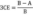
де А – кількість живих плодів;
В – кількість жовтих тіл вагітності.
2. Передімплантаційна смертність визначалась за формулою:
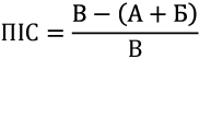
де А – кількість живих плодів;
Б – кількість загиблих (резорбованих) плодів;
В – кількість жовтих тіл вагітності.
3. Постімплантаційна смертність визначалась за формулою:
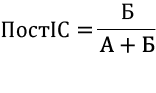
де А – кількість живих плодів;
Б – кількість загиблих (резорбованих) плодів.
4. Кількість плодів на 1 самку (середній показник у групах).
5. Середні значення вагових показників ембріонів (г).
Статистичну обробку даних здійснювали для порівняння показників, що характеризують ембріотоксичний ефект за різних умов експозиції. Оцінювання відмінностей між незалежними групами проводили із застосуванням t-критерію Стьюдента. Статистична обробка даних проводилась із застосуванням Microsoft Exel R версія 4.3.1 (https//www.r-project.org/) [16]. Аналіз виконували відповідно до сучасних стандартів прозорості та відтворюваності біостатистичних досліджень.
РЕЗУЛЬТАТИ ТА ЇХ ОБГОВОРЕННЯ
У нашому експерименті всі самиці вижили, впродовж дослідження залишались активні, добре споживали їжу та воду. Забір ембріонального матеріалу, підрахування кількості жовтих тіл вагітності в яєчниках проводився на 13-ту та 20-ту добу ембріогенезу. Хронічна інтоксикація хлоридом кадмію в дозі 3 мг/кг вже на 13-ту добу експерименту спровокувала порушення топографії ембріонів у рогах матки, а саме: ми визначали асиметричну імплантацію у 8,43% самиць. Для норми в поліестральних багатоплідних ссавців характерний рівномірний розподіл зародків в обох рогах матки. Фізіологічні інтервали між зонами імплантації зазвичай зумовлені поодинокими резорбціями ембріональних та плацентарних структур, що зрідка фіксується і в контрольній групі. Проміжки в маткових трубах між місцями прикріплення вказують на випадки резорбції плаценти чи плода, які поодиноко трапляються і в контролі. Проте в групі ізольованої дії кадмієвої солі поширеність часткової або повної топографічної асиметрії була значно вищою, а при ізольованому введенні хлориду кадмію частота випадків повної або часткової асиметрії розподілу ембріонів була суттєво вищою. Цей факт визначався нами і на 20-ту добу дослідження. Таким чином, хронічне введення хлориду кадмію призвело до того, що на 13-ту та 20-ту добу в самиць щурів спостерігалася девіація нормального розміщення ембріонів у вигляді асиметричної імплантації.
Як показало обчислення кількісних показників, середня кількість живих ембріонів у контрольній групі на 13-ту добу становила 12,50±0,28, тоді як у групі ізольованого впливу хлоридом кадмію цей показник знизився до 8,88±0,24 без достовірного зниження середньої кількості жовтих тіл у яєчниках в обох групах (рис. 1).
Статистична значущість за t-критерієм Стьюдента: * – p<0,05 порівняно з контролем; ** – p<0,05 порівняно з групою хлорид кадмію. Рис. 1. Кількість живих ембріонів на 1 самицю на 13-ту добу гестації в експериментальних групах (M±m) ↓
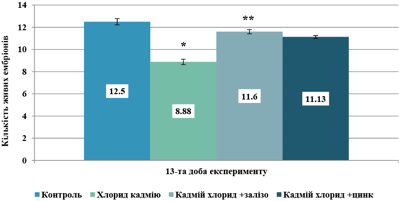
Аналіз та порівняння отриманих числових обрахувань продемонстрували, що пренатальний ізольований щоденний вплив хлориду кадмію в досліджуваній дозі знижує показник кількості живих ембріонів на 13-ту добу на 28,9%. Таким чином, вже на 13-ій добі експерименту виявлено достовірне зниження показника порівняно з контролем (p<0,05) Суттєве зниження кількості живих ембріонів порівняно з контролем підтверджує той факт, що кадмій є сильною ембріотоксичною отрутою, яка долає плацентарний бар’єр та здатна викликати високу ембріональну смертність.
У групах комбінованого застосування кадмію з металосукцинатами спостерігався чіткий модифікуючий ефект. У групі комбінації кадмію із сукцинатом заліза відмічалась тенденція до збільшення кількості живих ембріонів порівняно з групою ізольованого впливу – 11,6±0,19, що свідчило про зростання кількості ембріонів більше ніж на 25% відносно показника групи кадмію, хоча й залишалося на 11% нижче за контроль. Недостовірно нижчий показник протекторного впливу порівняно з групою комбінації кадмію із сукцинатом заліза зафіксовано в четвертій групі комбінованого надходження кадмію із сукцинатом цинку, де досліджуваний показник становив 11,13±0,13. У цій групі кількість живих ембріонів також наближалася до фізіологічної норми, що вказує на антитоксичну ефективність сукцинату цинку як антагоніста кадмію при їх одночасному надходженні в організм в експерименті на щурах.
На 20-ту добу ембріогенезу плоди вилучали оперативно разом з плацентою для подальшого морфологічного аналізу й верифікації їхньої відповідності стадіям розвитку за критеріями Гамбургера-Гамільтона (НН). На 20-ту добу гестації середня кількість живих ембріонів у групі ізольованого введення хлориду кадмію була найнижчою серед усіх експериментальних груп і становила 8,75±0,27, тоді як у контрольній групі цей показник становив 12,63±0,19 (достовірне зниження показника порівняно з контролем (p<0,05)). У групах комбінованого введення середня кількість живих ембріонів була вищою за групу ізольованого введення хлориду кадмію, але не мали достовірної різниці при їх порівнянні між собою: 11,00±0,20 у групі комбінації хлориду кадмію із сукцинатом заліза та 11,38±0,28 у групі одночасного введення хлориду кадмію та сукцинату цинку. Тобто, при комбінованому введенні кадмію із сукцинатами цинку або заліза спостерігається збереження показників середніх значень кількості ембріонів, що свідчить про антагоністичний характер дії сукцинатів на кадмієву інтоксикацію в підгострому експерименті на щурах при їх одночасному надходженні в досліджуваних дозах (рис. 2).
Статистична значущість за t-критерієм Стьюдента: * – p<0,05 порівняно з контролем; ** – p<0,05 порівняно з групою хлорид кадмію. Рис. 2. Кількість живих ембріонів на 1 самицю на 20-ту добу гестації в експериментальних групах (M±m) ↓
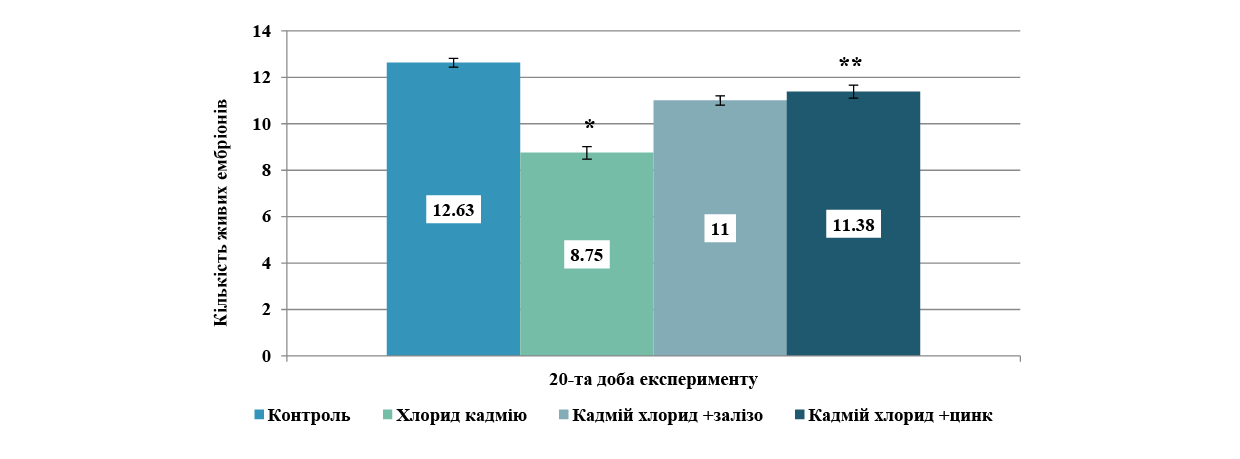
Таким чином, завершальний етап пренатального розвитку плодів дозволив кількісно оцінити ступінь ембріотоксичної дії хлориду кадмію та протекторну ефективність металосукцинатів упродовж усього гестаційного періоду. У групі ізольованого впливу хлоридом кадмію спостерігається, як і на попередньому терміні дослідження, критичне зниження показника кількості ембріонів у посліді – на 30,7% відносно контролю. Це підтверджує стійку антенатальну токсичність кадмію, яка зберігається протягом усього періоду гестації.
Поєднання токсиканту із сукцинатом заліза забезпечило часткове відновлення виживаності плодів, а саме: показник зріс на 23,9% порівняно з групою ізольованого введення кадмію. Застосування сукцинату цинку продемонструвало найвищий захисний ефект ембріотоксичності кадмію. Показник зріс на 30,1% відносно групи кадмієвої інтоксикації і максимально наблизився до рівня контрольної групи (відставання від норми становить лише 11,6%). Отримані результати доводять, що сукцинати цинку та заліза проявляють модифікуючий вплив на показники ембріотоксичності хлориду кадмію при їх комбінованому надходженні в підгострому експерименті на щурах.
Одним з важливих критеріїв ембріотоксичності є вагові показники ембріонів. У нашому дослідженні на 20-ту добу ембріогенезу в контрольній групі середня вага ембріона становила 2,89±0,06 г, що відповідає фізіологічному еталону, а ізольоване введення хлориду кадмію призвело до вірогідного зниження маси тіла плодів до 2,65±0,07 г, що на 8,30% менше порівняно з контрольними значеннями. Сумісне застосування кадмію із сукцинатом заліза продемонструвало позитивну динаміку: маса ембріонів зберігалася до 2,77± 0,05 г, перевищуючи показник ізольованої інтоксикації на 4,53% (рис. 3).
Статистична значущість за t-критерієм Стьюдента: * - p<0,05 порівняно з контролем. Рис. 3. Вагові показники ембріонів (г) на 20-ту добу гестації в експериментальних групах ↓
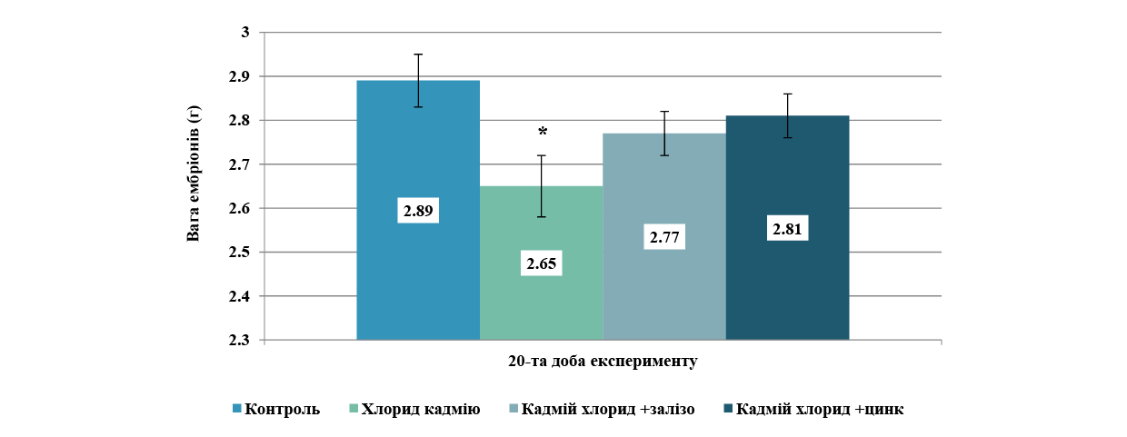
Найбільш виражений ростостимулюючий та антитоксичний ефект зафіксовано в групі поєднання хлориду кадмію із сукцинатом цинку. Середня маса плодів тут досягла 2,81±0,05 г, що на 6,04% вище за рівень ізольованого впливу і практично нівелює затримку розвитку, відстаючи від фізіологічної норми контролю лише на 2,77%.
Таким чином, хлорид кадмію викликає синдром затримки пренатального розвитку плодів, що проявляється зменшенням їхньої кількості та маси. Сукцинат цинку при одночасному надходженні з кадмієм в організм дослідних тварин демонструє вищу протективну здатність щодо збереження кількості та соматичного росту плодів, ніж сукцинат заліза, майже повністю відновлюючи масові параметри до рівня контролю.
Аналіз та порівняння кількісних та вагових даних по ембріонам доводить, що на 13-ту добу ембріогенезу ізольоване введення хлориду кадмію призводить до високопрезентативного зниження кількості живих ембріонів відносно контролю (p<0,001). Введення металосукцинатів на тлі інтоксикації забезпечує статистично значущий протективний ефект: у групі сукцинату заліза показник вірогідно зростає порівняно з ізольованим впливом важкого металу (p<0,055), аналогічна високозначуща тенденція до нормалізації реєструється і за умов застосування сукцинату цинку (p<0,01). Наприкінці гестаційного періоду (20-та доба ембріогенезу) ізольована дія хлориду кадмію призводила до глибокого та статистично значущого пригнічення виживаності плодів порівняно з контрольною групою – кількість живих ембріонів зменшилась до 8,75±0,27 проти 12,63±0,19 у контролі (p<0,05). Застосування досліджуваних металосукцинатів забезпечило стійку протекцію ембріонального матеріалу від ксенобіотичного пресингу. У групі сукцинату заліза показник живих ембріонів вірогідно зріс до 11,0±0,20 порівняно з ізольованою інтоксикацією (p<0,05). Найбільш виражений та статистично високорепрезентативний захисний ефект зафіксовано за умов введення сукцинату цинку, де кількість живих плодів досягла 11,38±0,28, що демонструє максимальний рівень значущості відновлення показника відносно групи інтоксикації (p<0,05).
Окрім кількісних та вагових показників ембріонів, нами для визначення ступеня ембріотоксичності досліджуваних речовин обчислювались відносні показники ембріональної смертності, які враховують не лише кількість ембріонів, а й кількість резорбцій, кількість жовтих тіл вагітності та дозволяють зіставити їх між собою. Найбільш значущим та доказовим показником ембріотоксичності речовини є показник загальної ембріональної смертності (ЗЕС). Обрахування цього показника на 13-ту добу ембріогенезу продемонструвало, що зниження кількості ембріонів у групі ізольованого введення хлориду кадмію відбувалось за рахунок високої ембріональної смертності – 0,26±0,03, тобто показник збільшувався у 8,6 раза (p<0,05) порівняно з контролем (0,03±0,01). Водночас у групах комбінованого введення показники ЗЕС були достовірно нижчі. У групі впливу кадмію разом із сукцинатом заліза цей показник становив 0,06±0,02, що було в 4,3 раза нижче за групу ізольованого впливу (p<0,05), а при комбінації з цинком – 0,09±0,02, що у 2,8 раза нижче (p<0,05), ніж у групі ізольованого впливу хлоридом кадмію (рис. 4).
Статистична значущість за t-критерієм Стьюдента: * – p<0,05 порівняно з контролем; ** – p<0,05 порівняно з групою хлорид кадмію.Рис. 4. Показник загальної ембріональної смертності на 13-ту добу гестації в експериментальних групах ↓
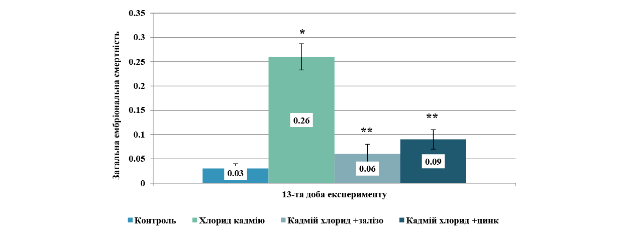
Таким чином, експозиція хлоридом кадмію під час вагітності має виражену ембріотоксичність уже на 13-ту добу ембріогенезу щурів, що проявляється стрімким зростанням загальної ембріональної смертності та активізацією процесу резорбції ембріонів. Сукцинати заліза та цинку виявляють протективні властивості: комбіноване застосування цих сполук разом із ксенобіотиком ефективно нівелює його негативний ембріотоксичний вплив. Одночасне надходження з хлоридом кадмію сукцинату заліза знижує показник ЗЕС порівняно з групою ізольованого впливу кадмію, повертаючи цей показник у бік до контрольних значень.
На 20-й добі експерименту показник ЗЕС збільшувався при ізольованому впливі кадмієм до 0,32±0,02, що перевищувало контрольні показники в 10,7 раза й було максимальним результатом серед усіх груп. У групах комбінованого введення із сукцинатом цинку показник ЗЕС становив 0,10±0,2, а із сукцинатом заліза дорівнював 0,13±0,03, такі дані були достовірно нижчими за групу ізольованого впливу. У групі кадмієвої інтоксикації наприкінці вагітності самиць щурів наростали темпи ембріональної смертності порівняно з попереднім терміном дослідження, а показник ЗЕС збільшився в 1,2 раза.
Привертала увагу статистично незначуща відмінність між показниками ЗЕС у групах комбінованого введення хлориду кадмію в поєднанні із сукцинатом заліза та хлориду кадмію в поєднанні із сукцинатом цинку на 13-ту добу експерименту, водночас на 20-ту добу експерименту різниця між середніми показниками ЗЕС також не є достовірною, хоча до контрольних показників наближається саме група комбінації із сукцинатом цинку.
Аналіз таких даних вказує, що на ранніх стадіях ембріогенезу сукцинат заліза має більш значущий антагоністичний ефект по відношенню до ембріотоксичності кадмію порівняно із цинком, водночас на 20-ту добу експерименту цинк та залізо мають приблизно однакову ефективність, проте показник ЗЕС у групі комбінації із цинком найнижчий серед експериментальних груп.
Сукцинати заліза та цинку виявляють виражений захисний ефект щодо ембріотоксичності кадмію вагітних щурів на 13-ту та 20-ту добу гестації при їх одночасному потраплянні в організм. Такі результати перегукуються з даними експериментальних досліджень, у яких проводили пошук біоантагоністів токсичності важким металам серед органічних солей біометалів – цитратів. У дослідженнях також аналізувались результати комбінованого впливу солей важких металів та цитратів мікроелементів (заліза, церію, германію, селену та ін.) і визначали зниження рівня загальної ембріональної смертності та збільшення кількості живих плодів. Експериментально доведено модифікуючий вплив цитратів мікроелементів на ембріотоксичні властивості кадмію [8, 12, 13]. Дослідники експериментально довели, що нутрієнтні втручання (зокрема залізо) можуть бути перспективним способом зменшення рівня інтоксикації кадмієм. Робота підкреслює важливість балансу мікроелементів у харчуванні та їхнього впливу на токсикологічні процеси [8]. Дослідниками-морфологами Дніпровського державного медичного університету проводилось також попередні дослідження з виявлення потенційних біоантагоністичних характеристик сукцинатів на тлі кадмієвої інтоксикації вагітних самиць, проте доза кадмію була менша. Вже в цих експериментах комбіноване введення хлориду кадмію із сукцинатами цинку або міді сприяло зниженню всіх показників ембріотоксичності хлориду кадмію та покращенню репродуктивних показників. Досліджувались також показники доімплантаційної, післяімплантаційної та загальної ембріональної смертності [14, 15], але в цих дослідженнях не вивчали захисні біопротекторні властивості сукцинату заліза, а доза кадмію була менша на третину.
ВИСНОВКИ
Внески авторів:
Нефьодова О.О. – концептуалізація, ведення, нагляд, методологія, адміністрування проєкту;
Шевченко І.В. – проведення дослідження, курація даних, формальний аналіз, написання: рецензування та редагування.
Фінансування. Дослідження не має зовнішніх джерел фінансування.
Конфлікт інтересів. Автори заявляють про відсутність конфлікту інтересів.
REFERENCES
Key words: visual disability, combat ophthalmic traumas, bilateral lesions, age-related vulnerability, foreign bodies
Ключові слова: порушення зору, бойові офтальмологічні травми, двосторонні ураження, вікова вразливість, сторонні тіла
Abstract
Gunshot and explosive injuries to the visual organs are one of the most severe and socially significant forms of ophthalmological pathology, especially in the context of armed conflicts, terrorist attacks, and man-made disasters. The anatomical complexity of the eye, its vulnerability to high-energy impact, and the vital importance of visual function determine the extreme severity of the consequences of such injuries. These injuries not only reduce the quality of life of the victims but also produce a substantial load on healthcare and social support systems due to the high incidence of disabling outcomes. The study aimed to investigate the clinical and functional characteristics of gunshot and explosive injuries of the visual organs with subsequent analysis of treatment outcomes in a specialised trauma centre. A retrospective study was conducted at the University Trauma Clinic in Tirana, Albania, between January 1995 and December 2006. The sample included 172 patients (240 eyes) admitted within the first 24 hours after injury. The methodology included the analysis of medical records and ophthalmological examination data, including visometry using Snellen tables, biomicroscopy using a slit lamp, ophthalmoscopy, and orbital radiography. In cases of suspected foreign bodies in the posterior segment, computed tomography was utilised. Statistical data processing was performed using IBM SPSS Statistics v.22.0, utilising Student's t-test for quantitative indicators and Pearson's χ² test for categorical variables. Epidemiological analysis revealed peaks in morbidity in 1997 (25%) and 1999 (22%), corresponding to periods of political crisis and armed conflict. The victims were predominantly male (92%) with an average age of 23.9±14.1 years. Notably, 54% of victims were minors, and 71% resided in rural areas. Explosions caused the largest proportion of cases (49%), followed by gunshot wounds (31%), indicating high-energy injury mechanisms. Clinical evaluation showed that severe eye injuries predominated (76%), often accompanied by combined injuries to other organs (75%). Foreign bodies were detected in 75% of eyes, and bilateral injuries occurred in 39% of patients. Despite treatment, severe anatomical outcomes (enucleation, atrophy) were recorded in 57% of patients, and irreversible loss of vision was recorded in 39% of eyes. The high level of disability identified highlights the need for systemic preventive measures and the readiness of ophthalmology services to receive seriously injured people on a massive scale. The practical significance of the data suggests that healthcare authorities must plan medical evacuation, train personnel, and create specialised ophthalmic trauma centres to manage complex, combined injuries effectively. Special attention must be paid to vulnerable groups, particularly children and rural residents, to prevent injuries during periods of social instability.
Реферат
Вогнепальні та вибухові травми очей. Барді Н. Поранення органів зору від вогнепальної зброї та вибухових пристроїв є однією з найтяжчих і соціально значущих форм офтальмологічної патології, особливо в умовах збройних конфліктів, терористичних актів та техногенних катастроф. Анатомічна складність ока, його вразливість до високоенергетичного впливу та життєво важливе значення зорової функції визначають надзвичайну тяжкість наслідків таких травм. Ці травми не тільки знижують якість життя потерпілих, але й створюють значне навантаження на системи охорони здоров'я та соціального забезпечення через високу частоту інвалідності. Мета дослідження полягала у вивченні клінічних та функціональних особливостей вогнепальних та вибухових травм органів зору з подальшим аналізом результатів лікування у спеціалізованому травматологічному центрі. Ретроспективне дослідження було проведено в Університетській травматологічній клініці в Тирані, Албанія, у період із січня 1995 року до грудня 2006 року. До вибірки увійшли 172 пацієнти (240 очей), госпіталізовані протягом перших 24 годин після травми. Методологія включала аналіз медичних карт та даних офтальмологічного обстеження, включаючи візометрію за допомогою таблиць Снеллена, біомікроскопію за допомогою щілинної лампи, офтальмоскопію та рентгенографію орбіти. У випадках підозри на наявність сторонніх тіл у задньому сегменті використовувалася комп'ютерна томографія. Статистична обробка даних проводилася за допомогою IBM SPSS Statistics v.22.0 з використанням t-критерію Стьюдента для кількісних показників та χ²-критерію Пірсона для категоріальних змінних. Епідеміологічний аналіз виявив піки захворюваності в 1997 (25%) та 1999 (22%) роках, що відповідало періодам політичної кризи та збройного конфлікту. Жертвами були переважно чоловіки (92%) із середнім віком 23,9±14,1 року. Примітно, що 54% жертв були неповнолітніми, а 71% проживали в сільській місцевості. Найбільшу частку випадків становили вибухи (49%), за ними йшли вогнепальні поранення (31%), що вказує на механізми травмування з високою енергією. Клінічна оцінка показала, що переважали тяжкі травми очей (76%), які часто супроводжувалися комбінованими травмами інших органів (75%). Чужорідні тіла були виявлені в 75% очей, а двосторонні травми траплялися в 39% пацієнтів. Незважаючи на лікування, тяжкі анатомічні наслідки (енуклеація, атрофія) були зафіксовані в 57% пацієнтів, а незворотна втрата зору – у 39% очей. Високий рівень інвалідності, виявлений у дослідженні, підкреслює необхідність проведення системних профілактичних заходів та готовності офтальмологічних служб до прийому великої кількості людей з тяжкими травмами. Практичне значення даних свідчить про те, що органи охорони здоров'я повинні планувати медичну евакуацію, навчати персонал та створювати спеціалізовані офтальмологічні травматологічні центри для ефективного лікування складних комбінованих травм. Особливу увагу слід приділяти вразливим групам населення, зокрема дітям та мешканцям сільської місцевості, з метою запобігання травмам у періоди соціальної нестабільності.
Gunshot and explosive injuries to the visual organs are one of the most severe and socially significant forms of ophthalmological pathology, especially in the context of armed conflicts, terrorist attacks and man-made disasters. The anatomical complexity of the eye, its vulnerability to high-energy impact and the vital importance of visual function determines the extreme severity of the consequences of such injuries. Between 1995 and 2006, the problem was significantly exacerbated by the growing number of localized armed conflicts, instability in border and rural areas, and the proliferation of improvised explosive devices. The high incidence of disabling outcomes, including complete loss of vision and eyeballs, not only reduces the quality of life of the victims but also produces a substantial load on healthcare and social support systems. In this context, the study of the clinical features and outcomes of ophthalmic injuries caused by firearms and explosives is of relevance to improve approaches to medical care, rehabilitation and prevention.
A substantial clinical problem is the high incidence of bilateral eye injuries in blast injuries, which significantly worsens the prognosis of restoration of visual function and reintegration of the victim into society. N. Bardhi [1] emphasised that the main pathogenetic mechanism of bilateral lesions is diffuse energy transfer through the bone structures of the facial skull, accompanied by multiple foreign bodies in the orbital tissues. Despite the proposed algorithms for diagnosis and treatment, approaches to emergency care remain fragmented, and the rate of outcomes with complete vision loss remains high, which indicates the need for a comprehensive review of protocols for the management of such patients. A significant obstacle to effective ophthalmic care for combat injuries is the high incidence of combined injuries, which makes early surgical intervention difficult. G. Warjri et al. [2] emphasised that ophthalmological care in patients with multiple organ trauma often falls by the wayside, leading to delayed treatment of ocular injuries. Despite the introduction of multidisciplinary teams, there is a lack of coordination between ophthalmologists and surgeons of other specialties, which reduces the effectiveness of rehabilitation measures.
High incidence of underage drinking, especially in rural areas where access to specialized medical care is limited, is notable. D. Zekaj et al. [3] highlighted that the lack of safety education programmes and affordable personal protective equipment for children and adolescents makes them particularly vulnerable. Despite the steps taken to prevent injuries among young people, the statistics of severe eye injuries in this age group grow, indicating the need for targeted prevention initiatives and training in basic safety skills in hazardous environments. The problem of preserving functional vision even with partial anatomical restoration of the eye remains unresolved. V. Kumar et al. [4] demonstrated that restoration of the eyeball anatomy does not guarantee the return of visual function, especially in cases of damage to the optic nerve and central retinal structures. Despite the development of microsurgery and reconstructive technologies, the rates of visual rehabilitation after blast injuries remain extremely low, which requires the search for new biotechnological solutions and methods of neurorehabilitation.
The absence of specialised ophthalmic trauma centres in most regions of Albania significantly reduces the efficiency and effectiveness of care. G. Nallbani and M. Krasniqi [5] addressed the shortage of staff, insufficient equipment and low readiness of institutions to receive mass casualties. Even if medical evacuation is available, the lack of standardised patient routes leads to delays in care and, consequently, to worsening outcomes. This highlights the need for strategic planning of resource provision and development of a network of specialised institutions. The high degree of disability after ophthalmic injuries is also associated with psychological consequences, which are often ignored in the treatment process. Y. Zhang et al. [6] noted that vision loss is accompanied by the development of post-traumatic stress disorders, depression and social isolation, especially in young patients. At the same time, comprehensive psychological support is practically not integrated into standard rehabilitation protocols, which limits the effectiveness of returning victims to active life.
There is no systematic database reflecting the structure, causes, and outcomes of ophthalmic injuries in the context of hostilities. D. Prat et al. [7] emphasized that the fragmentation of statistical sources does not provide an accurate assessment of the scale of the problem and makes it difficult to forecast and plan resources. In the context of growing instability and the probability of mass admission of severely injured people, this situation is unacceptable, which requires the development of a centralised information system and registers of ophthalmic injuries. The mechanisms for providing emergency ophthalmological care in the first hours after an injury are also insufficiently developed. A. Mohamed et al. [8] highlighted that the effectiveness of treatment largely depends on the time of transport and the availability of standard operating protocols. However, in most cases, victims are delivered late, and the initial examination is conducted by untrained specialists. These factors significantly limit the potential for preserving vision and restoring ocular structures, which requires regulatory regulation and improved training of medical staff.
The combination of these problems suggests that the existing system of ophthalmological care for gunshot and blast injuries requires significant modernisation at both the organisational and clinical levels. The study aimed to investigate the clinical and functional characteristics of gunshot and explosive injuries of the visual organs with subsequent analysis of treatment outcomes in a specialised trauma centre. The objectives of the study included a retrospective analysis of medical records, identification of the main factors influencing the severity of injuries, and evaluation of the effectiveness of the care provided.
MATERIALS AND METHODS OF RESEARCH
The study was conducted at the Trauma University Hospital (formerly Central Military University Hospital) [9] in Tirana, Albania, between January 1995 and December 2006. The study is a retrospective design based on the analysis of medical records and diagnostic materials of patients admitted with gunshot and blast injuries of the eye. The sample included all patients admitted to the hospital within the first 24 hours after the injury, with a documented injury to the eye or orbit due to gunshot or explosive exposure and a complete medical record. Cases with isolated soft tissue injuries of the eyelids without involvement of the eyeball, patients with incomplete documentation and those who died before admission were excluded. Thus, the study included 172 patients (158 men and 14 women) with a total of 240 injured eyes. The average age of the victims was 23.9±14.1 years. For each case, socio-demographic characteristics (age, gender, occupation, place of residence), circumstances of injury, type of injury (bullet, shell and mine fragments, explosives), severity of eye injury and the presence of combined injuries to other organs and systems were recorded. The latter were classified into five main groups: maxillofacial area, brain, limbs, thorax and abdomen. This study was considered by the members of the Research Ethics Council of the University Hospital of Trauma. Based on the vote of the Commission's members, a decision was made: All procedures performed in the study were in accordance with the Declaration of Helsinki [10] and the rules of Good Clinical Laboratory Practice (GCLP) [11]. The scientific research can be implemented in its current form, No. 258101.
The ophthalmological examination included visometry using Snellen tables [12] (Germany), biomicroscopy using a Haag-Streit BM 900 slit lamp (Switzerland), ophthalmoscopy (Welch Allyn 3.5V, USA), orbital radiography using a Siemens Multix Pro (Germany), and, if necessary, ultrasound A- and B-scan (Tomey UD-6000, Japan). Intraocular pressure was measured by Goldmann applanation tonometry in eyes with preserved globe integrity. In patients with suspected or confirmed open-globe injuries, tonometry was not performed before primary wound closure because external pressure could aggravate the injury; intraocular pressure was assessed after restoration of globe integrity when clinically indicated. In case of suspected foreign bodies in the posterior segment of the eye, computed tomography (GE HiSpeed CT/i, USA) was used. The injuries were classified into three degrees: mild (contusions, superficial injuries), moderate (penetrating wounds without massive destruction of the eyeball), and severe (destruction of the eyeball, massive injuries with large foreign bodies). Bilateral injuries were addressed separately. The treatment included both conservative methods (antibiotic therapy, anti-inflammatory drugs, antihypertensive therapy, and local wound treatment) and surgical interventions. In patients with clinical signs of purulent complications, material from the ocular wound and, when clinically obtainable, aqueous humor or vitreous samples collected during surgery were submitted for microbiological culture and identification of the causative microorganism. Antibiotic susceptibility testing was performed for the isolated microorganisms, and antibacterial therapy was subsequently adjusted according to the obtained results. Depending on the clinical situation, primary surgical treatment of wounds, removal of foreign bodies, corneal and scleral reconstructive surgery, orbital reconstruction, and, in cases of irreversible damage, enucleation of the eye were performed.
The outcomes were assessed comprehensively. The dynamics of visual acuity on admission and discharge were measured, and anatomical results (preservation of the eyeball, development of atrophy or subatrophy), and the incidence of complications (endophthalmitis, panophthalmitis, secondary glaucoma) were analyzed. Visual outcomes were classified into four levels: preserved vision (≥0.3), moderate reduction (0.1-0.29), severe reduction (0.01-0.09) and functional blindness (≤0.01 or enucleation). Statistical data processing was performed using IBM SPSS Statistics v.22.0 [13] (USA). Student's t-test was used for quantitative indicators, and Pearson's χ² test for categorical variables. Multivariate logistic regression was used to identify factors associated with unfavorable outcomes (eye loss, functional blindness). The level of statistical significance was assumed to be p<0.05. Based on the retrospective analysis, practical recommendations were formulated to optimize the care of victims of gunshot and blast eye injuries. Timeliness of interventions, the sequence of diagnostic and therapeutic measures, and the factors associated with the most adverse visual outcomes were emphasized.
Statistical data processing was performed using IBM SPSS Statistics v.22.0 [13] (USA) [License No. 3022941]. The normality of the distribution of quantitative data was assessed using the Kolmogorov-Smirnov test. Quantitative data following a normal distribution are presented as Mean ± Standard Deviation (SD), while categorical variables are presented as absolute numbers (n) and percentages (%). Since the quantitative data met the assumption of normality, Student's t-test [14] was used for comparative analysis. Pearson's χ² test [15] was utilized for categorical variables. Multivariate logistic regression [16] was used to identify factors associated with unfavorable outcomes (eye loss, functional blindness). The level of statistical significance was assumed to be p<0.05. Based on the retrospective analysis, practical recommendations were formulated to optimize the care of victims of gunshot and blast eye injuries. Timeliness of interventions, the sequence of diagnostic and therapeutic measures, and the factors associated with the most adverse visual outcomes were emphasised.
RESULTS AND DISCUSSION
The issue of annual dynamics of eye injuries caused by gunshot and explosive impact was a key element of the epidemiological assessment of the war and post-war period. The chronological distribution of cases was used to identify peaks in morbidity associated with specific political and social events, including internal conflicts, armed clashes and massive weapons proliferation (Table 1). The analysis by year reflected the duration of the impact of crises on the civilian population and the emergency eye care system. In times of instability, such data was used to plan the allocation of resources, staff training and rapid response of clinics. In addition, temporal patterns in the admission of patients with severe eye injuries formed the basis for subsequent prevention measures and legal regulation in the field of civilian security.
Years Number of cases (damaged persons) Percentage 1995 1 0.5 1996 6 3.5 1997 43 25.0 1998 14 8.1 1999 37 21.5 2000 19 11.0 2001 15 8.7 2002 15 8.7 2003 6 3.5 2004 8 4.7 2005 7 4.1 2006 1 0.5 Sum 172 100Table 1. Distribution of eye injuries by year (1995-2006) ↓
The annual analysis demonstrated a pronounced irregularity in the frequency of eye injuries caused by gunshot and explosive impact. The highest number of cases was recorded in 1997 – 43 patients (25% of the total number over 12 years). This sharp increase corresponded to the political crisis in Albania, which was accompanied by massive arms seizures and spontaneous violence. The second peak occurred in 1999, with 37 cases (22%), of which a significant number came from Kosovo amid the armed conflict. In total, 80 incidents (47%) occurred in these two years, which is almost half of all recorded cases. In the remaining years, the number of victims ranged from one to nineteen, showing sporadic but significant spikes in injuries.
The study of the gender of victims of gunshot and blast eye injuries established socio-behavioural and demographic patterns typical of conflict periods (Table 2). In the context of military or near-military activity, gender differences in morbidity reflected not only biological but also socio-cultural factors such as participation in hostilities, access to weapons, lifestyle characteristics, and risks associated with certain professions. Notably, in the context of this study, the gender imbalance was not only statistically significant, but also preventive: it indicated risk groups requiring targeted intervention and special attention in educational and restrictive programmes.
Gender (sex) Number of cases (persons) % Male (M) 158 92 Female (F) 14 8 M + F 172 100 Notes: χ²(1)=120.6; p<0.001. Table 2. Distribution of cases by gender ↓
The gender distribution of the victims showed a pronounced predominance of men: 158 (92%) of the 172 patients were men, and only 14 (8%) were women. This imbalance was consistent with the nature of the armed incidents and the typical social role of men in situations of instability. Men, especially younger men, were significantly more likely to be involved in or witness confrontations, to have access to weapons, and to be exposed to the risks associated with hostilities, illicit arms trafficking and domestic conflicts involving explosives. Women, on the other hand, were more likely to be outside the epicentre of active hostilities, which explains the extremely low percentage of casualties. This indicator also highlighted the disproportionate involvement of men in the events that led to injuries and was used as a substantial indicator of social vulnerability. The data suggested that in conditions of increased militarisation of society or social destabilization, men were the main risk group for severe eye injuries, which should be considered when developing targeted prevention and rehabilitation strategies.
The study of the age characteristics of victims of gunshot and blast injuries to the eye identified the most vulnerable age groups and to identify demographic shifts typical of periods of armed instability (Table 3). The average age of patients and gender disaggregation were central for research of the structure of the affected population, their level of activity, involvement in armed conflict or its consequences. Age analysis was key in terms of assessing the severity of injuries, prognosis of recovery of visual functions and the possibility of further rehabilitation. Data in this category was also central in planning medical care and preventive measures aimed to protect the most at-risk age groups.
Number of cases (persons-patients) Average age (years) Gender male female 172 23.94±14.11 158 14 Notes: data are presented as Mean±Standard Deviation (SD); t≈–1.30; p=0.21 (not significant). Table 3. Average age of victims ↓
The average age of patients with eye injuries caused by gunshot or explosive impacts was 23.94±14.11 years, indicating a predominance of young people among the victims. Men represented the absolute majority in this sample, which was also reflected in their dominance by age. This age structure corresponded to the social reality of the period, when young men were more likely to be involved in armed conflicts or to become accidental victims.
An analysis of the distribution of victims by age group provided a more detailed assessment of the extent to which different generational categories were involved in the armed conflict (Table 4). This breakdown was substantial for the identification of the most at-risk groups, especially among children and adolescents, where the consequences of trauma had not only medical but also serious social impact. Determining age-related vulnerability was also of practical importance in developing primary prevention programs, planning educational campaigns, and organising eye care and long-term rehabilitation. In the context of the study period, which included years of political crisis and military conflicts, the age distribution reflected the overall dynamics of violent events and the extent of civilian casualties.
Age -group Number of cases (persons) % Note 0-10 years 19 11 54 11-20 years 74 43 21-30 years 35 20 46 >30 years 44 26 Sum 172 100 100Table 4. Distribution of victims by age group ↓
The age structure data revealed the highest concentration of ocular injuries in young people: 74 patients (43%) belonged to the age group of 11-20 years, and 19 cases (11%) were children under 10 years of age. Thus, more than half of the victims (54%) were minors. The next largest group was 21-30 years of age (20%), while the proportion of patients over 30 years of age was only 26%. These results reflected the high degree of involvement of children and adolescents in traumatic events that occurred in a context of social instability and uncontrolled proliferation of weapons. It is noteworthy that a significant number of victims were among the younger age groups, indicating violations of the public security system and insufficient preventive measures. Such a young age structure also indicated a high risk of permanent disability with severe consequences for decades to come. Given the shortage of specialized assistance in the post-conflict period, these findings highlighted the importance of developing and implementing multisectoral strategies to protect children and adolescents in times of crisis.
The professional and social status of the victims assessed the structure of involvement of different categories of the population in situations involving gunshot and blast injuries to the organ of vision (Table 5). This information was substantial for identification of vulnerable social groups that were at the highest risk in the context of civil conflict, armed clashes and an unstable social environment. The comparative analysis of the professional composition of the victims also made it possible to establish indirect links between the type of activity, age, place of residence and the nature of the injuries sustained. In practical terms, this information served as a basis for targeting prevention resources and developing educational programmes aimed for certain social groups at increased risk of injury.
Children and schoolchildren accounted for the largest share of the victims – 70 people (41%). The second largest group consisted of people without official employment, pensioners and the temporarily unemployed, with 66 cases (38%). This structure reflected on the one hand, the low social protection and high degree of vulnerability of these categories, and, on the other hand, their potential involvement in informal or illegal activities, including the use of weapons. Military personnel accounted for only 21% of the total number of victims (36 people), indicating the predominantly civilian nature of most of the cases reported.
Profession and occupation Number of cases (persons) % Children -pupils 70 41 Military persons 36 21 Out of work, others 66 38 Sum 172 100Table 5. Distribution of victims by professional status ↓
The analysis of the victims’ place of residence was a substantial aspect of the study of the epidemiology of ocular injuries caused by gunshot and blast exposure (Table 6). Comparison of urban and rural populations in terms of injury rates identified territorial risk areas and determined the impact of infrastructural, economic and cultural factors on the prevalence of severe eye injuries. The spatial distribution could also indicate the availability of weapons, population density and level of medical care in different regions. This data was of strategic importance in the development of preventive measures, including targeted information and crisis response programmes adapted to the specifics of urban and rural environments.
Residence (social origin) No of cases % Odds Ratio (OR) 95% CI p-value Country-village 122 71 2.66 1.36-5.17 0.005 City 50 29 1.00 1.00-1.00 1.000 Sum 172 100 2.66 1.36-5.17 0.005Table 6. Distribution of victims by residence ↓
Of the 172 victims, the majority (122 people, or 71%) lived in rural areas, while only 50 patients (29%) were urban residents. This ratio reflected both the territorial structure of Albania's population at that time and the specific nature of access to weapons and explosives. In rural areas, especially during the crisis years, there was a wider distribution of firearms stored outside official control. In addition, inadequate medical infrastructure and safety education also contributed to the increase in the number of serious injuries. Despite higher population density, urban areas had a lower proportion of victims, which may have been due to better public safety regulations and more rapid access to medical care. These data highlighted the need to strengthen preventive measures in rural areas, including restricting unauthorised access to weapons and conducting educational campaigns to prevent domestic and non-combat injuries among the rural population.
The study of the immediate causes of eye injuries associated with gunshot and blast exposure made it possible to classify the damaging factors and assess their relative contribution to the structure of injuries (Table 7). The etiological classification was substantial in the research of the mechanism of injury, its potential severity and the probability of concomitant injuries. In addition, the data on the types of weapons and explosives that caused eye injuries reflected both the nature of the armed conflict and the level of uncontrolled proliferation of dangerous objects among the civilian population. This information was directly relevant to the development of legal and prevention strategies aimed to limit access to the most traumatic munitions.
The analysis showed that the most common cause of eye injuries was explosions of various origins, accounting for 49% of cases (84 victims). This category included injuries caused by TNT, dynamite and other detonating substances used both in the military and in the home. Injuries caused by firearms bullets, fragments and cartridges were the second most frequent (31%, 53 cases), reflecting the effects of both hostilities and careless handling of weapons. In third place were injuries caused by grenades, mines and bombs (20%, 35 cases), characterized by an extremely high degree of tissue destruction and frequent bilateral lesions. This structure confirmed that the most dangerous for the organ of vision were explosive impacts with significant kinetic and thermal energy, leading to multiple injuries to both the eyeball and the orbital region. Moreover, most of the incidents occurred outside of organised hostilities, indicating the civilian nature of the injuries and the critical situation with explosives control in society.
Cause of injury Number of cases (persons) Sum % Odds Ratio (OR) 95% CI p-value Bullets and shells Bullets (pieces of them) 48 53 31 3.69 1.79-7.58 0.001 Shells 5 1.00 1.00-1.00 1.000 Explosions TNT, dynamite 26 84 49 4.86 2.01-11.75 0.001 Other explosions 58 3.69 1.79-7.58 0.001 Bombs, Grenades and Mines Bombs, Grenades 20 35 20 3.69 1.79-7.58 0.001 Mines 15 1.00 1.00-1.00 1.000 Sum 172 172 100 4.86 2.01-11.75 0.001Table 7. Distribution of cases by type of injury factor ↓
The analysis of combined injuries to the organ of vision with injuries to other body systems assessed the overall severity of the victims' condition and the complexity of managing such patients in a clinic setting (Table 8). Combined injuries significantly increased the risk of adverse outcomes, prolonged treatment time and required the participation of multidisciplinary teams. The presence of multiple injuries, especially in the context of blast or gunshot exposure, reflected not only the mechanisms of injury but also the level of overall lethality of the weapon used. These data were of practical importance for both prognostic assessment and planning of medical evacuation, surgical care and subsequent rehabilitation in conditions of limited resources.
Total number of cases (persons)with injuries to the eyes Number of cases (persons)with eye injuries and other organs % 172 129 75 Notes: χ²(1)=43.0; p<0.001. Table 8. Distribution of cases by the presence of combined injuries ↓
Of the 172 victims, 129 (75%) had combined injuries to the eye and other body systems. Only 43 cases (25%) were isolated eye injuries, indicating the high-energy nature of most injuries, especially those resulting from explosions, where the damage was systemic. Injuries to the skull, brain, maxillofacial area, chest and abdomen were common. The high proportion of combined injuries required ophthalmologists to work with neurosurgeons, maxillofacial surgeons, traumatologists and anaesthetists. In addition, the presence of multiple injuries significantly complicated both the treatment tactics and prognosis to save the patient's vision and life. These data pointed to the need to organize specialized multidisciplinary teams in clinics that receive victims of war and post-war trauma, as well as the importance of early comprehensive diagnosis on admission. A correlation between the severity of ophthalmic lesions and the prevalence of concomitant injuries was noted, especially in blast injuries, which emphasizes the systemic damage to the body that was typical for this period.
Assessment of the severity of ocular injuries was a key parameter in stratifying patients into clinical categories and the prognosis of visual function recovery (Table 9). Classification of injuries into mild, moderate and severe made it possible not only to standardise treatment approaches but also to compare results between different groups based on the initial level of damage. This gradation was crucial for the rapid triage of victims during mass admissions, as well as for long-term planning of surgical interventions and rehabilitation. In addition, the severity of the injury was directly correlated with the need for radical intervention, the incidence of complications and the likelihood of permanent disability.
Number of damage cases (persons) Degree (Step) of eye injuries Sum light {I} medium {II} hard {III} 172 22 19 131 172Table 9. Distribution of victims by severity of eye injuries ↓
Most patients had severe eye injuries, 131 people, which accounted for 76% of the total number of victims. Medium-severity injuries were recorded in 19 patients (11%), and mild injuries in only 22 (13%). These data underlined the highly destructive nature of the injuries, mainly caused by blast exposure and proximity to the epicentre of the injury. Severe injuries were usually accompanied by severe destruction of the eyeball, foreign bodies, haemophthalmos and optic nerve damage. Injuries of moderate severity were more often characterized by penetrating wounds without massive structural damage. Minor injuries, such as contusions or superficial lesions, were much less common and probably did not always lead to hospitalization, which could also distort the overall statistics towards more severe cases. Thus, the study sample primarily reflected the most severe segment of ophthalmic injuries requiring inpatient and often surgical treatment. These data also confirmed that the clinic where the study was conducted received the most severe patients in the context of the military and post-crisis strain on the healthcare system.
Comparison of the severity of ocular injuries with the presence of concomitant injuries to other organs revealed correlations between the anatomical distribution of the lesion and the clinical severity of the condition (Table 10). This cross-assessment was indicative of the formation of clinical and tactical teams and the identification of priorities in the provision of assistance. The high severity of ophthalmic injuries was often accompanied by multiorgan injuries, which required a comprehensive approach to treatment. Establishment of these links can be used not only to predict the outcome, but also to clarify the nature of the injury mechanism, especially in the context of blast injuries.
Eye injury degree (Step) Number of cases (injured person) % Combined injuries Chi-Square Value df p-value No. % Light {I} 22 13 18 14 0.903 2 0.637 Medium {II} 19 11 15 12 0.903 2 0.637 Hard {III} 131 76 96 74 0.903 2 0.637 {I+II+III} 172 100 129 100 0.903 2 0.637Table 10. Extent of eye injuries depending on the presence of combined injuries ↓
Of the 131 patients with severe eye injuries, 96 (74%) had simultaneous injuries to other organs, confirming the systemic nature of the lesion due to high-energy mechanisms. In the moderate severity group, 15 out of 19 patients (79%) had combined injuries, and in the mild injury category, 18 out of 22 (82%) had combined injuries. Thus, regardless of the degree of ophthalmic damage, the presence of combined injuries remained significant in all clinical groups. However, the absolute numbers and the proportion of severe cases among the total number of victims emphasized that severe injuries were most closely associated with multiple injuries. These results concluded that eye injuries sustained in conditions of gunshot and especially explosive exposure were almost always accompanied by widespread damage to the body. This required not only specialized ophthalmological care, but also the constant readiness of multidisciplinary teams to provide comprehensive medical support. This systematic nature of injuries was not only clinically but also organisationally relevant, affecting the allocation of resources, the stages of treatment and medical triage.
The analysis of the location of ocular injuries on the side of the lesion, whether unilateral or bilateral, was substantial for assessment of the anatomical severity of the injury and prognosis of visual function recovery (Table 11). Injury to one eye often resulted in functional vision, while bilateral injuries were associated with a higher risk of total blindness, disability and severe psychosocial consequences. The frequency of bilateral injuries also reflected the specifics of the impact mechanism, where blast injuries had the greatest potential for symmetrical damage to the face and eyes. This data was used to develop emergency care standards, plan prosthetic care and psychological support for patients.
Eyes Injuries Number of cases(persons) % In one eye (R or L) 104 61 Both eyes (R +L) 68 39 Sum 172 100 Notes: χ²(1)=7.54; p=0.006. Table 11. Distribution of victims by the nature of eye damage (one or both eyes) ↓
Of the 172 patients, 104 (61%) were diagnosed with unilateral eye injuries, while 68 (39%) had bilateral injuries. Thus, bilateral damage was observed in almost every third case. These data emphasized the high intensity of the traumatic factor, especially in patients with blast injuries, where the radial spread of energy and the presence of multiple fragments contributed to the damage to both orbits. At the same time, patients with unilateral injuries had prospects for maintaining their ability to work and autonomy in everyday life, which could not be said about victims with bilateral injuries requiring long-term rehabilitation support. The high proportion of cases with bilateral injuries, especially in peacetime, indicated a deep systemic security crisis and uncontrolled access to powerful traumatic means. This further strengthened the requirements for reforming the system of public control over the circulation of weapons and explosives, as well as the development of ophthalmological services capable of providing care to patients with complete visual deficits.
A detailed analysis of the number of affected eyes, depending on the nature of the injury, unilateral or bilateral, was used to assess the distribution of the burden on ophthalmological services and to clarify the extent of damage in the clinical sample (Table 12). In contrast to the analysis by the patient, consideration of the eyes as separate anatomical units gave a more accurate picture of the extent of the injury, the degree of involvement of the visual organs and the prognosis for the restoration of visual function. These data were substantial for both clinical assessment and statistical modelling of outcomes in mass trauma, especially in the context of crises.
Among the 240 injured eyes, 104 (44%) were in patients with unilateral injuries, with 54 eyes being right and 50 being left. The remaining 136 eyes (56%) were injured in patients with bilateral injuries, with an equal proportion of 68 right and left eyes. These results confirmed the high frequency of symmetrical lesions characteristic of explosive trauma mechanisms, especially at close range or in confined spaces. The predominance of bilateral lesions in terms of the number of eyes compared to the number of patients indicated a severe degree of damage in such injuries and a potential threat of complete vision loss. In addition, the even distribution between the right and left eyes confirmed the absence of selectivity in the mechanism of injury, which is typical for non-selective damaging factors, such as fragments from explosive devices. From a practical point of view, this meant that a significant number of patients required symmetrical restoration or prosthetics, as well as comprehensive psychological support in cases of complete or almost complete loss of vision.
Injuries Number of cases (persons) % Cases of injured eyes R L Sum % In one eye (R or L) 104 61% 54 50 104 44% Both eyes (R + L) 68 39% 68 68 136 56% Sum 172 100% 122 118 240 100%Table 12. Distribution of injured eyes by side and nature of the lesion ↓
The presence of foreign bodies in the eyes after trauma was a substantial indicator of the mechanism of injury and determined the specifics of treatment tactics and the outcome of ophthalmological interventions (Table 13). Intraocular and orbital foreign bodies posed a substantial threat to both anatomical integrity and functional prognosis, increasing the risk of infectious complications, retinal detachment and permanent visual loss. Their frequency served not only as a marker of injury severity but also as a central diagnostic criterion for triaging patients requiring emergency surgery. These data were also significant for the organizational structure of the trauma service, requiring the availability of high-tech imaging diagnostics and microsurgical equipment.
Nominations of cases damaged by Gunshots and Explosions No. of cases (persons & eyes) No. of cases (with foreign ocular bodies) % Damaged persons (patients) 172 129 75 Injured eyes 240 179 75 Notes: OR=6.1, 95% CI 2.8-13.1; p<0.001. Table 13. Presence of foreign bodies in the eyes of victims ↓
Among the 172 victims, foreign bodies were found in the eyes of 129 people (75%), and a total of 179 eyes out of 240 (also 75%) were affected. This meant that in many cases, foreign bodies were present in both eyes of the same patient. Such a high frequency indicated the predominance of an explosive mechanism of injury, in which fragments of metal, stone or other materials penetrated the tissues of the eye and orbit at high speed. The presence of foreign bodies required urgent imaging studies using X-ray, ultrasound and, if necessary, computed tomography. In addition, given the limited time and resources available, their presence determined the need for priority surgical intervention. Clinical experience showed that the longer foreign bodies remained in the structures of the eye, the higher the risk of developing endophthalmitis, choroidoretinal inflammation, and secondary glaucoma. This indicator also emphasised the extremely traumatic nature of the weapons used and the complexity of managing patients, who often required several stages of surgical removal of fragments, followed by restoration of the integrity of the eyeball or prosthetics.
Assessment of the effectiveness of surgical removal of foreign bodies from the eye was key to analysing the quality of care provided and the level of technical equipment of the clinic (Table 14). The ability to remove the foreign body at an early stage significantly reduced the risk of infectious complications and increased the probability of preserving residual vision. At the same time, the presence of residual fragments in the eye or orbit indicated either the severity of anatomical damage that prevented complete removal or limited imaging and microsurgery capabilities in the acute period. These data are also substantial for the research of the long-term prognosis and the incidence of late complications.
Among 129 patients with registered foreign bodies, surgical removal was performed in 78 patients (60%). Accordingly, in 51 patients (40%), the fragments remained in the eye tissue. A similar proportion was observed at the eye level: out of 179 affected eyes, only 104 (58%) had the bodies removed, while 75 (42%) remained. This indicated a high proportion of cases where removal was either technically impossible, or the risk of intervention exceeded the potential benefit. Such situations often arose when foreign bodies were in the posterior segment of the eye, close to the optic nerve or surrounded by destroyed structures, where surgery could lead to complete anatomical loss. In addition, the retention of foreign bodies in the eye implied the need for long-term follow-up due to the risk of late inflammation or a reactive response. The high percentage of non-explanted fragments also raised the issue of the need to upgrade surgical equipment and implement reoperation protocols aimed for secondary removal of the bodies after the patient's condition stabilised. The data emphasised the complexity of the early surgical stage in ophthalmic injuries in the context of blast trauma.
Nomination of cases Number of cases with ocular foreign bodies Exited or moved % Remained yet % χ² p-value Damaged persons 129 78 60 51 40 5.65 0.017 Injured eyes 179 104 58 75 42 4.70 0.030Table 14. Removal and preservation of foreign bodies in victims ↓
Determination of the severity of ocular injuries with a focus on cases resulting in anatomical or functional loss of the organ of vision identified the most severe clinical subgroup (Table 15). This category included patients with destruction of the eyeball, enucleation, atrophy of the eyeball and complete blindness. This stratification was substantial for assessment of the extent of disability, planning prosthetics and rehabilitation, and analyzing the effectiveness of the treatment measures applied. In addition, bilateral cases of severe injuries were of pronounced psychosocial significance, as they resulted in complete loss of vision and complete dependence on others.
Number of damaged or injured persons Very Hard Injuries, in one eye (R or L) Very Hard Injuries in both eyes (R+L) Total sum % descruction enucleation (anophthalmia) atrophia, subatrophia, b.auli vizusi sum % blinded in both eyes dead sum % 172 32 16 30 78 45% 16 4 20 12% 98 57%Table 15. Incidence of severe and extremely severe eye injuries (in one or both eyes) ↓
Of the 172 victims, 78 (45%) had extremely severe damage to one eye, while 20 (12%) had similar damage in both eyes. In total, this amounted to 98 people (57%) of the entire sample. These injuries included destruction of the eyeball, its surgical removal (enucleation), severe atrophy or subatrophy, and persistent functional blindness. These data indicated that more than half of the patients who had suffered gunshot or blast injuries to the eyes were left with permanent damage incompatible with the preservation of visual function. The fact that 20 patients were completely blind due to bilateral lesions was indicative. This circumstance required a comprehensive approach to subsequent rehabilitation, including prosthetics, psychological support and restoration of basic life skills.
The re-systematization of data on extremely severe ocular injuries with clarification of anatomical outcomes and distribution by side of the lesion contributed to the research of the extent of disability in the study cohort (Table 16). The presentation of such data was necessary to substantiate clinical and social rehabilitation decisions, including those related to referral of patients for prosthetics, disability registration, social support and specialized care. In addition, the detailing of the types of anatomical damage improved the accuracy of recording of cases of anopthalmia, atrophy and total blindness, which is relevant for long-term medical and epidemiological monitoring.
Number of damaged (injured) persons Very hard injuries in one eye(R^L) Very hard injuries in both eyes(R+L) Total sum % descruction enucleation (anophthalmia) atrophia, subatrophia, b.auli vizusi blinded in both eyes dead 172 32 16 30 16 4 98 57%Table 16. Number of all casualties (injured) with very severe eye injuries (VSI) ↓
Among 172 patients, 98 (57%) had extremely severe anatomical consequences of trauma. This included 32 people who had their eyeballs destroyed and then removed (enucleation), 16 who had anophthalmia due to destruction, and 30 who had severe eyeball atrophy with complete loss of function. Sixteen patients with complete bilateral blindness were identified separately, four of whom also had concomitant fatal outcomes or irreversible CNS damage. These data demonstrated that in most cases the outcome was irreversible and accompanied by a significant reduction in quality of life. Such a high concentration of severe anatomical outcomes in a single clinical sample reflected not only the high traumatic nature of the damaging factors used, but also the fact that the study included mainly patients with the most severe forms of damage. This category of patients required the most comprehensive approach to further management, including prosthetics, spatial orientation training and psychological adaptation, as well as systematic state support.
The study of the severity of eye injuries on admission was key to determining the treatment tactics and prognosis (Table 17). At the initial examination stage, doctors had to distinguish between mild, moderate and severe injuries, as this determined both the extent of surgical intervention and the likelihood of preserving vision. Given the massive number of casualties typical of military conflicts and explosive situations, this classification can be used for prompt allocation of resources and prioritisation of treatment. In addition, the statistical distribution of cases by severity was used to assess the real burden on the ophthalmic traumatology system and identify which forms of injuries prevailed in clinical practice.
No. Degree of eye injuries Evaluation of eye injuries Injured eyes Sum % OD (R) OS (L) 1 I Light (L) 12 11 23 10% 2 II Medium (M) 11 13 24 10% 3 III Hard (H) 99 94 193 80% 4 (I+II+III) (L+M+H) 122 118 240 100%Table 17. Number of cases (eye injuries), by G&E, depending on the degree of eye damage, on admission ↓
On admission, the 240 ocular injuries were distributed as follows: mild injuries were observed in 23 eyes (10%), moderate injuries in 24 eyes (10%), and most severe injuries were in 193 eyes (80%). Thus, every fifth patient was admitted with a relatively mild injury, while 4 out of 5 cases were associated with devastating damage to the eyeball. This emphasized the specificity of military and explosive injuries, where the probability of severe trauma was extremely high. The prevalence of severe injuries was explained by both the power of the traumatic factor and the proximity of the victims to the epicentre of the explosion or the line of fire. Clinically, these patients required immediate surgical interventions, often in conditions of time and resource constraints. Such a high proportion of severe injuries also meant that ophthalmological care in these conditions almost always dealt with critical cases, where the prognosis was unfavorable from the outset. Later, the dynamics of these patients' outcomes were compared with those at discharge, which assessed the effectiveness of the treatment measures applied.
The analysis of the dynamics of visual functions at the time of discharge was of value for assessing the effectiveness of the treatment provided (Table 18). In contrast to the data on admission, where severe damage was significantly prevalent, the results after conservative and surgical interventions traced positive changes. Comparison of these indicators gave an idea of how realistic it was to improve the condition of the eyeball at different degrees of severity. Notably, even in the case of an unfavourable outcome, the treatment was substantial in preventing complications and preserving the anatomical integrity of the organ of vision. Thus, the dynamics of the indicators reflected not only the medical but also the social significance of ophthalmological care provided in the context of war trauma.
No. Degree of eye injuries Evaluation of eye injuries Injured eyes Sum % OD (R) OS (L) 1 I Light (L) 40 43 83 34% 2 II Medium (M) 8 13 21 9% 3 III Hard (H) 73 63 136 57% 4 (I+II+III) (L+M+H) 121 119 240 100%Table 18. Number of cases (Eye injuries), by G&E, according to the degree of eye injury, at discharge ↓
At the time of discharge, the distribution of eyes by severity had changed significantly. Mild injuries were already observed in 83 eyes (34%), which represented a significant increase compared to the baseline data. Moderate injuries accounted for 21 eyes (9%), which was slightly lower than at admission. Severe injuries decreased to 136 eyes (57%) but remained dominant. This shift indicated that some of the eyes initially classified as severe had moved into the milder or moderate category after treatment. Clinically, this was due to successful surgical treatment of wounds, removal of foreign bodies and restorative interventions that preserved the structure and function of the eye. Despite this, more than half of the eyes remained severely injured, reflecting the limited therapeutic options available for destructive injuries. Nevertheless, positive dynamics were recorded, confirming the effectiveness of timely comprehensive treatment in reducing the number of severe outcomes.
Comparative analysis of the frequency of severe and extremely severe eye injuries identified a general trend and determined the most devastating injuries among all victims (Table 19). This indicator was of fundamental importance, as severe and extremely severe injuries were the main causes of disability and loss of vision among the victims. The high proportion of these injuries indicated the specifics of combat and explosive injuries, where the mechanical energy of a projectile or shrapnel caused damage far beyond mild or moderate injuries. Thus, the table demonstrated the real clinical severity of trauma cases that came to the clinic.
Total number of cases (eyes), injured Number of cases (eyes), Number of cases (eyes), Total % 240 52 94 146 61Table 19. Injured eyes with severe and very severe injuries in proportion to the total number of (injured) eyes ↓
with hard injuries
with very hard injuries
Of the 240 eyes affected, 146 (61%) were classified as severe to extremely severe. Among them, 52 eyes (36%) were classified as severe, while 94 eyes (64%) were considered extremely severe. Thus, every six out of ten affected eyes had devastating damage, and most of it was irreversible. These data underlined that ophthalmologist were dealing mainly with the most severe category of injuries, where the prognosis for restoring vision was extremely limited. Clinically, this meant that a significant proportion of patients required radical interventions, including enucleation or treatment of complications such as subatrophy or endophthalmitis. Such a high proportion of extremely severe injuries also indicated that the conditions in which the injuries occurred were associated with exposure to powerful traumatic factors of explosions and close-range gunfire.
Determination of the clinical diagnosis and residual visual acuity in patients with severe and extremely severe injuries was a key step in assessing the outcomes of treatment (Table 20). This information was not only medically but also socially relevant, as it reflected the level of disability of the victims and the possibility of their rehabilitation. In the context of military ophthalmic traumatology, it was necessary not only to preserve the anatomical integrity of the eye but also to assess the functional outcome, expressed in the preservation of at least minimal visual function. The data presented divided patients based on clinical outcomes from enucleation to various degrees of residual vision, which gave a holistic view of the consequences of severe and extremely severe injuries.
Clinic diagnosis and visual acuity Number of cases (eyes) with hard and very hard injuries OD OS sum final % note Enucleation, excision (destruction of the boculi), Anophthalm 17 17 34 94 64 VH injuries Atrophia,Subatrophia b.oculi 8 12 20 Visual acuity vizus-zero Ø 23 17 40 Visual acuity vizus-0.01-0.2 27 25 52 52 36 H injuries Sum 75 71 146 146 100Table 20. Clinical diagnosis and visual acuity (vizus) of injured eyes in relation to severe and very severe injuries ↓
Among the 146 eyes with severe and extremely severe injuries, a significant proportion (34 eyes) resulted in anatomical loss by enucleation or destruction of the eyeball, which corresponded to extremely severe injuries. In 20 eyes, atrophy or subatrophy developed, which also led to irreversible loss of visual function. In 40 cases, zero visual acuity (vizus Ø) was recorded, while 52 eyes retained residual vision within 0.010.2. Thus, about 64% of the eyes were classified as extremely severe damage, and 36% as severe. These results demonstrated that even with active surgical and conservative therapy, there was a high probability of functional blindness. Clinically, this meant that most patients were left without visual capabilities, while only a small proportion had a chance of minimal visual recovery.
The assessment of the outcomes of very severe eye injuries determined the actual degree of disability of the victims (Table 21). This analysis was of fundamental importance, as it was the “very severe injuries” category that reflected the most severe consequences of war-related eye injuries. In contrast to the previous tables, which addressed both severe and extremely severe injuries, the focus was exclusively on eyes with irreversible changes that led to complete loss of visual function. These data represented the final picture of outcomes for patients whose injuries could not be effectively treated even with the use of the full range of surgical and conservative methods.
Of the total 240 injured eyes, 94 (39%) were found to have extremely severe injuries. Among them, 34 eyes resulted in anatomical loss due to enucleation or destruction of the eyeball, 20 cases resulted in atrophy or subatrophy, and 40 eyes had zero visual acuity. Thus, almost four out of ten eyes had an outcome of functional or anatomical blindness. These figures demonstrated that extremely severe injuries accounted for a significant proportion of all cases and determined the overall level of disability among the victims. Clinically, this meant that even with timely treatment and active surgical interventions, it was impossible to preserve visual function in a significant proportion of patients. Such a high proportion of irreversible outcomes underscored the devastating nature of war trauma and pointed to the need to develop strategies for prevention and improve the protection of military personnel and civilians in combat.
Total number of cases (eyes)injured Number of cases (eyes) with Very Hard Injuries Sum % anophthalm atrophia b.oculi visual acuity (zero)ø 240 34 20 40 94 39Table 21. Number of cases (injured eyes with very severe injuries (VSI) – outcome ↓
The study demonstrated a clear dependence of the frequency of eye injuries on socio-political instability and armed conflict. The most pronounced peaks in morbidity occurred in 1997 and 1999, which correlated with large-scale crises and internal clashes. Similar patterns are described in the studies by M. Laubscher et al. [17] and R. Abaza et al. [18], highlighting a correlation between the intensity of armed incidents and the growth of ophthalmic injuries. However, in contrast to these studies, the present study focuses on the long-term effect of instability, covering a wider timeframe, which provides improved objectivity in the conclusions. The predominance of males among the victims (92%) underlines the gender specificity of participation in armed events. A. Straszewski et al. [19] achieved similar results, highlighting the increased involvement of men in hostilities. At the same time, J. Jabara et al. [20] offer a different interpretation, insisting on the equal exposure of women and men in urban conflict. However, the data do not support this conclusion, as the percentage of women among the victims was extremely low, which makes the hypothesis of J. Jabara et al. [20] controversial in the context of the region and period under study.
The average age of the victims (23.94 years) and the high proportion of young people in the overall structure of injuries confirm the social vulnerability of the younger generation in the context of political and social instability. The age indicator indicates that most patients were in the most active phase of their lives, both socially and professionally, which increases the significance of the consequences of their injuries. These results are confirmed by A. Hussein et al. [21] and S. Maqungo et al. [22], noting that people under the age of 30 are the main risk group in conflicts. According to these authors, young people, especially men, are more likely to be involved in armed episodes either by virtue of their status as participants or as direct witnesses and victims. These authors attribute this to their high mobility, social instability, and lack of awareness of potential risks. In addition, young people often lack sustainable forms of employment and support, which makes them more susceptible to the impact of an aggressive environment. These observations are fully consistent with the patterns identified in the study and highlight the need to develop age-appropriate prevention and social adaptation programs.
Children and adolescents (under 20 years of age) accounted for more than half of all victims, a demographic structure that indicates not only the physical vulnerability of this age group, but also significant gaps in the system of public and institutional protection. The involvement of minors in situations involving gunshot and explosive injuries may be due to their direct presence in combat zones and the lack of effective measures to restrict access to weapons in civilian environments. H. Qi and K. Li [23] emphasised the importance of age stratification in assessing the consequences of trauma, but do not include minors in a separate category, which limits the value of their analysis, especially in the context of conflict and post-conflict areas. In contrast, D. Bieler et al. [24] identified children as a critically vulnerable group, highlighting the need to include special mechanisms of protection, prevention and long-term support, which is confirmed by the results of the study. This proportion of child and adolescent injuries requires the development of targeted educational programmes, strengthening of interagency control and the creation of sustainable channels of interaction between healthcare, education and social services to effectively identify and support minors at risk. In terms of occupational status, the largest proportion was children and schoolchildren (41%), as well as people without formal employment (38%). These data demonstrate the insufficient protection of the most vulnerable social categories. Similar findings are presented by A. Żyluk et al. [25] and H. Ghoraba et al. [26], emphasizing the correlation between low employment and the risk of injury in conflict.
The distribution of cases by place of residence showed a predominance of rural residents (71%). This can be explained by both the level of access to weapons and the limited medical infrastructure. S. Eksert et al. [27] identified similar trends in post-conflict regions, confirming the importance of the geographical factor. At the same time, K. Likhari [28] highlighted that there are no differences between urban and rural areas, which is not consistent with the results of the present analysis and probably reflects different urbanisation conditions in the study sample. The analysis of the causes of injuries revealed the dominance of blast injuries (49%). S. Stewart et al. [29] emphasise that explosive devices cause the most severe ophthalmological consequences, which is consistent with the results presented. On the contrary, M. Kalaycı et al. [30] believe that firearms are the leading cause of severe injuries, but this hypothesis was not confirmed in the present study. The high proportion of combined injuries (75%) indicates the systemic nature of the traumatic impact. Similar data were previously reported by J. Menezes et al. [31], describing multiorgan damage as a frequent occurrence in blast injuries. The results obtained complement their findings, emphasizing the need for a multidisciplinary approach in the treatment of victims [32].
Most injuries (76%) were classified as severe, requiring high clinical preparedness. J.S. Daniels et al. [33] noted similar severity values in armed conflict zones. H. Baker et al. [34] noted that symmetrical lesions are characteristic of shrapnel wounds, which is confirmed by the data presented here. Thus, the results of the study contributed to the research on the specifics of the damaging factors in mass traumatism.
The presence of foreign bodies in 75% of eyes emphasizes the destructive nature of the injury and indicates a high-energy mechanism of injury typical of blast exposure. The presence of metal, glass or other fragments in the structures of the eye and orbit significantly increases the risk of secondary complications, including infectious processes, endophthalmitis, chronic inflammation and mechanical damage to the internal structures of the eye. Similar data are presented by J. Scherer et al. [35], describing a high incidence of shrapnel wounds involving foreign bodies, emphasising their key role in the formation of late ophthalmic complications and disability. However, the efficiency of their removal remains insufficient (60%), which indicates technical limitations, especially when the fragments are in the posterior segment of the eye or near the optic nerve. This points to the need for technical re-equipment and staff training, as noted by M. Bakhiet et al. [36] in a study on surgical ophthalmic traumatology, emphasizing the importance of using high-precision equipment, improved imaging techniques and a multi-stage approach to the management of patients with intraocular and orbital foreign bodies.
The final category of patients with very severe injuries, accounting for 57% of the sample, demonstrates the extent of disability and a high probability of complete loss of visual function. In these patients, enucleation, anatomical destruction of the eyeball, severe atrophy, and persistent blindness, including bilateral lesions, were recorded. This clinical picture indicates a significant deterioration in the quality of life, long-term disability and the need for comprehensive rehabilitation. These data correlate with the analysis of A. Baig et al. [37] emphasise the high level of blindness and anopthalmia in post-conflict regions, linking them to the use of unguided high-explosive munitions. Therefore, the present study not only confirms but also extends these findings, demonstrating the systemic inability of early response infrastructures to provide timely intervention. This fact highlights the need for a systematic response to mass ophthalmic trauma, including the development of specialised ophthalmology centres, training of multidisciplinary teams and the creation of effective triage and follow-up protocols.
Taken together, these findings highlight the critical importance of implementing multisectoral prevention and rehabilitation programs in the context of armed instability. The high proportion of severe and extremely severe eye injuries, the identified age, gender and social characteristics, and the limited treatment options in post-crisis settings require proactive preparation and coordination with other civilian security institutions.
CONCLUSIONS
Funding. There is no funding for the study.
Conflict of interests. The author declares no conflict of interest.
REFERENCES
Key words: bronchial asthma, anxiety, autonomic dysfunction, autonomic nervous system, heart rate variability
Ключові слова: бронхіальна астма, тривожність, автономна дисфункція, автономна нервова система, варіабельність серцевого ритму
Abstract
This analytical cross-sectional study investigated the complex relationship between anxiety severity, autonomic nervous system (ANS) status, heart rate variability (HRV), and clinical control in patients with bronchial asthma. The study enrolled 60 asthmatic patients aged 35-65 years (30 men and 30 women) undergoing inpatient treatment at the Allergology Center in Dnipro, Ukraine, during 2024-2025. Participants were symmetrically allocated into four age- and sex-matched factorial cells (15 patients each) based on their asthma control status (controlled versus uncontrolled) and the presence or absence of clinically significant anxiety assessed via the Hospital Anxiety and Depression Scale (HADS). Pulmonary function was evaluated by spirometry (Spirolab III®, MIR) to determine forced expiratory volume in 1 second (FEV1). Autonomic status was quantified using 5-minute electrocardiographic recordings captured with a Polar H10 chest sensor and processed via Kubios HRV Scientific software to extract key time-domain (MHR, SDNN, RMSSD) and frequency-domain (LF, HF, LF norm, HF norm, LF/HF ratio) parameters. Factorial analysis using a rank-based Aligned Rank Transformation (ART) ANOVA revealed strong, independent main effects of asthma control on pulmonary function and major spectral HRV measures, alongside a profound independent main effect of anxiety on total heart rate variability indexed by SDNN (F=80.20, p<0.001). Patients with comorbid anxiety demonstrated a significant shift toward sympathetic dominance driven primarily by the severe suppression of parasympathetic activity. In patients with controlled asthma and anxiety, the LF/HF ratio reached a peak of 1.96 (1.39-2.55), while RMSSD and SDNN decreased to 10.60 (8.10-13.20) ms and 23.50 (17.90-29.30) ms, respectively. Conversely, uncontrolled asthma in the absence of anxiety was characterized by a distinct hyper-vagal state, exhibiting the highest RMSSD value of 45.60 (34.80-56.90) ms and the lowest LF/HF ratio of 0.64 (0.49-0.79). Crucially, significant interaction effects were detected for RMSSD and LF norm, whereas the interaction for HF norm was not statistically significant, indicating that the association of anxiety with these outcomes was not uniform across asthma-control strata. Secondary analyses revealed a strong inverse correlation between anxiety severity and total HRV (Spearman's ρ= -0.882 for the entire cohort) and a powerful synergistic relationship between psychological distress and autonomic imbalance regarding overall asthma controllability (multiple correlation coefficient =0.914). In conclusion, clinically significant anxiety in bronchial asthma induces a profound autonomic dysregulation that disrupts the predictable physiological links between the lungs and the cardiorespiratory axis, leading to a consistent decline in clinical asthma control. Short-term HRV assessment using validated wearable sensors represents a practical, non-invasive digital tool for routine clinical settings, and systematic psychological screening should be integrated into comprehensive asthma management strategies to optimize therapeutic outcomes.
Реферат
Тривожність та автономна дисфункція в пацієнтів з астмою. Височина І.Л., Березуцький І.В. Це аналітичне поперечне дослідження присвячене вивченню комплексного взаємозв'язку між рівнем тривожності, станом автономної нервової системи (АНС), варіабельністю серцевого ритму (ВСР) та клінічним контролем у пацієнтів з бронхіальною астмою. У дослідження було включено 60 пацієнтів з бронхіальною астмою віком 35-65 років (30 чоловіків і 30 жінок), які проходили стаціонарне лікування в Алергологічному центрі м. Дніпро, Україна, протягом 2024-2025 років. Учасники були симетрично розподілені на чотири зіставлені за віком та статтю факторіальні групи (по 15 пацієнтів у кожній) відповідно до рівня контролю астми (контрольована або неконтрольована) та наявності чи відсутності клінічно значущої тривожності, яку оцінювали за Госпітальною шкалою тривоги та депресії (HADS). Функцію зовнішнього дихання оцінювали методом спірометрії (Spirolab III®, MIR) з визначенням об'єму форсованого видиху за 1-шу секунду (FEV₁). Статус автономної нервової системи оцінювали за даними 5-хвилинної електрокардіографії, зареєстрованої за допомогою нагрудного датчика Polar H10 з подальшим аналізом у програмі Kubios HRV Scientific для визначення ключових часових (MHR, SDNN, RMSSD) та спектральних (LF, HF, LF norm, HF norm, співвідношення LF/HF) показників. Факторіальний аналіз із застосуванням непараметричного методу ART-ANOVA виявив сильні незалежні головні ефекти контролю астми на функцію легень та основні спектральні параметри ВСР, а також глибокий незалежний головний ефект тривожності на загальну варіабельність серцевого ритму, індексовану показником SDNN (F=80,20, p<0,001). У пацієнтів із коморбідною тривожністю виявлено значний зсув вегетативного балансу в бік симпатикотонії, зумовлений насамперед вираженим пригніченням парасимпатичної активності. У групі контрольованої астми з тривожністю співвідношення LF/HF досягало пікового значення 1,96 (1,39-2,55), тоді як показники RMSSD та SDNN суттєво знижувалися до 10,60 (8,10-13,20) мс і 23,50 (17,90-29,30) мс відповідно. Навпаки, неконтрольована астма за відсутності тривожності характеризувалася вираженим гіпервагусним станом з найвищим значенням RMSSD 45,60 (34,80-56,90) мс та найнижчим коефіцієнтом LF/HF 0,64 (0,49-0,79). Важливо, що для RMSSD та LF norm було виявлено статистично значущі ефекти взаємодії факторів, тоді як взаємодія для HF norm не була статистично значущою, що доводить здатність тривожності активно пригнічувати вихідний парасимпатичний тонус незалежно від рівня контролю захворювання. Вторинний аналіз продемонстрував тісний обернений кореляційний зв'язок між тяжкістю тривоги та загальною ВСР (коефіцієнт Спірмена ρ= -0,882 для всієї когорти), а також потужну синергетичну взаємодію між психологічним стресом й автономним дисбалансом щодо загального контролю перебігу астми (коефіцієнт множинної кореляції становив 0,914). На завершення, клінічно значуща тривожність при бронхіальній астмі індукує глибоку автономну дисрегуляцію, яка порушує передбачувані фізіологічні зв’язки легень з кардіореспіраторною віссю, призводячи до стійкого погіршення клінічного контролю астми. Короткочасне оцінювання ВСР за допомогою валідованих портативних сенсорів є практичним неінвазивним цифровим інструментом для повсякденної клінічної практики, а систематичний психологічний скринінг має бути інтегрований у комплексні стратегії ведення пацієнтів з астмою для оптимізації результатів лікування.
Asthma is frequently accompanied by emotional disorders, primarily anxiety. Recent data indicate that this comorbidity occurs in 60-70% of cases and consistently worsens the clinical course of asthma [1]. Research has shown that the relationship between anxiety and asthma is bidirectional: anxiety can lead to the development of bronchospastic syndrome, while asthma can trigger anxiety symptoms [2]. Additionally, an imbalance in the autonomic nervous system (ANS) is known to play a significant role in the development of both conditions. The dysfunction of the ANS in anxiety disorders has been extensively studied, with most research showing increased activity in the sympathetic nervous system (SNS) and reduced activity in the parasympathetic nervous system (PNS) [3]. However, regarding ANS dysfunction in asthma, researchers do not have a unanimous perspective. Some studies indicate increased PNS activity in patients with uncontrolled asthma (UA), while others suggest a predominance of SNS activity [5]. The pathogenesis of ANS dysfunction in the context of asthma and anxiety has only been partially studied [6, 7, 8, 9]. Understanding how the ANS contributes to the interaction between asthma and anxiety could lead to the development of more effective treatment strategies for these patients.
The aim of this study is to investigate the ANS status in patients with asthma and anxiety.
MATERIALS AND METHODS OF RESEARCH
This study utilized an analytical cross-sectional design and was conducted at Dnipro State Medical University, Ukraine. We selected 60 asthmatic patients aged between 35 and 65 years, 30 men and 30 women, undergoing inpatient treatment in the allergological center in Dnipro. Participants were stratified according to asthma control and clinically significant anxiety status, resulting in a balanced allocation across four factorial cells of 15 patients each. All 60 patients received the same treatment regimen, which included a combination of inhaled corticosteroids (ICS) and long-acting beta-agonists (LABAs). The four factorial cells were clinically defined as follows: the first group included patients with CA who were at risk for clinical stress and anxiety (CASA); the second group consisted of patients with controlled asthma without risk of clinical stress and anxiety (CA); the third group was made up of patients with UA at risk for clinical stress and anxiety (UASA); and the fourth group comprised patients with UA who did not have risk factors for clinical stress and anxiety (UA) [10].
The HADS (Hospital Anxiety and Depression Scale) questionnaire was given individually to each patient. This allowed for the assessment of anxiety using the following criteria: 0-7 points – normal; 8-10 points – subclinical anxiety (mild impairment); 11 points and above – clinically significant anxiety [11]. Lung function was assessed using a computerized spirograph (Spirolab III® MIR). Forced Expiratory Volume in 1 second (FEV1) was determined.
ANS status was assessed using Heart Rate Variability (HRV). The ANS supplies nerves to both the heart and lungs. It regulates the heart rate and the force of contraction in the heart, as well as bronchial smooth muscle constriction and mucus hypersecretion in the lungs. There is a connection between these two components. Therefore, HRV is recognized as a reliable method for diagnosing autonomic dysfunction in both the myocardium and respiratory systems. Non-invasive assessments of the ANS are conducted using HRV based on established measurement standards, physiological interpretations, and clinical uses outlined in the guidelines from the working group of the European and American Society of Cardiology and Electrophysiology [12]. In 2024, the Psychophysiological Research Societies Committee released an updated report on Heart Rate and Heart Rate Variability. This report provides guidelines for methodology, interpretation, and reporting in HRV research. It supports the use of mobile HRV recording devices and encourages the application of short-term HRV measurements [13]. Recent studies have demonstrated the validity and reliability of short-term heart rate variability (HRV) parameters in assessing the status of ANS [14, 15, 16].
HRV parameters were derived from 5-min electrocardiogram recordings by the Polar H10 sensor chest strap device (Polar Electro Oy, Kempele, Finland) in a seated position after ensuring clean ECG signals, absence of movement artifacts and comfortable breathing. After extraction of the 1-channel ECG data and RR data (exported from the Elite HRV app) import into Kubios HRV Scientific Software version 3.5.0 (Biosignal Analysis and Medical Imaging Group, Department of Physics, University of Kuopio, Finland) was conducted. For the HRV analysis, we used both the time domain and frequency domain methods: heart rate (MHR), the square root of the variance of RR intervals (SDNN), the square root of the mean squared differences of successive RR intervals (RMSSD); very low frequency (VLF), low frequency (LF), high frequency (HF), normalized low frequency (LF Norm), normalized high frequency (HF Norm), and the LF/HF ratio.
The study procedure was explained to the patients. We only included research participants who were willing to provide written Patient Informed Consent for the Study (Code: 22253/2024). The patients were selected based on their clinical history. We excluded patients with other diseases like hypertension, diabetes, thyroid disorders, and ischemic heart disease. Smokers, alcoholics and pregnant women also were excluded from our study. All patients underwent clinical examination and investigations during the period from 09.00 AM to 12.00 PM to avoid confounding effects of circadian rhythm on HRV. On the day of the HRV study, patients did not receive inhalation therapy. Statistical evaluation was performed using the Microsoft Office Excel and IBM SPSS Statistics (version 27.0.1.0) [17].
Continuous variables were summarized as median (Q1-Q3). The two categorical factors were asthma control (controlled vs uncontrolled) and clinically significant anxiety (present vs absent). Because the outcomes were analyzed without assuming normality, a rank-based aligned-rank transformation (ART) procedure was applied to the full 2×2 factorial structure. The ART approach permits separate testing of the main effect of asthma control, the main effect of anxiety, and the asthma-control × anxiety interaction while retaining the factorial design. No Student's t-test was used, because the analysis did not assume normally distributed continuous outcomes.
For each factorial effect, F statistics with 1 and 56 degrees of freedom, two-sided p values, and partial eta-squared (partial η²) were reported. P values for factorial outcomes were unadjusted for multiplicity and were interpreted at α=0.05. Spearman rank correlations were used as secondary analyses to quantify associations involving HADS-A and selected HRV measures. The sign of each coefficient was interpreted according to the direction of the association; negative coefficients indicate inverse relationships and positive coefficients indicate direct relationships.
The study protocol was approved by the Biomedical Ethics Committee of Dnipro State Medical University (Extract from the Minutes No. 33, dated 17 December 2025). The study was conducted in accordance with the ethical principles of the Declaration of Helsinki (World Medical Association, 2013 revision). Written informed consent was obtained from every participant before enrollment. Clinical and imaging data were analysed in anonymised form to protect confidentiality.
RESULTS AND DISCUSSION
A total of 60 patients with bronchial asthma were included in the study. According to asthma control and anxiety status, participants were allocated into four age- and sex-matched factorial cells comprising 15 patients in each factorial cell.
Descriptive characteristics
The four factorial cells were balanced. Table 1 presents the baseline clinical, pulmonary, heart-rate, and HRV variables represented as median and interquartile range (Q1-Q3). HADS-A values were substantially higher in the clinically significant anxiety groups, whereas ACQ-7 and FEV₁ differentiated controlled from uncontrolled asthma as expected from the factorial classification. ACQ-7 and HADS-A are shown descriptively because they were used to define the corresponding experimental factors.
Factorial Analysis
The Aligned Rank Transformation (ART) analysis of the pulmonary and HRV outcomes is shown in Table 2. The principal findings revealed strong main effects of asthma control on pulmonary function and several key HRV measures, together with a strong main effect of anxiety on SDNN (F=80.20, p<0.001). Significant interactions were detected for RMSSD and LF norm, whereas the interaction for HF norm was not statistically significant, indicating that the association of anxiety with these outcomes was not uniform across asthma-control strata.
Variable CA + anxiety (CASA) CA + no anxiety (CA) UA + anxiety (UASA) UA + no anxiety (UA) ACQ-7 0.65 (0.43–0.86) 0.55 (0.50–0.61) 4.82 (4.18–5.47) 3.78 (3.34–4.22) HADS-A 15.50 (12.80–18.40) 4.30 (3.80–4.90) 16.10 (12.90–19.50) 5.20 (4.40–6.00) FEV1 (%) 76.60 (72.40–81.20) 81.80 (76.60–87.30) 64.20 (59.70–68.80) 68.20 (64.30–72.10) MHR (bpm) 87.60 (81.20–94.30) 80.10 (75.30–85.00) 71.20 (66.40–76.00) 61.40 (57.80–65.00) SDNN (ms) 23.50 (17.90–29.30) 46.50 (35.80–56.70) 21.50 (16.20–26.80) 44.20 (36.40–52.30) RMSSD (ms) 10.60 (8.10–13.20) 16.80 (13.40–20.10) 25.40 (20.50–30.20) 45.60 (34.80–56.90) LF (ms²) 838.50 (646.20–1034.10) 762.70 (588.30–939.80) 435.60 (324.50–546.80) 321.00 (196.40–444.20) HF (ms²) 421.50 (295.30–546.60) 386.50 (270.80–503.70) 698.70 (556.80–840.50) 835.60 (634.70–1037.20) LF norm (n.u.) 40.60 (30.20–51.40) 50.40 (38.60–62.10) 43.80 (31.40–56.20) 33.20 (22.70–43.60) HF norm (n.u.) 21.40 (15.80–27.20) 32.50 (24.90–40.30) 38.20 (27.50–48.90) 58.80 (46.50–71.40) LF/HF ratio 1.96 (1.39–2.55) 1.52 (1.09–1.95) 1.04 (0.74–1.32) 0.64 (0.49–0.79) Notes: CA, controlled asthma; UA, uncontrolled asthma; HADS-A, Hospital Anxiety and Depression Scale Anxiety subscale; FEV1, forced expiratory volume in 1 second; MHR, mean heart rate; SDNN, standard deviation of normal-to-normal intervals; RMSSD, root mean square of successive differences; LF, low-frequency power; HF, high-frequency power; n.u., normalized units. Table 1. Clinical, pulmonary, and HRV characteristics by factorial cell ↓
(n=15)
(n=15)
(n=15)
(n=15)
Autonomic and correlation patterns
Several distinct autonomic patterns across the factorial cells become evident from the consolidated data:
Anxiety and total HRV suppression: A notable finding across the factorial cells is the low SDNN in patients with clinically significant anxiety (CASA: 23.50 ms; UASA: 21.50 ms), reflecting a restricted range of beat-to-beat variation and an unadaptable autonomic nervous system (ANS). Statistical analysis revealed a strong inverse correlation between SDNN and HADS-A scores: Spearman's rank correlation coefficient for all 60 patients combined reached -0.882.
Parasympathetic regulation and vagal shifts: RMSSD and HF parameters-both specific indices of parasympathetic nervous system (PNS) tone-demonstrated unique behavior across asthma strata. Contrary to simplified models, PNS tone was higher in patients with uncontrolled asthma (UA branches) compared to those with controlled disease (CA branches), reflecting a pattern of increased vagal modulation in uncontrolled asthma. However, within both strata, the presence of anxiety was associated with reduced vagal activity. For instance, HF norm decreased from 32.50 n.u. (CA) to 21.40 n.u. (CASA), and from 58.80 n.u. (UA) to 38.20 n.u. (UASA).
Autonomic balance and disease control: The LF/HF ratio confirmed a marked predominance of sympathetic activity in patients with controlled asthma, which was further amplified by concurrent anxiety (LF/HF =1.96 in CASA). In contrast, the uncontrolled asthma group without anxiety showed a clear parasympathetic shift (LF/HF =0.64). Vagal suppression induced by anxiety shifted the autonomic balance toward a relative sympathetic increase in both groups, showing a strong direct correlation with HADS-A scores (Spearman's ρ=0.769 in Group 1; Spearman's ρ=0.875 in Group 3).
Interaction of control and autonomic status: Statistical evaluation exploring the relationship between asthma control (overall ACQ-7 score) and ANS status (LF/HF ratio) revealed a strong correlation. In the anxiety factorial cells (1 and 3), Spearman's ρ coefficient was 0.775, while in the non-anxiety factorial cells (2 and 4), it was even higher at 0.894, suggesting that anxiety shifts or confounds the direct relationship between asthma clinical control and autonomic regulation.
Outcome Effect F-value p-value Partial η² FEV1 Asthma control 85.63 <0.001 0.605 MHR Asthma control 74.10 <0.001 0.570 SDNN Asthma control 0.29 0.595 0.005 RMSSD Asthma control 218.27 <0.001 0.796 LF norm Asthma control 13.74 <0.001 0.197 HF norm Asthma control 63.06 <0.001 0.530 LF/HF Asthma control 54.86 <0.001 0.495 Table 2. Aligned rank transformation (ART) ANOVA results for factorial effects ↓
Anxiety
Interaction
1.45
0.37
0.234
0.546
0.025
0.007
Anxiety
Interaction
0.45
4.07
0.507
0.048
0.008
0.068
Anxiety
Interaction
80.20
0.00
<0.001
0.989
0.589
0.000
Anxiety
Interaction
20.13
16.84
<0.001
<0.001
0.264
0.231
Anxiety
Interaction
4.18
9.74
0.046
0.003
0.069
0.148
Anxiety
Interaction
11.06
2.79
0.002
0.101
0.165
0.047
Anxiety
Interaction
4.96
0.61
0.030
0.437
0.081
0.011
Given the complexity of the relationships among asthma controllability, anxiety, and ANS parameters, a single correlation does not fully capture the nature of their interaction. Therefore, a multiple correlation coefficient was calculated to describe the combined relationship. The high multiple correlation coefficient of 0.914 indicates a strong joint association of anxiety severity (HADS-A) and autonomic balance (LF/HF ratio) with asthma controllability.
The principal finding of this study is the presence of a profound autonomic nervous system (ANS) imbalance in patients with bronchial asthma, which is compounded and structurally altered by the presence of clinically significant anxiety. By utilizing a rigorous Aligned Rank Transformation (ART) factorial design, we were able to isolate the independent main effects of asthma control and anxiety, as well as their significant interactions, on specific time- and frequency-domain heart rate variability (HRV) parameters.
A central finding across all asthma factorial cells is the severe suppression of total heart rate variability, indexed by low SDNN values, in patients with concomitant anxiety (CASA: 23.50 ms; UASA: 21.50 ms). Factorial ANOVA confirmed a highly significant main effect of anxiety on SDNN (F=80.20, p<0.001), while asthma control showed no significant independent impact on this parameter (F=0.29, p=0.595). This pattern is further supported by the secondary correlation analysis, which demonstrated a remarkably strong inverse relationship between total HRV and anxiety severity, both within the anxious cohort (Spearman's ρ= -0.856) and across the entire study cohort of 60 patients (Spearman's ρ= -0.882). These data align with compelling evidence from previous psychophysiological research indicating that low SDNN serves as a reliable and robust digital biomarker for clinical anxiety disorders, reflecting an unadaptable, rigid ANS unable to flexibly respond to environmental and internal stressors [3, 14].
However, evaluating ANS status solely through total variability provides an oversimplified view of the pathogenic mechanisms at play. Our analysis of parasympathetic nervous system (PNS) modulations via RMSSD and normalized high-frequency power (HF norm) revealed a far more complex, bi-directional dynamic. In contrast to traditional paradigms suggesting uniform autonomic suppression in chronic diseases, our data demonstrated a strong independent main effect of asthma control on both RMSSD (F=218.27, p<0.001) and HF norm (F=63.06, p<0.001). Patients with uncontrolled asthma without anxiety (the UA group) exhibited a marked predominance of parasympathetic modulation, characterized by the highest RMSSD (45.60 ms) and HF norm (58.80 n.u.) values, alongside the lowest mean heart rate (61.40 bpm). Physiologically, this hyper-vagal state represents the classic autonomic hallmark of uncontrolled airway inflammation, driving bronchoconstriction, airway hyperresponsiveness, and mucus hypersecretion, which is consistent with several respiratory and functional studies [4, 19, 20].
The critical clinical insight, however, emerges from the significant interaction effects detected for RMSSD (F=16.84, p<0.001) and LF norm (F=9.74, p=0.003). These statistics indicate that the impact of anxiety on autonomic outcomes is not uniform across different asthma severity strata. Within both controlled and uncontrolled asthma branches, the presence of clinically significant anxiety actively dampens and suppresses the vagal component [3, 9, 10]. This is explicitly visible in the drop in HF norm from 32.50 n.u. (CA) to 21.40 n.u. (CASA), and from 58.80 n.u. (UA) to 38.20 n.u. (UASA).
This finding challenges simplified clinical assumptions regarding autonomic interactions [6, 7, 8, 9]. Theoretically, a shift toward sympathetic dominance (such as the highest LF/HF ratio of 1.96 observed in the CASA group) should promote bronchodilation via β1adrenergic pathways. However, in the clinical reality of comorbid anxiety, this protective mechanism is absent. Instead, anxiety-induced sympathetic activation coupled with simultaneous parasympathetic withdrawal appears to cause a profound autonomic dysregulation or "arrhythmia" of the respiratory-cardiac axis. This mechanism is reflected in the ACQ-7 values: the score increased from 0.55 in the CA group to 0.65 in CASA, and from 3.78 in the UA group to 4.82 in UASA. This underscores that while anxiety increases SNS activity, it consistently leads to a simultaneous decline in asthma controllability, emphasizing the bidirectional relationship between the two conditions [1, 2].
The tight physiological coupling between autonomic balance and asthma control is further highlighted by the strong inverse correlation between the ACQ-7 overall score and the LF/HF ratio, which was particularly prominent in the non-anxiety groups (Spearman's ρ= -0.894) compared to the anxiety groups (ρ= -0.775). This discrepancy suggests that while autonomic tone directly mirrors asthma control under stable psychological conditions, the presence of clinical anxiety acts as a profound confounding and disruptive factor that distorts the predictable physiological relationship between the lungs and the ANS.
Ultimately, because chronic asthma controllability, psychological distress, and autonomic tone are inherently interwoven, a simple double-factor analysis cannot fully capture the clinical picture. A multiple rank correlation analysis showed a strong combined association of HADS-A and the LF/HF ratio with ACQ-7 (multiple correlation coefficient, R=0.914), indicating that anxiety severity (HADS-A) and autonomic imbalance (the LF/HF ratio) are jointly and strongly associated with overall asthma controllability [5, 19, 20].Consequently, optimizing therapeutic outcomes in bronchial asthma requires a holistic approach that moves beyond basic bronchodilation to actively screen for and manage comorbid anxiety, thereby stabilizing the underlying autonomic equilibrium.
Limitations. The present study has several limitations. First, it was a single-center study with a relatively small sample size and no control group, which may have influenced the results. Additionally, the respiratory rate, as it is known to impact HRV analysis, was not recorded in this study. Efforts were made to reduce any potential effects, but this remains a limitation. Furthermore, although the non-parametric Aligned Rank Transformation (ART) ANOVA design is highly robust for non-normal data, it evaluates significance based on rank-based transformations rather than the original physical metrics (raw milliseconds or percentages). Furthermore, although the non-parametric Aligned Rank Transformation (ART) ANOVA design is highly robust for non-normal data, it evaluates significance based on rank-based transformations rather than the original physical metrics (raw milliseconds or percentages). This approach establishes the statistical presence of factorial interactions but inherently limits direct parametric modeling of raw effect sizes. On the positive side, the strength of this study was the formation of age- and sex-matched groups of asthma patients, categorized according to their level of asthma control and anxiety. When analyzing the HRV assessment results, we attempted to minimize the influence of inhalation therapy: patients refrained from using ICS, LABA or LAMA for several hours before the study. However, patients with UA were allowed to use salbutamol. The existing data on the effects of inhaled LABA and LAMA on HRV are contradictory. For instance, Sarah Elhage et al. concluded that LABA and LAMA do not significantly affect HRV in asthma patients [21]. In contrast, Hajar Ali et al. reported that the administration of salbutamol within two hours prior to HRV testing or the use of LABA within two weeks prior resulted in decreased PNS activity and increased SNS activity in asthma patients [20]. Furthermore, Ching-Hui Tsou et al. found prolonged SNS activation following β2-agonist inhalation [22]. In their review of recent studies, Lorena Soto-Retes et al. concluded that, currently, there is no reliable evidence regarding the impact of LABA and LAMA on the ANS in asthma patients [7].
CONCLUSIONS
Acknowledgement. This research is not currently receiving funding from any source. We would like to thank the authors who contributed to this work
Contributors:
Vysochyna I.L. – conceptualization, methodology, formal analysis;
Berezutskyi I.V. – resources, data curation, investigation, software.
Funding.Thisresearchreceivednoexternalfunding.
Conflict of interests. The authors declare no conflict of interest.
REFERENCES
 UK
 EN