Ключові слова: селезінка, харчові добавки, оксидативний стрес, морфологія, проліферація клітин,
В-лімфоцити, експеримент, щури
Abstract
Morphofunctional characteristics of remodeling processes in the structural components of the rat spleen during prolonged exposure to a mixture of food additives. Mamai O.V., Bilash S.M., Pronina O.M., Bieliaieva O.M., Pirog-Zakaznikova A.V., Koptev M.M., Bilash V.P., Bilanov O.S., Oliinichenko Ya.O., Donchenko S.V., Oleksiienko V.V., Kolomiiets S.V., Mazur B.R. The aim of the study was to determine the morphofunctional characteristics of spleen remodeling in rats during prolonged oral administration of a mixture of food additives at subtoxic doses. The study included 70 Wistar rats, with equal numbers of males and females, divided into seven groups of 10 animals each: one week-0 group and six experimental groups. The animals received monosodium glutamate at a dose of 20 mg/kg, Ponceau 4R at a dose of 5 mg/kg, and sodium nitrite at a dose of 0.6 mg/kg and were euthanized after 1, 4, 8, 12, 16, or 20 weeks by sodium thiopental overdose under ether anesthesia. Superoxide dismutase and catalase activities and malondialdehyde and nitrite concentrations were measured spectrophotometrically in a 10% spleen homogenate; Ki-67-positive and CD20-positive cells were evaluated immunohistochemically. Data were presented as mean±standard deviation and analyzed using the Shapiro-Wilk test, Welch analysis of variance, the Kruskal-Wallis test, Holm-adjusted pairwise comparisons, quadratic regression, and Spearman rank correlation. At week 12, superoxide dismutase and catalase activities were 58.5% and 57.8% lower than in the week-0 group, respectively, whereas malondialdehyde concentration increased 7.23-fold. Nitrite concentration peaked at week 8 and was 83.3% above the level in the week-0 group. The number of Ki-67-positive cells was lowest at week 16, (by 90.7% below that in the week-0 group); the number of CD20-positive cells increased by 56.6% at week 8 and decreased by 28.1% at week 20, with Holm-adjusted p<0.05 for all reported comparisons. Thus, prolonged exposure to the mixture of food additives was accompanied by oxidative-nitrosative stress and exposure duration-dependent changes in proliferative activity and the B-cell component of the spleen.
Реферат
Морфофункціональні особливості процесів ремоделювання структурних компонентів селезінки щурів при тривалому впливі комплексу харчових добавок. Мамай О.В., Білаш С.М., Проніна О.М., Бєляєва О.М., Пирог-Заказнікова А.В., Коптев М.М., Білаш В.П., Біланов О.С., Олійніченко Я.О., Донченко С.В., Олексієнко В.В., Коломієць С.В., Мазур Б.Р. Метою дослідження було встановити морфофункціональні особливості ремоделювання селезінки щурів за умов тривалого перорального введення комплексу харчових добавок у субтоксичних дозах. Дослідження виконано на 70 щурах лінії Вістар обох статей порівну, розподілених на сім груп по 10 тварин: групу 0-го тижня та шість експериментальних. Тварини отримували глутамат натрію в дозі 20 мг/кг, Понсо 4R у дозі 5 мг/кг та нітрит натрію в дозі 0,6 мг/кг; їх евтаназували після 1, 4, 8, 12, 16 або 20 тижнів шляхом передозування тіопенталу натрію під ефірним наркозом. У 10% гомогенаті селезінки спектрофотометрично визначали активність супероксиддисмутази й каталази, концентрацію малонового діальдегіду та нітритів; кількість Ki-67- і CD20-позитивних клітин оцінювали імуногістохімічно. Дані наведено як середнє±стандартне відхилення; застосовано критерії Шапіро–Вілка, Уелча та Краскела–Уолліса, попарні порівняння з поправкою Холма, квадратичну регресію і рангову кореляцію Спірмена. На 12-му тижні активність супероксиддисмутази й каталази була нижчою за показник групи нульового тижня відповідно на 58,5% і 57,8%, а концентрація малонового діальдегіду зросла в 7,23 раза. Найвищу концентрацію нітритів зареєстровано на 8-му тижні (на 83,3% вище рівня групи нульового тижня). Кількість Ki-67-позитивних клітин була найнижчою на 16-му тижні (на 90,7% нижче за групу нульового тижня); кількість CD20-позитивних клітин на 8-му тижні зросла на 56,6%, а на 20-му тижні зменшилася на 28,1% (для всіх наведених порівнянь скориговане за Холмом p<0,05). Отже, тривалий вплив комплексу харчових добавок супроводжувався оксидативно-нітрозативним стресом і залежними від тривалості експозиції змінами проліферативної активності та В-клітинного компонента селезінки.
Natural and synthetic food additives are widely used in modern food production to improve the taste, color, consistency, shelf life, and commercial appearance of products [1, 2]. In a usual diet, a person consumes several foods at the same time, each of which may contain its own set of technological ingredients. Consequently, the body is exposed not only to individual compounds, but also to their combinations.
The safety of most food additives is assessed primarily when they are used separately in regulated amounts. At the same time, the toxicological effects of mixtures may differ from those of the individual components and depend on composition, dose, and duration of intake [3, 4]. Current reviews and experimental studies also indicate that some food additives may affect the intestinal barrier, microbiota, redox status, and immune response [23, 24]. Prolonged combined exposure to low doses of additives with different functional purposes requires particular attention because gradual metabolic and cellular changes may develop without signs of acute intoxication.
The spleen is the largest peripheral organ of the immune system and combines filtration, circulatory, and immunoregulatory functions. Prolonged metabolic or pro-oxidant load may alter the state of its lymphoid tissue, cellular proliferative activity, and tissue redox balance. Oxidative-nitrosative stress is regarded as one of the universal mechanisms of cellular injury and impaired immune response [5, 6].
Previous studies using this experimental model separately established impaired antioxidant defense in the splenic parenchyma and changes in its macrophage component [7, 8]. However, it remains insufficiently understood how biochemical manifestations of oxidative-nitrosative stress are combined with changes in proliferative activity and the B-cell component of lymphoid tissue at different durations of food additive mixture administration.
Therefore, a comprehensive study of biochemical and immunohistochemical changes in the spleen during prolonged combined exposure to food additives is relevant for clarifying the characteristics of remodeling in peripheral organs of the immune system.
The study parameters were selected to permit simultaneous assessment of the pro-oxidant-antioxidant balance and cellular changes in the spleen. Superoxide dismutase and catalase activities characterize the enzymatic component of antioxidant defense, malondialdehyde concentration reflects the intensity of lipid peroxidation, and nitrite concentration is an integrated indicator of changes in nitric oxide metabolism [5]. Ki-67 was used to assess the proliferative activity of cells in the splenic red pulp, and CD20 to characterize the B-cell component of lymphoid tissue in the splenic red pulp [20, 21]. Combined measurement of these parameters made it possible to compare redox changes with the dynamics of proliferative activity and the B-cell component of the splenic red pulp.
The aim of this study was to determine the morphofunctional characteristics of remodeling in the structural components of the rat spleen during prolonged exposure to a mixture of food additives at subtoxic doses, based on biochemical parameters and immunohistochemical expression of Ki-67 and CD20.
MATERIALS AND METHODS OF RESEARCH
The study was conducted on 70 white laboratory Wistar rats, with equal numbers of both sexes, aged 6 months at the beginning of the experiment and weighing (190±1.34) g. The animals were kept under standard conditions in the vivarium of Poltava State Medical University. The rats were housed four per standard cage measuring 90×60×120 cm on corn bedding. Room temperature was maintained at (22±2)°C, relative humidity at (50±10)%, and the light cycle at 12 h light/12 h dark. The study was conducted from March to September 2024.
The animals were divided into seven groups of 10 animals each: one week-0 group and six experimental groups. Every morning before the main feeding, animals in the experimental groups received 0.5 mL of a freshly prepared solution of the food additive mixture orally through a soft gavage tube for 1-20 weeks. The animals had free access to drinking water and the standard vivarium diet. The solution contained monosodium glutamate (E621) at a dose of 20 mg/kg (Meihua Holdings Group Co., Ltd., China), Ponceau 4R (E124) at a dose of 5 mg/kg (Ajanta Chemical Industries, India), and sodium nitrite (E250) at a dose of 0.6 mg/kg (BASF, Germany). These substances were selected because they belong to different functional classes: monosodium glutamate is a flavor enhancer, Ponceau 4R is a synthetic colorant, and sodium nitrite is a preservative. The regulatory basis for selecting the initial levels comprised Order No. 45 of the Ministry of Health of Ukraine dated 08.01.2024 [13] and the Requirements for Food Additives approved by this order [14], which define the general conditions of use, functional classes, and maximum levels of food additives in foods. The experimental doses were 50% of the corresponding initial levels adopted in the approved study model. Interspecies conversion to rat body weight was performed using biological activity coefficients according to the Rybolovlev formula: D_rat = D_human ×3.62/0.57=D_human × 6.35 [15]. The animals were euthanized after 0, 1, 4, 8, 12, 16, or 20 weeks by sodium thiopental overdose under ether anesthesia. Taking the duration of the experiment into account, age at euthanasia ranged from 6 months in the week-0 group to 10.5 months in the week-20 group. No separate concurrent age-matched control groups or gavage control groups receiving water or saline at the corresponding time points were formed. After euthanasia, the spleen was removed, rinsed with saline, and used for biochemical and immunohistochemical examination.
The experimental studies were conducted in accordance with the General Ethical Principles for Experiments on Animals (Kyiv, 2001), Law of Ukraine No. 3447-IV dated 21.02.2006, On the Protection of Animals from Cruelty, as amended, and the Procedure for Conducting Experiments on Animals by Scientific Institutions, approved by Order No. 249 of the Ministry of Education and Science, Youth and Sports of Ukraine dated 01.03.2012. The study design was approved by the Biomedical Ethics Commission of Poltava State Medical University, protocol No. 219 dated 21.09.2023.
The following were determined in a 10% spleen homogenate: superoxide dismutase activity, based on the ability of the sample to inhibit epinephrine auto-oxidation in an alkaline medium at 26°C, with the result expressed in arbitrary units [16]; catalase activity, based on the amount of hydrogen peroxide decomposed in the presence of the sample after reaction with ammonium molybdate at a wavelength of 405 nm, with the result expressed in μkat/g [17]; malondialdehyde concentration, determined spectrophotometrically by reaction with 1-methyl-2-phenylindole at 586 nm, with the result expressed in μmol/g tissue [18]; nitrite concentration, determined by the Griess-Ilosvay reaction with photometry at 530 nm, with the result expressed in nmol/g tissue [19].
Immunohistochemical examination was performed on 3-4 μm formalin-fixed, paraffin-embedded spleen sections using a standard indirect immunoperoxidase method and antibodies validated by the manufacturer for immunohistochemistry of paraffin-embedded rat tissues [20, 21]. After standard preparation of the sections, recombinant rabbit Anti-CD20 antibody, clone SP32 (ab64088, Abcam, Cambridge, UK) [20], and Anti-Ki-67 antibody, clone SP6 (ab16667, Abcam, Cambridge, UK) [21], were applied. Visualization was performed using the Mouse/Rabbit PolyVue™ HRP/DAB polymer system (Diagnostic BioSystems, USA), and the sections were counterstained with Mayer's hematoxylin (Biognost, Croatia). Microscopy was performed using an Olympus C 3040-ADU light microscope with Olympus DP-Soft software. Ki-67-positive and CD20-positive cells were counted in 10 non-overlapping fields of the red pulp using a ×40 objective. For each animal, the mean number of positive cells in the red pulp was calculated in10 fields of view.
Statistical analysis was performed at the level of the individual rat as the experimental unit using IBM SPSS Statistics 26.0 (IBM Corp., Armonk, NY, USA, 2020; subscription identifier 515892337). Distribution normality was assessed using the Shapiro-Wilk test. Data are presented as mean±standard deviation (M±SD). Differences between groups were assessed using Welch analysis of variance for seven independent groups, and the Kruskal-Wallis test was used under nonparametric conditions. Pairwise comparisons of each experimental group with the week-0 group were performed using Welch's t-test for two independent samples, with Holm correction of p-values [22]. To assess the pattern of temporal dynamics, exposure duration was analyzed as an ordered quantitative variable (0, 1, 4, 8, 12, 16, and 20 weeks) in models that included linear and quadratic terms. Exploratory associations between parameters were assessed using Spearman's rank correlation coefficient [22]. All statistical tests were two-sided, and differences were considered statistically significant at p<0.05.
RESULTS AND DISCUSSION
For all study groups, mean biochemical values and mean numbers of positive cells in 10 non-overlapping fields of view in the splenic red pulp were calculated (Table 1).
{slider title="Table 1. Changes in biochemical and immunohistochemical parameters over the course of the experiment (* – p<0.05 after Holm correction) ↓" class="tbl-articles my-figure" open="false"}
|
Outcome |
Week 0 |
Week 1 |
Week 4 |
Week 8 |
Week 12 |
Week 16 |
Week 20 |
|
SOD, a.u. |
10.42±0.76 |
8.55±0.71* |
6.79±0.45* |
7.50±0.51* |
4.32±0.24* |
5.77±0.35* |
5.10±0.90* |
|
Catalase, μkat/g |
0.500±0.002 |
0.487±0.002* |
0.453±0.004* |
0.474±0.002* |
0.211±0.004* |
0.297±0.002* |
0.248±0.004* |
|
MDA, μmol/g |
0.43±0.14 |
0.81±0.14* |
1.66±0.12* |
1.38±0.16* |
3.11±0.33* |
1.72±0.16* |
2.27±0.17* |
|
Nitrites, nmol/g |
0.042±0.004 |
0.069±0.013* |
0.054±0.009* |
0.077±0.018* |
0.071±0.004* |
0.050±0.005* |
0.061±0.011* |
|
Ki-67+cells in 10 fields of view |
77.1±22.1 |
38.2±11.8* |
30.5±6.5* |
18.5±6.9* |
46.5±11.0* |
7.2±2.4* |
46.6±10.6* |
|
CD20+cells in 10 fields of view |
24.2±3.5 |
32.3±4.2* |
27.3±4.5 |
37.9±2.8* |
30.4±1.7* |
21.3±1.9 |
17.4±2.7* |
Notes: data are presented as M±SD, where M is the mean and SD is the standard deviation; n=10 in each group; SOD – superoxide dismutase; MDA – malondialdehyde; Ki-67+ - Ki-67-positive cells; CD20+ - CD20-positive cells; * – p<0.05 versus the week-0 group after Holm correction for multiple comparisons. {/sliders}
The overall between-group effect was statistically significant for all study parameters according to Welch analysis of variance and was confirmed by the Kruskal-Wallis test (Table 2).
{slider title="Table 2. Overall between-group tests and models of temporal dynamics ↓" class="tbl-articles my-figure" open="false"}
|
Outcome |
Welch |
Welch |
Kruskal- |
Kruskal- |
R² |
β |
p |
β |
p |
|
SOD, a.u. |
160.28 |
<0.001 |
64.03 |
<0.001 |
0.752 |
-0.5724 |
<0.001 |
0.0178 |
<0.001 |
|
Catalase, μkat/g |
15590.61 |
<0.001 |
67.63 |
<0.001 |
0.757 |
-0.0188 |
<0.001 |
0.0002 |
0.180 |
|
MDA, μmol/g |
190.75 |
<0.001 |
65.49 |
<0.001 |
0.653 |
0.2489 |
<0.001 |
-0.0085 |
<0.001 |
|
Nitrites, nmol/g |
50.34 |
<0.001 |
44.48 |
<0.001 |
0.159 |
0.0032 |
<0.001 |
-0.0002 |
<0.001 |
|
Ki-67+ cells |
66.36 |
<0.001 |
55.25 |
<0.001 |
0.354 |
-6.9477 |
<0.001 |
0.3027 |
<0.001 |
|
CD20+ cells |
62.95 |
<0.001 |
56.37 |
<0.001 |
0.585 |
1.6925 |
<0.001 |
-0.1124 |
<0.001 |
Notes: F – Welch analysis of variance statistic; H – Kruskal-Wallis statistic; p – significance level; R² – coefficient of determination; β– regression coefficient; week² – quadratic term of the model; p<0.05 – statistically significant result. {/sliders}
All experimental groups showed lower superoxide dismutase and catalase activities and higher malondialdehyde and nitrite concentrations than the week-0 group. The most pronounced changes in the antioxidant system were found at week 12 – superoxide dismutase and catalase activities were by 58.5% and 57.8% lower than in the week-0 group, respectively, and malondialdehyde concentration increased 7.23-fold. The highest nitrite concentration was recorded at week 8, 83.3% above the level in the week-0 group. At weeks 16 and 20, the biochemical parameters remained significantly altered, indicating persistent oxidative-nitrosative disturbances.
Immunohistochemical examination revealed divergent dynamics of the two markers. The number of Ki-67-positive cells was lower than in the week-0 group at all observation times, and the lowest value was recorded at week 16 – 90.7% below the value in the week-0 group.
The number of CD20-positive cells changed in a phase-dependent manner. It increased at the early time points, reaching a maximum at week 8 (+56.6% compared with week 0), and subsequently decreased. At week 20, the value was 28.1% lower than at week 0.
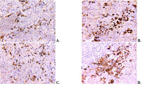
The results of light microscopic analysis of sections stained for Ki-67 and CD20 were consistent with the quantitative data. In the series of CD20 micrographs, denser clusters of positive cells were observed in the red pulp at the early time points (Fig. 1A, B), whereas their number decreased at the later time points (Fig. 1C, D).
{slider title="Fig. 1. Representative micrographs of CD20-positive cells in the red pulp of the rat spleen: A – week 4; B – week 8; C – week 16; D – week 20. Magnification: ×10 eyepiece, ×40 objective ↓" class="tbl-articles my-figure" open="false"}
{/sliders}
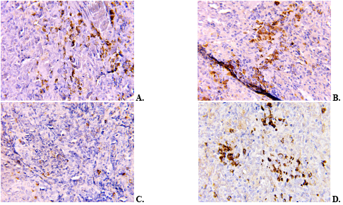
The series of Ki-67 micrographs showed an overall decrease in the number of positive nuclei (Fig. 2A, B), most pronounced at week 16 (Fig. 2C). At week 20, the number of positive cells was higher than at week 16, but remained lower than in the week-0 group (Fig. 2D).
{slider title="Fig. 2. Representative micrographs of Ki-67-positive cells in the red pulp of the rat spleen: A – week 4; B – week 8; C – week 16; D – week 20. Magnification: ×10 eyepiece, ×40 objective ↓" class="tbl-articles my-figure" open="false"}
{/sliders}
Quadratic models of temporal dynamics showed a statistically significant quadratic component for superoxide dismutase activity, malondialdehyde and nitrite concentrations, and the numbers of Ki-67-positive and CD20-positive cells in the red pulp of the rat spleen. This confirmed the nonlinear pattern of changes during the experiment. The heatmap of standardized mean values reflected the sequential change in the relationships among the study parameters (Fig. 3).
{slider title="Fig. 3. Heatmap of standardized group means ↓" class="tbl-articles my-figure" open="false"}
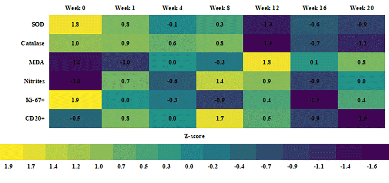
SOD – superoxide dismutase; MDA – malondialdehyde; Ki-67+ - Ki-67-positive cells; CD20+ - CD20-positive cells; Z-score - standardized deviation of the group mean from the overall mean, expressed in standard deviation units. Positive values indicate a parameter level above the overall mean, and negative values indicate a level below it. {/sliders}
Exploratory analysis using Spearman's rank correlation coefficient at the level of individual animals revealed direct relationships between the measures of enzymatic antioxidant defense and inverse associations between antioxidant enzyme activities, concentrations of lipid peroxidation products, and immunohistochemical parameters. Given the nonlinear temporal dynamics, these correlations were regarded as an additional description of the relationships rather than independent evidence of causality.
A synthesis of the data made it possible to provisionally distinguish three consecutive periods of change. In the early period (weeks 1-8), the development of oxidative-nitrosative disturbances was accompanied by an increase in the number of CD20-positive cells. The most pronounced biochemical changes were observed at week 12. In the late period (weeks 16-20), antioxidant defense remained reduced, the number of CD20-positive cells decreased, and proliferative activity was most markedly suppressed.
The observed decrease in superoxide dismutase and catalase activities against a background of increased malondialdehyde concentration is consistent with reports of redox-dependent injury to lymphoid organs after exposure to monosodium glutamate. Das et al. showed that four weeks of monosodium glutamate administration was accompanied by oxidative changes and signs of immune dysfunction in the rat thymus and spleen [9]. The similar direction of the changes supports the assumption that impairment of enzymatic antioxidant defense is one of the factors involved in remodeling of the cellular composition of the spleen.
Recent studies of Ponceau 4R also describe biological effects associated with impaired cell proliferation and redox balance. Kara et al. found a decrease in the mitotic index with increasing Ponceau 4R concentration, whereas signs of genotoxicity were detected mainly at high concentrations [10]. In an experimental model using Artemia franciscana, E124 altered the activity of mitochondrial respiratory chain components and the redox state [11]. Differences between the models preclude direct extrapolation of these results to the rat spleen, although they are consistent with the combination of redox disturbances and a lower number of Ki-67-positive cells observed in our study.
Sodium nitrite can enhance the formation of reactive nitrogen species and secondary lipid peroxidation. In the study by Ahmed et al., its administration to rats was accompanied by an increase in malondialdehyde concentration and a decrease in antioxidant enzyme activities in the tissues examined [12]. In our study, the peak nitrite concentration at week 8 preceded the maximum increase in malondialdehyde at week 12, which may reflect a temporal sequence of nitrosative and lipid peroxidation changes.
The time difference between the maximum manifestations of oxidative-nitrosative stress at week 12 and the lowest number of Ki-67-positive cells at week 16 may reflect delayed suppression of proliferation due to accumulated redox-dependent injury. The increase in this parameter at week 20 was probably associated with compensatory and adaptive renewal of cell populations [9].
The effect of the mixture should not be regarded as a simple sum of the effects of its components. Studies of food additives in combination show that the cellular response to a mixture may differ from the response to individual substances [3], and combined intake of common additives in an animal model was accompanied by simultaneous changes in oxidative status, the inflammatory response, and immune cells [4]. Reviews of the effects of food additives on intestinal microecology and experimental data on interactions between the intestinal barrier and the immune system confirm that the effects of some technological ingredients may extend beyond the organ of initial contact [23, 24]. The results of this study complement previously established changes in antioxidant defense [7] and the macrophage component of the spleen [8] in the same experimental model by linking them to the dynamics of Ki-67-positive and CD20-positive cells.
The combination of reduced proliferative activity and a late decrease in the number of CD20-positive cells may indicate functional suppression and remodeling of the B-cell component of the spleen. These dynamics may be associated with disruption of the stromal-follicular microenvironment that supports the organization of B-cell follicles [25]. At the same time, lower CD20 expression may reflect not only a decrease in the number of mature B cells, but also altered marker expression or their differentiation into CD20-negative plasma cells [26].
A separate limitation of this study is the absence of daily gavage control groups. Because animals of the same initial age were euthanized at different time points, their age at euthanasia differed. All study time points fell within the adult period of the rats' lifespan; however, in the absence of concurrent age-matched control groups, the contribution of age to the observed biochemical and immunohistochemical changes cannot be fully excluded.
Repeated restraint and gavage administration of the solution may potentially induce a short-term stress response, the magnitude of which depends on the technical performance of the procedure and the volume and properties of the solution. In a separate study in male Sprague-Dawley rats acclimated to handling, daily orogastric administration of water at a volume of 5 mL/kg for 28 days, when the procedure was performed properly, was not accompanied by signs of chronic stress [27]. In our study, the initial dosing volume was 2.6 mL/kg; however, exposure lasted up to 20 weeks, and specific markers of the stress response were not measured. Therefore, the contribution of repeated gavage and associated handling stress cannot be qualitatively or quantitatively separated from other factors.
CONCLUSIONS
- Prolonged oral administration of a mixture of food additives (monosodium glutamate, Ponceau 4R, and sodium nitrite) at subtoxic doses was accompanied by the development of persistent oxidative-nitrosative stress in rat spleen tissue, manifested by lower superoxide dismutase and catalase activities and higher malondialdehyde and nitrite concentrations.
- The most pronounced changes in free radical oxidation and lipid peroxidation were observed at week 12 of administration of the food additive mixture, whereas the highest nitrite concentration was recorded at week 8.
- The number of cells representing the B-cell component changed in a phase-dependent manner: the increase at the early time points was followed by a subsequent decrease, most pronounced at week 20, which may indicate functional suppression or reduced marker expression during differentiation into plasma cells.
- The number of proliferatively active cells was lower than the control value at all time points of the experiment; the most pronounced decrease in proliferative activity was observed at week 16 as a result of delayed redox-dependent injury, with partial compensatory and adaptive recovery at week 20.
- Splenic remodeling during prolonged exposure to the food additive mixture followed a nonlinear pattern dependent on exposure duration and combined early reactive changes with a subsequent decrease in proliferative activity and the B-cell component at the later time points.
Contributors:
Mamai O.V. – conceptualization, investigation, writing – original draft;
Bilash S.M. – conceptualization, project supervision, methodology;
Pronina O.M. – project administration;
Bieliaieva O.M. – writing, review and editing;
Pirog-Zakaznikova A.V. – methodology;
Koptev M.M. – data curation;
Bilash V.P., Donchenko S.V. – investigation;
Bilanov O.S., Mazur B.R. – visualization;
Oliinichenko Ya.O. – resources;
Oleksiienko V.V. – validation;
Kolomiiets S.V. – formal analysis;
Funding.The study was conducted as part of the investigator-initiated research project of the Department of Anatomy with Clinical Anatomy and Operative Surgery, Poltava State Medical University, entitled “Pathogenetic mechanisms of post-stress disorders under exogenous and wartime influences and the search for methods of their correction” (state registration No. 0124U003313).
Conflict of interests. The authors declare no conflict of interest.
REFERENCES
- Amchova P, Siska F, Ruda-Kucerova J. Food safety and health concerns of synthetic food colors: an update. Toxics. 2024;12(7):466. doi: https://doi.org/10.3390/toxics12070466
- de Oliveira ZB, Silva da Costa DV, da Silva dos Santos AC, da Silva Júnior AQ, de Lima Silva A, de Santana RCF, et al. Synthetic colors in food: a warning for children’s health. Int J Environ Res Public Health. 2024;21(6):682. doi: https://doi.org/10.3390/ijerph21060682
- Recoules C, Touvier M, Pierre F, Audebert M. Evaluation of the toxic effects of food additives, alone or in mixture, in four human cell models. Food Chem Toxicol. 2025;196:115198. doi: https://doi.org/10.1016/j.fct.2024.115198
- Gomaa S, Mohamed N, Nassef M. Frequently utilized food additives contribute to immune system dysfunction, chronic inflammation and oxidative stress. Drug Chem Toxicol. 2026 Jun 8:1-14. Online ahead of print. doi: https://doi.org/10.1080/01480545.2026.2681174
- Shannon OM, Easton C, Shepherd AI, Siervo M, Bailey SJ, Clifford T. Dietary nitrate and population health: a narrative review of the translational potential of existing laboratory studies. BMC Sports Sci Med Rehabil. 2021;13(1):65. doi: https://doi.org/10.1186/s13102-021-00292-2
- Pant A, Dasgupta D, Tripathi A, Pyaram K. Beyond antioxidation: Keap1-Nrf2 in the development and effector functions of adaptive immune cells. Immunohorizons. 2023;7(4):288-98. doi: https://doi.org/10.4049/immunohorizons.2200061
- Bilash SM, Mamai OV, Pronina OM, Koptev MM, Pirog-Zakaznikova AV, Oliinichenko YaO, et al. State of antioxidant defence of the spleen parenchyma of rats under the influence of a complex of chemical food additives. World Med Biol. 2025;21(92):155-9. doi: https://doi.org/10.26724/2079-8334-2025-2-92-155-159
- Mamai OV, Bilash SM, Pronina OM, Koptev MM, Pirog-Zakaznikova AV, Kolomiiets SV, et al. Morphological and functional state of the macrophagal link of the spleen under conditions of oxidative-nitrosative stress caused by prolonged administration of a complex of chemical food additives. World Med Biol. 2026;22(95):199-202. doi: https://doi.org/10.26724/2079-8334-2026-1-95-199-202
- Das D, Banerjee A, Manna K, Sarkar D, Shil A, Sikdar M, et al. Quercetin counteracts monosodium glutamate to mitigate immunosuppression in the thymus and spleen via redox-guided cellular signaling. Phytomedicine. 2024;126:155226. doi: https://doi.org/10.1016/j.phymed.2023.155226
- Kara SG, Yuzbasioglu D, Avuloglu-Yilmaz E, Unal F. Do the azo food colorings carmoisine and Ponceau 4R have a genotoxic potential? Toxicol Res (Camb). 2025;14(2):tfaf033. doi: https://doi.org/10.1093/toxres/tfaf033
- Fasciolo G, Napolitano G, Muscari Tomajoli MT, Geremia E, Petito A, Gravato C, et al. Synthetic and natural red food dyes affect oxidative metabolism and the redox state in the nauplii of brine shrimp Artemia franciscana. Antioxidants (Basel). 2025;14(6):634. doi: https://doi.org/10.3390/antiox14060634
- Ahmed EH, Abdeen MA, Soliman S. Impact of chlorogenic acid on submandibular salivary gland and liver of albino rats exposed to sodium nitrite. BMC Oral Health. 2024;24:911.
doi: https://doi.org/10.1186/s12903-024-04661-4 - [On approval of the requirements for food flavorings, food additives and food enzymes. Order Ministry of Health of Ukraine No. 45 of 2024 Jan 8]. [Internet]. 2024 [cited 2026 Jul 14]. Ukrainian. Available from: https://zakon.rada.gov.ua/go/z0119-24
- [Requirements for food additives. Order of the Ministry of Health of Ukraine dated 2024 Jan 8 No. 45]. [Internet]. 2024 [cited 2026 Jul 14]. Ukrainian. Available from: https://zakon.rada.gov.ua/go/z0120-24
- Rybolovlev YuR, Rybolovlev RS. [Dosage of substances for mammals according to biological activity constants]. Dokl Akad Nauk SSSR. 1979;247(6):1513-6. Russian.
- Berkalo LV, Bobovych OV, Bobrova NO, Heyko OO, Kaydashev IP, Kutsenko LO, et al. [Methods of clinical and experimental research in medicine]. Poltava: Polimet; 2003. p. 96-99. Ukrainian.
- Korolyuk MA, Ivanova LI, Mayorova IG, Tokarev VE. [Method for determining catalase activity]. Laboratornoe delo. 1988;1:16-19. Russian.
- Gérard-Monnier D, Erdelmeier I, Régnard K, Moze-Henry N, Yadan JC, Chaudière J. Reactions of 1-methyl-2-phenylindole with malondialdehyde and 4-hydroxyalkenals: analytical applications to a colorimetric assay of lipid peroxidation. Chem Res Toxicol. 1998;11(10):1176-83. doi: https://doi.org/10.1021/tx9701790
- Akimov OYe, Kostenko VO. Functioning of nitric oxide cycle in gastric mucosa of rats under excessive combined intake of sodium nitrate and fluoride. Ukr Biochem2016;88(6):70-75. doi: https://doi.org/10.15407/ubj88.06.070
- Anti-CD20 antibody [SP32] (ab64088) [Internet]. Cambridge: Abcam; [cited 2026 Jul 14]. Available from: https://www.abcam.com/en-us/products/primary-antibodies/cd20-antibody-sp32-ab64088
- Anti-Ki67 antibody [SP6] (ab16667) [Internet]. Cambridge: Abcam; [cited 2026 Jul 14]. Available from: https://www.abcam.com/en-us/products/primary-antibodies/ki67-antibody-sp6-ab16667
- Field A. Discovering statistics using IBM SPSS Statistics. 6th ed. London: SAGE Publications; 2024. 1144 p.
- Wang H, Bai J, Miao P, Wei Y, Chen X, Lan H, et al. The key to intestinal health: a review and perspective on food additives. Front Nutr. 2024;11:1420358. doi: https://doi.org/10.3389/fnut.2024.1420358
- Lamas B, Evariste L, Houdeau E. Dysregulation along the gut microbiota-immune system axis after oral exposure to titanium dioxide nanoparticles: a possible environmental factor promoting obesity-related metabolic disorders. Environ Pollut. 2023;330:121795. doi: https://doi.org/10.1016/j.envpol.2023.121795
- Heesters BA, van Megesen K, Tomris I, de Vries RP, Magri G, Spits H. Characterization of human FDCs reveals regulation of T cells and antigen presentation to B cells. J Exp Med. 2021;218(10):e20210790. doi: https://doi.org/10.1084/jem.20210790
- Kläsener K, Jellusova J, Andrieux G, Salzer U, Böhler C, Steiner SN, et al. CD20 as a gatekeeper of the resting state of human B cells. Proc Natl Acad Sci U S A. 2021;118(7):e2021342118. doi: https://doi.org/10.1073/pnas.2021342118
- Turner PV, Vaughn E, Sunohara-Neilson J, Ovari J, Leri F. Oral gavage in rats: animal welfare evaluation. J Am Assoc Lab Anim Sci. 2012 Jan;51(1):25-30. PMID: 22330864; PMCID: PMC3276962.
]]>
Ключові слова: експеримент, щури, важкі метали, кадмій, печінка, накопичення, сукцинат цинку, сукцинат заліза, сукцинат міді
Abstract
The study aimed to determine the dynamics of changes in the accumulation of cadmium and biogenic metals in the liver of experimental pregnant female rats under chronic isolated administration and when combined with biometal succinates. Based on the results of polyelement analysis, the degree of dyselementosis in the liver microelement composition and the bioprotective properties of zinc, iron, and copper succinates were evaluated. The effect of cadmium chloride on the accumulation levels of zinc, iron, and copper in the rat liver was assessed during their simultaneous entry into the body throughout pregnancy. An exceptionally high capacity for cadmium accumulation was established, with a critical excess over the control group by 67-fold on day 13 and by 87-fold at the end of the experiment. Iron, zinc, and copper succinates exhibit a pronounced protective effect, significantly reducing cadmium accumulation in the liver of pregnant rats on days 13 and 19 of the study. In terms of efficacy in limiting toxicant accumulation, the compounds form the following descending order: iron succinate > zinc succinate > copper succinate. Chronic cadmium intoxication was found to disrupt zinc metabolism in the liver of pregnant rats, abnormally increasing its level by 1.9-fold (day 13) and 1.4-fold (day 19) as a protective response for metallothionein biosynthesis. The administration of biogenic metal succinates effectively modulates this process; however, their normalizing effect depends on the gestational age. For the correction of zinc metabolism disorders, copper succinate is priority in the first half of pregnancy, while iron succinate is preferred in the second half. It was established that cadmium chloride intoxication profoundly disrupts iron metabolism and blocks its transport mechanisms, causing a rapid and significant (p<0.05) accumulation of iron in the liver parenchyma by more than 2-fold. This indicates a pronounced toxic effect of the xenobiotic on the iron-storing function of the liver at both gestational stages. The most pronounced protective effect was demonstrated by zinc succinate, which maintained the iron concentration at a level closest to the control values. This confirms the high efficiency of zinc succinate in supporting microelement homeostasis at both periods of the study. The effect of cadmium chloride on copper homeostasis disruption in the liver parenchyma of pregnant females was evaluated. It caused a rapid and significant increase in copper concentration throughout the entire observation period, indicating profound cadmium-induced alterations in the microelement balance and the retention of essential metals in the maternal depot.
Реферат
Динаміка змін мікроелементного балансу печінки щурів за умов підгострого впливу хлоридом кадмію в комбінації із сукцинатами біометалів. Шаторна В.Ф., Захаров С.В., Закут І.М. Метою дослідження було визначення динаміки змін накопичення кадмію та біогенних металів в печінці дослідних вагітних самиць щурів при підгострому ізольованому та комбінованому із сукцинатами біометалів введенні. За результатами поліелементного аналізу визначали ступінь дизелементозу мікроелементного складу печінки та біопротекторні властивості сукцинатів цинку, заліза та міді. Оцінено вплив хлориду кадмію на рівень накопичення цинку, заліза та міді печінкою щурів при їх одночасному надходженні в організм упродовж вагітності. Встановлено надзвичайно високу здатність накопичення кадмію з критичним перевищенням контролю в 67 разів на 13-ту добу та у 87 разів наприкінці експерименту. Сукцинати заліза, цинку та міді чинять виражений захисний ефект, значно знижуючи накопичення кадмію в печінці вагітних щурів на 13-ту та 19-ту добу дослідження. За ефективністю обмеження акумуляції токсиканта сполуки утворюють такий спадний ряд: сукцинат заліза > сукцинат цинку > сукцинат міді. Виявлено, що підгостра інтоксикація кадмієм порушує метаболізм цинку в печінці вагітних щурів, аномально підвищуючи його рівень в 1,9 раза (13-та доба) та в 1,4 раза (19-та доба) як захисну реакцію для біосинтезу металотіонеїнів. Застосування сукцинатів біогенних металів ефективно модулює цей процес, проте їхня нормалізація залежить від терміну гестації. Для корекції порушень обміну цинку пріоритетним у першій половині вагітності є сукцинат міді, а в другій половині – сукцинат заліза. Встановлено, що інтоксикація хлоридом кадмію глибоко порушує метаболізм заліза та блокує його транспортні механізми, викликаючи стрімке й достовірне (p<0,05) накопичення заліза в паренхімі печінки більше ніж у 2 рази, що свідчить про виражений токсичний вплив ксенобіотика на залізодепонуючу функцію печінки на обох термінах гестації. Найбільш виражений протективний ефект продемонстрував сукцинат цинку, де спостерігалося утримання концентрації заліза на рівні, максимально наближеному до контрольних значень, що підтверджує високу ефективність сукцинату цинку для підтримки мікроелементного гомеостазу на обох термінах дослідження. Оцінено вплив хлориду кадмію на порушення гомеостазу міді в паренхімі печінки вагітних самиць, який викликає стрімке й достовірне збільшення її концентрації впродовж усього періоду спостереження, що свідчить про глибокі кадмій-індуковані зміни мікроелементного балансу та затримання есенціальних металів у материнському депо.
Сучасний стан біосфери характеризується зростанням антропогенного навантаження, серед якого особливе місце посідає забруднення важкими металами. Хлорид кадмію є одним з найбільш небезпечних екотоксикантів з вираженою кумулятивною дією. Тривалий період напіввиведення з біологічних систем, який становить від 15 до 30 років, у поєднанні з високою хімічною стійкістю зумовлює персистенцію цього металу в навколишньому середовищі без втрати первинних властивостей [1]. Його тривале надходження до організму призводить до накопичення в організмі та хронічної інтоксикації, що супроводжується деструктивними змінами на клітинному та тканинному рівнях. Одним з ключових механізмів патогенезу кадмієвої токсичності є дезорганізація метаболізму есенціальних елементів.
Функціонування мікроелементів у живих системах ґрунтується на принципах жорсткого синергізму та антагонізму. Біохімічний гомеостаз є настільки взаємозалежним, що відхилення від норми навіть одного компонента неминуче запускає каскадний дисбаланс усієї мікроелементної системи [2, 3]. З огляду на це, об'єктивним критерієм функціонального стану організму є не лише абсолютний вміст конкретних мікроелементів, а, насамперед, їхнє коректне міждіапазонне співвідношення. Розвиток мікроелементозів, викликаний браком життєво необхідних сполук або інтерференцією з боку ксенобіотиків, суттєво модифікує та маскує клінічну картину отруєнь [3, 4]. Це значно ускладнює диференційну діагностику хронічних інтоксикацій.
Після абсорбції в шлунково-кишковому тракті та потрапляння вв системний кровотік мікроелементи розподіляються в органах украй нерівномірно. Найвища концентрація більшості як ендогенних елементів, так і екзогенних токсикантів (зокрема кадмію) реєструється в гепатобіліарній системі [5, 6]. Оскільки печінка виконує роль головного функціонального депо мікроелементів в організмі, вона стає основним органом-мішенню та першою зазнає деструктивного впливу за умов тривалої хімічної агресії [7, 8]. Це дозволяє розглядати печінку як головне функціональне депо мікроелементів, яке першим зазнає системного впливу за умов постійного довготривалого антропогенного навантаження.
Взаємодія мікроелементів в організмі ґрунтується на складних процесах синергізму та антагонізму [4, 5]. Через це дефіцит або надлишок навіть одного компонента неминуче руйнує загальний елементний гомеостаз за принципом ланцюгової реакції. Отже, фізіологічний стан організму визначається не стільки абсолютними значеннями окремих мікроелементів, скільки їхнім збалансованим співвідношенням.
Таким чином, актуальність теми зумовлена широким поширенням кадмієвої інтоксикації в промислових регіонах та її згубним впливом на організм вагітних і плода. Вивчення міжіонних взаємодій кадмію з есенціальними мікроелементами відкриває нові можливості для патогенетичної корекції метаболічних порушень. Застосування біометал-сукцинатів є перспективним напрямком для створення ефективних, безпечних і доступних вітчизняних засобів антенатального метаболічного захисту.
Мета дослідження – визначити динаміку змін накопичення кадмію та біогенних металів у печінці дослідних вагітних самиць щурів при підгострому ізольованому та комбінованому із сукцинатами біометалів введенні. Визначити особливості механізму комбінованого антенатального впливу хлориду кадмію і сукцинатів мікроелементів та ступінь дизелементозу мікроелементного складу печінки.
МАТЕРІАЛИ ТА МЕТОДИ ДОСЛІДЖЕНЬ
Експериментальна частина дослідження була виконана на молодих вагітних самицях щурів лінії Wistar віком 2,5-3 місяці, яких отримували з ліцензованого розплідника «Далі 2001» (м. Київ). Тварин утримували у віварії Дніпровського державного медичного університету в стандартних пластикових клітках (по 3-4 особини) за контрольованих умов: t=20-25°C, вологість ≥50%, регулярна вентиляція, світловий режим «день/ніч» (12/12 год). Раціон був стандартний, з вільним доступом до води.
Дослідження схвалені комісією з питань біомедичної етики Дніпровського державного медичного університету (протокол № 39 від 17.06.2026 р.) та проведені відповідно до вимог Закону України «Про захист тварин від жорстокого поводження» від 21.02.2006 р. № 3447-IV та Наказу Міністерства освіти і науки, молоді та спорту України від 01.03.2012 р. № 249 «Про затвердження Порядку проведення науковими установами дослідів, експериментів на тваринах», а також з урахуванням принципів Директиви 2010/63/ЄС Європейського парламенту і Ради від 22.09.2010 р. про захист тварин, які використовуються в наукових цілях.
Для проведення експерименту у віварію Дніпровського державного медичного університету тварин було розподілено на чотири дослідні групи та контрольну. Введення препаратів здійснювали внутрішньошлунково, щоденно впродовж усього терміну гестації самиць. Група I (контроль, 10 самиць): тварини отримували 0,5 мл ізотонічного 0,9% розчину NaCl для встановлення фонових показників. Група II (модель інтоксикації, 18 самиць): застосовували ізольований вплив хлориду кадмію в дозі 3,0 мг/кг. Застосована доза хлориду кадмію становила 3% від LD₅₀ для щурів при пероральному введенні, що відповідало низькому токсичному рівню. Такий рівень експозиції є достатнім для моделювання гестаційної інтоксикації та порушень гомеостазу металів без розвитку летальних наслідків, що дозволяло проводити тривалий щоденний вплив протягом усієї вагітності. Група III (корекція цинком, 18 самиць): на фоні щоденного введення 3,0 мг/кг хлориду кадмію тварини отримували сукцинат цинку в дозі 5 мг/кг для оцінювання його протективних властивостей. Група IV (корекція залізом, 18 самиць): схема передбачала поєднання кадмію із сукцинатом заліза в дозі 5,0 мг/кг з метою дослідження його коригувальної дії. Група V (корекція купрумом, 18 самиць): схема передбачала поєднання кадмію із сукцинатом міді в дозі 0,1 мг/кг з метою дослідження його коригувальної дії. Дози сукцинатів біметалів вибрані як фізіологічно прийнятні та безпечні для вагітних щурів, проте достатні для впливу на системний пул заліза, цинку для корекції кадмієво-індукованих порушень без розвитку залізо-індукованої токсичності.
Для оцінювання специфіки акумуляції кадмію, цинку та заліза в паренхімі печінки вагітних самиць щурів на 13-ту та 19-ту добу гестації за умов ізольованої інтоксикації хлоридом кадмію та супутньої корекції сукцинатами есенціальних металів застосовували метод атомно-емісійної спектрометрії з електродуговою атомізацією. Цей аналітичний підхід дозволяє здійснювати прецизійний якісний і кількісний поліелементний скринінг біосубстратів різного походження. Елементний аналіз зразків виконували на базі ДП «Український науково-дослідний інститут медицини транспорту МОЗ України» (м. Одеса) в рамках договору про науково-практичне співробітництво. Кількісне визначення мікроелементів у печінці проводили методом атомно-емісійної спектрометрії з дуговою атомізацією на приладі Емас‑200 CCD відповідно до принципів, описаних Кучеруком Д.Д., Головком А.К. [9]. Для порівняння кількісних показників вмісту металів у тканинах печінки щурів на різних термінах вагітності застосовували t-критерій Стьюдента для незалежних вибірок. Статистичний аналіз проводили з метою порівняння кількісних показників вмісту металів у тканинах печінки щурів на різних термінах вагітності. Для оцінювання різниці між незалежними вибірками застосовували t-критерій Стьюдента. Статистична обробка даних проводилась із застосуванням Microsoft Exel R версія 4.3.1 (https//www.r-project.org/) [13]. Аналіз виконували відповідно до сучасних стандартів прозорості та відтворюваності біостатистичних досліджень.
РЕЗУЛЬТАТИ ТА ЇХ ОБГОВОРЕННЯ
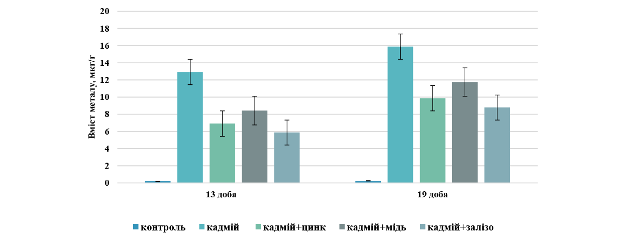
Аналіз вмісту кадмію в печінці вагітних самиць щурів за умов щоденної інтоксикації хлоридом кадмію виявив значне накопичення цього металу в органі впродовж усього періоду спостереження (рис. 1). За результатами поліелементного аналізу встановлено, що в печінці самиць контрольної групи вміст кадмію впродовж експерименту залишався на стабільно низькому рівні (близько 0,15-0,18 мкг/г), що відповідало фізіологічній нормі. На 13-ту добу експерименту вміст кадмію в печінках самиць групи ізольованого впливу хлоридом кадмію зріс у десятки разів порівняно з контролем.
Внутрішньошлункове введення хлориду кадмію призвело до стрімкого накопичення металу в печінці, а саме: на 13-ту добу його концентрація зросла до 12,9±1,5 мкг/г, а на 19-ту добу спостерігалося подальше достовірне збільшення показника до 15,9±1,5 мкг/г, що було статистично значущою різницею (p<0,05) та свідчило про високу кумулятивну здатність кадмію в гепатоцитах протягом вагітності. Таким чином, у першій половині вагітності зростання рівня кадмію відбулося порівняно з контролем у 67 разів, а наприкінці експерименту – у 87 разів. Активне накопичення кадмію печінкою порівняно з контролем під час підгострого експерименту зумовлене специфічними фізіологічними, біохімічними та транспортними механізмами печінки щурів. При внутрішньошлунковому введенні печінка першою приймає кров від шлунково-кишкового тракту через ворітну вену, тобто портальну циркуляцію. Орган працює як головний фільтр, затримуючи ксенобіотики для захисту інших систем для детоксикації. Кадмій у печінці індукує масштабний синтез білків металотіонеїнів (МТ) у гепатоцитах, які міцно зв'язують іони Cd, і утворюється тканинне депо. Загальновідомо, що утворені комплекси Cd-MT мають довгий період напіввиведення і затримуються в печінці. Такий рівень кумуляції кадмію ми пояснюємо бар'єрною роллю печінки, яка виступає головним детоксикаційним фільтром організму, першою затримуючи токсикант з портальної циркуляції шлунково-кишкового тракту для захисту інших систем.
{slider title="Рис. 1. Динаміка накопичення кадмію в паренхімі печінки вагітних щурів на 13-й та 19-й добі гестації (мкг/г) ↓" class="tbl-articles my-figure" open="false"}
{/sliders}
У групі комбінованого введення хлориду кадмію із сукцинатом цинку виявився високий протективний ефект. На 13-ту добу експерименту вміст кадмію в цій групі становив 6,9±1,5 мкг/г, що на 46,5% менше, ніж у групі з ізольованим введенням токсиканта. На 19-ту добу рівень накопичення паренхімою печінки кадмію дещо підвищився до 9,9±1,4 мкг/г, проте він залишався на 37,7% нижчим порівняно з групою інтоксикації. Це підтверджує виражений антагонізм іонів цинку та їх здатність блокувати депонування кадмію.
Проте в групі комбінованого введення хлориду кадмію із сукцинатом заліза ми визначали найбільш виражений протекторний ефект (рис. 1). У цій групі експерименту був найнижчий показник рівня накопичення кадмію, хоча він не завжди мав достовірну різницю з рівнем накопичення групою комбінації хлориду кадмію із сукцинатом цинку.
У групі комбінованого введення хлориду кадмію із сукцинатом міді також визначалось зниження рівня токсиканта, але цей вплив був менш ефективним порівняно з цинком та залізом. На 13-ту добу рівень кадмію становив 8,4±1,6 мкг/г, тобто спостерігалось зниження на 34,9% відносно групи кадмію. На 19-ту добу показник зріс до 11,8±1,7 мкг/г, що на 25,8% менше за показник групи без корекції. Порівнюючи дані про накопичення кадмію печінкою самиць у всіх групах корекції, можна констатувати, що сукцинат цинку та сукцинат заліза ефективніше за сукцинат міді обмежують накопичення кадмію печінкою на обох термінах дослідження в зазначених дозах та способі введення в експерименті на щурах.
Цинк – це мікроелемент, який є життєво важливим для імунної системи, оскільки його дефіцит пригнічує вроджені та набуті імунні механізми, підвищуючи схильність до інфекційних, автоімунних й онкологічних процесів. Крім того, оптимальний рівень цинку відіграє фундаментальну роль в ендокринній системі: він регулює синтез і структуру гормонів щитоподібної залози та забезпечує повноцінну роботу підшлункової залози, беручи безпосередню участь у синтезі та зберіганні інсуліну і глюкагону. Найбільше цинку накопичується в скелетних м'язах та кістковій тканині, проте печінка займає третє місце в ступені накопичення після опорно-рухового апарату та шкіри й волосся.
У нашому експерименті аналіз накопичення цинку паренхімою печінки показав таке. У контрольній групі на 13-ту та 19-ту добу експерименту рівень цинку збільшувався, що ми пояснюємо метаболічними змінами в організмі у зв’язку з перебігом вагітності самиць. На 13-й добі рівень цинку в печінці самиць становив 24,31±3,61 мкг/г, а наприкінці вагітності вже добігав 35,92±4,70 мкг/г, достовірність різниці p<0,05. Ізольоване введення хлориду кадмію змінювало рівень цинку в досліджуваних зразках (рис. 2).
Вже на 13-й добі експерименту в групі ізольованого введення кадмію рівень цинку підвищувався в 1,9 раза, перевищував контрольні дані й становив 45,21±2,40 мкг/г, а на 19-ту добу перевищення відбувалось в 1,4 раза, що становило 48,76±4,31 мкг/г. Порівняння отриманих результатів щодо накопичення цинку не було показовим для групи комбінації кадмію і цинку, тому що рівень цього мікроелемента хоч і був найвищим, але тварини цієї групи його отримували щоденно. Тому основні порівняння нами проводились між іншими групами комбінованого введення.
Неочікуваними виявились кількісні показники цинку в групі комбінованого введення хлориду кадмію із сукцинатом міді. На 13-ту добу експерименту рівень цинку (31,83±4,91 мкг/г) був зіставним з контрольними значеннями, як і в групі комбінації із залізом 33,51± 3,18 мкг/г. Ситуація на 19 -й добі експерименту змінювалась у межах цих груп комбінації. Саме в групі комбінованого введення кадмію із залізом рівень цинку становив 39,87±4,20 мкг/г, що не мало достовірної різниці з контролем, а в групі комбінації із сукцинатом міді показник дорівнював такому в групі кадмієвої інтоксикації. Таким чином, у першій половині вагітності рівень цинку підтримувався введенням сукцинату міді, а в другій половині – сукцинатом заліза.
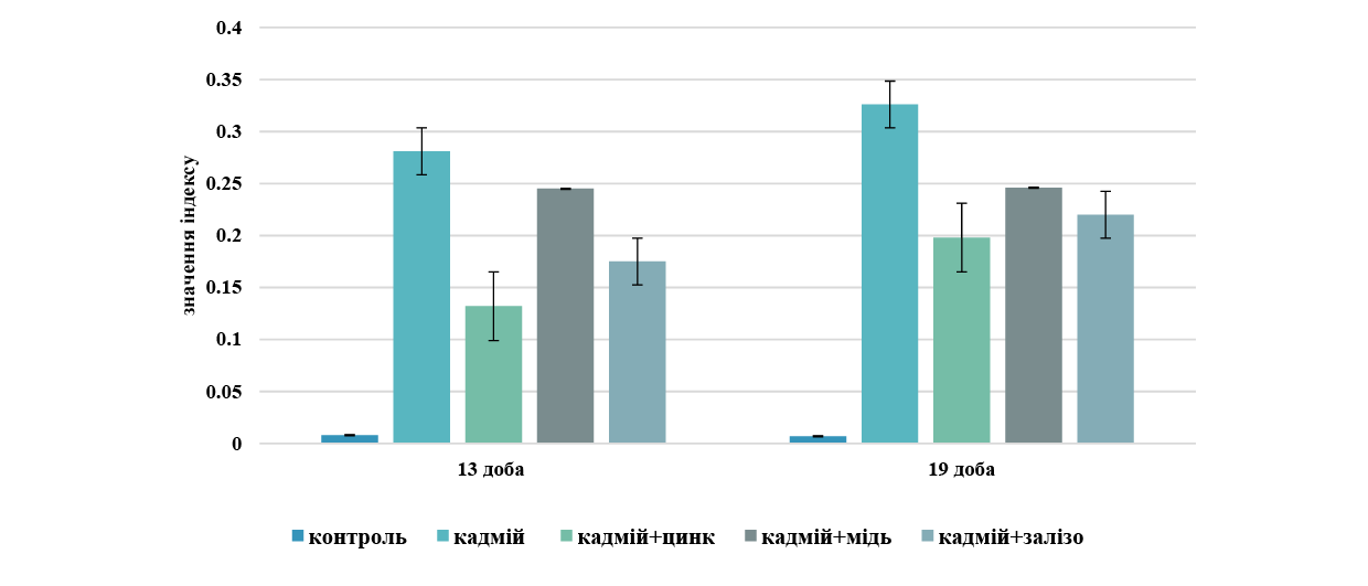
Нами також розраховувався показник співвідношення кадмію до цинку, який наочно демонструє рівень токсичного навантаження організму важкими металами та ступінь порушення мінерального обміну, оскільки цинк та кадмій є біологічними антагоністами, вони конкурують за одні й ті самі рецептори та транспортні білки в організмі (рис. 2).
{slider title="Рис. 2. Динаміка індексу співвідношення кадмію до цинку в печінці вагітних щурів на 13-й та 19-й добі гестації ↓" class="tbl-articles my-figure" open="false"}
{/sliders}
На початковому терміні спостереження показник у контрольній групі залишався мінімальним і становив близько 0,01. Введення кадмію призвело до різкого зростання індексу до 0,28 (p<0,05), що свідчить про гостре накопичення токсичного металу в печінці та порушення балансу мікроелементів.
Застосування біогенних металів у комбінації з тією самою дозою кадмію на 13-ту добу показало різну ефективність досліджуваних сполук. Цинк найбільш ефективно знижував індекс до 0,13, що майже вдвічі менше за групу ізольованого впливу кадмію. Залізо виявляло помірний захисний ефект, знижуючи показник до 0,17, а мідь виявилась найменш ефективною, оскільки індекс зменшувався лише до 0,24.
На 19-й добі експерименту в контрольній групі індекс зберігався на вихідному стабільно низькому рівні (~0,01). Проте в групі з ізольованим введенням кадмію спостерігалося його подальше зростання до пікового значення 0,33, що підтверджувало прогресуючу інтоксикацію. При оцінюванні потенційної корекції сукцинатами біометалів на 19-ту добу виявлено такі зміни. У групі кадмій+цинк індекс зростав порівняно з 13-ю добою до ~0,20, проте цинк і надалі надійно утримував лідерство за рівнем захисту печінки. У групі комбінованого введення хлориду кадмію і сукцинату заліза показник також підвищувався до 0,22, що вказує на поступове виснаження захисного ресурсу заліза за умов тривалого токсичного впливу. Комбіноване введення кадмію з міддю залишало індекс на попередньому рівні (~0,245), демонструючи найслабший ефект серед усіх досліджуваних металів-антагоністів.
Динаміка накопичення заліза (мг/г) у паренхімі печінки вагітних самиць дослідних тварин у розрізі всіх експериментальних даних дозволяє оцінити особливості метаболізму та депонування феруму в організмі під впливом досліджуваних чинників. У тварин контрольної групи рівень заліза в печінці впродовж всього періоду дослідження перебував у межах стабільної фізіологічної норми для вагітних самиць. На 13-ту добу експерименту рівень заліза в паренхімі печінки становив 44,37±3,61 мкг/г, а на 19-ту добу – 45,42±4,20 мкг/г. За результатами поліелементного аналізу визначалась тенденція помірного та прогнозованого коливання концентрації, що свідчить про адекватне забезпечення плодів залізом без виснаження материнського депо.
Ізольоване введення хлориду кадмію призвело до стрімкого достовірного підвищення рівня заліза в паренхімі печінки порівняно з контролем (p<0,05). На 13-ту добу експерименту рівень заліза в печінці тварин кадмієвої інтоксикації збільшувався до 86,29±4,41 мкг/г, що було в 1,9 раза вище за контрольні показники, а на 19-ту добу зростав до 98,66± 5,30 мкг/г, що перевищувало контроль у 2,14 раза. Це вказує на активну мобілізацію депонованого заліза на потреби фетоплацентарного комплексу або на порушення його метаболізму. При цьому в групі комбінованого впливу кадмію із сукцинатом цинку спостерігалось утримання концентрації заліза до рівня майже контрольних значень. Це підтверджує ефективність одночасного надходження кадмію із сукцинатом цинку для підтримки мікроелементного балансу організму на обох термінах дослідження.
Комбіноване введення хлориду кадмію із сукцинатом міді також підвищувало рівень заліза в зразках порівняно з контролем, проте порівняно з групою кадмієвої інтоксикації знижувало, хоча і в меншому ступені, ніж комбіноване введення кадмію з цинком. Рівень заліза в паренхімі печінки в групі комбінованого введення кадмій+сукцинат заліза не є показовим, бо тварини отримували залізо щоденно, тому ці результати накопичення не підлягали порівнянню.
Мідь є життєво важливим мікроелементом, який виступає кофактором багатьох металоферментів, що регулюють дихання, кровотворення та антиоксидантний захист. Цей мікроелемент входить до складу цитохром-с-оксидази як ключового ферменту мітохондрій, що забезпечує синтез АТФ, відповідаючи за клітинне дихання та енергію. У процесах кровотворення та обміну заліза важливу роль відіграє фермент церулоплазмін, який є головним білком-переносником міді, що окиснює залізо, дозволяючи йому зв'язуватися з трансферином і брати участь у синтезі гемоглобіну. Антиоксидантний захист організму також залежить від міді, яка разом із цинком утворює фермент супероксиддисмутазу, що нейтралізує вільні радикали.
Мідь відіграє критичну роль у функціонуванні печінки, оскільки цей орган є головним центром метаболізму, депонування та екскреції цього мікроелемента. Як центральний орган обміну, печінка захоплює мідь з кровотоку після її всмоктування в кишківнику та розподіляє її для потреб усього організму. При цьому гепатоцити синтезують церулоплазмін – головний мідьвмісний білок крові, який забезпечує безпечний транспорт міді та бере участь в обміні заліза. У групі контролю рівень міді в паренхімі печінки достовірно (р=0,05) зростав з 13-ї до 19-ї доби експерименту в 1,3 раза, що ми пояснюємо фізіологічними перебудовами та зміною балансу мікроелементів в організмі вагітної самиці. Це є фізіологічним процесом, зумовленим необхідністю забезпечити плід, що активно розвивається, цим мікроелементом, а також гормональними змінами в організмі матері. Вагітність супроводжується значним зростанням рівня церулоплазміну в крові матері, що, у свою чергу, активує підвищене всмоктування міді в кишківнику та знижує її виведення, що призводить до кумуляції міді в печінці та її подальшого транспортування. Мідь також необхідна для активізації ферментів, що включають залізо в порфіринове кільце, що важливо для посиленого біосинтезу гемоглобіну. Мідь як мікроелемент є життєво важливою для формування тканин плоду, зокрема печінки та мозку. У другій половині вагітності (після 13-ї доби) зростає потреба у синтезі фетальних білків (наприклад, металотіонеїнів), які потребують міді. Організм вагітної самиці накопичує мідь, щоб потім ефективно переносити її через плаценту, тобто відбувається активний транспорт мікроелемента до плоду через плаценту.
У групі ізольованого впливу кадмієм рівень міді збільшувався порівняно з контрольними показниками впродовж експерименту таким чином. На 13-ту добу експерименту рівень міді становив 1,904±0,08 мкг/г, що у 2,3 раза перевищувало контроль, а на 19-й добі експерименту становив 2,347±0,09 мкг/г, що перевищувало контрольні дані у 2,1 раза.
Таким чином, ізольована інтоксикація організму вагітної самиці хлоридом кадмію призводила до дизелементозу по міді в паренхімі печінки як на 13-ту добу вагітності, так і наприкінці ембріогенезу. У групах комбінованого введення визначалось зниження рівня міді в бік до контрольного показника при поєднанні кадмію з цинком та поєднанні із залізом на обох термінах експериментального дослідження. Вимірювання в групі поєднання кадмію із сукцинатом міді зі зрозумілих причин не проводилось. Найбільш ефективне відновлення рівня міді визначалось у групі комбінованого впливу кадмієм із сукцинатом цинку. Загалом отримані дані демонструють біоантагоністичний характер сукцинатів досліджуваних біометалів, що дозволяє їх розглядати як потенційні протектори гепатотоксичності кадмію в організмі дослідних тварин у підгострому експерименті введення зазначених доз та способу введення.
Сукцинати заліза, цинку та міді виявляли виражений захисний ефект акумуляції кадмію в печінці вагітних щурів на 13-ту та 19-ту добу гестації. За здатністю знижувати депонування токсиканта сполуки утворюють спадний ряд: сукцинат заліза > сукцинат цинку > сукцинат міді. Аналогічні спрямування визначення нових потенційних біоантагоністів кадмію визначались у попередніх дослідженнях Дніпровської морфологічної школи ДДМУ, тільки на інших органах: серце [10], нирки [11], статеві залози, хоча доза кадмію в тій серії досліджень була значно нижчою [12]. Результати мали досить різноспрямований характер упродовж етапів усього експериментального періоду.
Уперше проведено комплексне порівняльне дослідження протективної ефективності сукцинатів заліза, цинку та міді щодо депонування кадмію в паренхімі печінки на різних етапах гестаційного періоду. Встановлено чітку залежність модифікаційного впливу досліджуваних сполук на метаболізм цинку та заліза. Доповнено наукові дані щодо механізмів біологічного антагонізму та захисної ролі сукцинатів есенціальних мікроелементів за умов поєднаного надходження ксенобіотика. Уперше деталізовано характер порушень залізодепонуючої функції печінки вагітних самиць під впливом щоденної кадмієвої інтоксикації.
ВИСНОВКИ
- Вміст кадмію в паренхімі печінки вагітних самиць при ізольованому впливі розчином хлориду кадмію в дозі 3,0мг/кг зростав у першій половині вагітності в 67разів, а наприкінці експерименту – у 87 разів (p<0,0001) порівняно з контрольною групою, що свідчить про високу кумулятивну здатність кадмію в організмі.
- Комбіноване надходження з хлоридом кадмію сукцинатів заліза, цинку та міді знижувало депонування токсиканта в паренхімі печінки вагітних самиць щурів на 13-ту та 19-ту добу гестації. За ступенем протективної ефективності досліджувані сполуки утворювали такий спадний ряд: сукцинат заліза>сукцинат цинку > сукцинат міді. Найбільш стабільну дію впродовж усього експерименту виявив сукцинат заліза.
- Довготривале кадмієве навантаження порушує фізіологічний баланс цинку, аномально підвищуючи його рівень у паренхімі печінки, тобто провокує дизелементоз по цинку. Нормалізуючий вплив сукцинатів на обмін цинку має чітку етапну залежність: у першій половині вагітності ефективними є сукцинати міді та заліза (утримують показники в межах норми), тоді як у другій половині стабільний ефект зберігає виключно сукцинат заліза, а дія сукцинату міді повністю нівелюється.
- Ізольований вплив хлориду кадмію блокував транспортні механізми феруму в організмі та глибоко порушував залізодепонуючу функцію печінки вагітних самиць. Це проявлялося стрімким і достовірним (p<0,05) накопиченням заліза в паренхімі органа порівняно зі стабільною фізіологічною нормою контрольної групи: в 1,9 раза на 13-ту добу та у 2,14 раза на 19-ту добу експерименту.
- Уперше доведено етапну залежність захисної дії сукцинатів біометалів в умовах кадмієвої інтоксикації вагітних. Обґрунтовано доцільність диференційованого підходу до корекції мікроелементозів: застосування сукцинату міді є пріоритетним на ранніх термінах гестації, тоді як сукцинат заліза забезпечує надійний комплексний захист (як щодо обмеження кумуляції ксенобіотика, так і щодо стабілізації обміну есенціальних металів) упродовж усього періоду вагітності.
Внески авторів:
Шаторна В.Ф. – концептуалізація, ведення, методологія, адміністрування проєкту;
Захаров С.В. – концептуалізація, нагляд, дослідження, курація даних, перегляд та редагування;
Закут І.М. – методологія, дослідження, формальний аналіз, написання: рецензування та редагування.
Фінансування. Дослідження не має зовнішніх джерел фінансування.
Конфлікт інтересів. Автори заявляють про відсутність конфлікту інтересів.
REFERENCES
- Qu F, Zheng W. Cadmium exposure: mechanisms and pathways of toxicity and implications for human health. Toxics.2024;12(6):388. doi: https://doi.org/10.3390/toxics12060388
- Wang M, Chen Z, Song W, Hong D, Huang L, Li Y. A review on cadmium exposure in the population and intervention strategies against cadmium toxicity. Bull EnvironContam 2021;106(1):65-74. doi: https://doi.org/10.1007/s00128-020-03088-1
- Zhang X, Reynolds М. Cadmium exposure in living organisms: A short review. Sci Total Environ. 2019;678:761-7. doi: https://doi.org/10.1016/j.scitotenv.2019.04.395
- Erdem O, Yazihan N, Kocak MK, Sayal A, Akcil E. Influence of chronic cadmium exposure on the tissue distribution of copper and zinc and oxidative stress parameters in rats. Toxicol Ind Health. 2016;32(8):1505-14. doi: https://doi.org/10.1177/0748233714566875
- Kusak R, Nasiadek M, Stragierowicz J, Hanke W, Kilanowicz A. Changes in Endogenous Essential Metal Homeostasis in the Liver and Kidneys during a Six-Month Follow-Up Period after Subchronic Cadmium Exposure. IntJMol 2024;25(7):3829. doi: https://doi.org/10.3390/ijms25073829
- Chen J, Wang Z, Li W, Ying S. Research advances on cadmium-induced toxicity in hepatic macrophages. Toxics.2026;14(1):57. doi: https://doi.org/10.3390/toxics14010057
- Apykhtina OL, Kozlov KP. [Micro- and macroelements content in rats' liver and kidneys after exposure to cadmium sulfide nanoparticles and cadmium chloride]. Ukr zh probl med pratsi. 2017;3(52):60-66. Ukrainian. doi: https://doi.org/10.33573/ujoh2017.03.060
- Shatorna VF, Tymchuk KM. [Influence of chronic isolated administration of cadmium chloride on morphological structures of the small intestine wall in pregnant female rats]. Aktual probl suchas med. 2023;23(4):252-6. doi: https://doi.org/10.31718/2077–1096.23.4.252
- Kucheruk DD, Holovko AK. [Atomic emission spectroscopy: methodological recommendations]. Kyiv: NTUU "KPI"; 2010. 28 р. Ukrainian.
- Shatorna VF, Lomyha LL. Analysis of changes in the microelement status of the heart of rats during chronic cadmium intoxication. Visnyk Problem Biolohii i Medytsyny.2023;4(171):107-16. doi: https://doi.org/10.29254/2077-4214-2023-4-171-107-116
- Shatorna VF, Krasnov OO. [Dynamics of accumulation of cadmium, zinc and copper in the kidneys and blood of rats with isolated administration of cadmium and combined with zinc and copper succinate]. Visnyk Problem Biolohii i Medytsyny. 2022;4(167):242-50. Ukrainian. doi: https://doi.org/10.29254/2077-4214-2022-4-167-242-250
- Nefodova OO, Hruzd VV. [Changes in the microelement composition of the gonads of male rats under the influence of cadmium intoxication and correctors according to polyelement analysis]. Perspekt Innov Nauky. 2023;15(33):1192-204. Ukrainian. doi: https://doi.org/10.52058/2786-4952-2023-15(33)-1192-1204
Ключові слова: експеримент, ембріони, щури, важкі метали, кадмій, цинк, залізо, ембріотоксичність, ембріональна смертність, резорбція ембріонів
Abstract
The aim of the study was to determine the embryotoxicity of cadmium chloride under the conditions of its long-term isolated administration and in combination with zinc or iron succinates in an experiment on rats to assess the modifying effect of succinates on the embryotoxicity of cadmium. To determine the degree of embryotoxicity and calculate the types of embryonic mortality, the number of litters, the weight of fetuses, the number of corpora lutea in the ovaries of females, as well as live and resorbed embryos in the uterine horns of rats were analyzed. The effect of zinc and iron succinates on the level of embryotoxicity of cadmium chloride in rats with their simultaneous long-term daily intake into the body during pregnancy was experimentally evaluated. The results obtained by us demonstrate a significant increase in all indicators of embryonic mortality in the group of isolated exposure to cadmium chloride compared to the control group both on the 13th and 20th days of the experiment. It was established that isolated chronic exposure to cadmium chloride has a pronounced embryotoxic effect: on the 13th day of the experiment, which is manifested both by a significant decrease in the number of live fetuses compared to the control (p<0.05) and by a 4-fold increase in the indicator of total embryonic mortality against the background of active fetal resorption. At the end of the gestation period (20th day), the toxic pressure of cadmium in the studied dose reaches a maximum: the number of live fetuses critically decreases, and the indicator of total embryonic mortality exceeds the level of the control group by 10.7 times. Iron and zinc succinates exhibit pronounced protective properties as cadmium bioantagonists, significantly reducing the levels of embryonic death on both terms of gestation. On the 13th day, iron succinate reduces the total embryonic mortality by 2.8 times compared to the cadmium intoxication group, mainly due to preimplantation protection, and zinc succinate – by 2.9 times, bringing the indicators closer to the control. On the 20th day of gestation in the groups of combined administration with zinc and iron succinates, the level of total embryonic mortality decreases to 0.05±0.02 and 0.06±0.02, respectively. With regard to postimplantation mortality, both succinates demonstrate equally high protective efficacy. The nature and features of the stage-specific protective effect of iron succinate and zinc succinate compounds under conditions of antenatal cadmium chloride intoxication have been established for the first time.
Реферат
Модифікація ембріотоксичності хлориду кадмію за умов комбінованого введення із сукцинатами металів. Шевченко І.В., Нефьодова О.О. Метою дослідження було визначення показників ембріотоксичності хлориду кадмію за умов його довготривалого ізольованого введення та в комбінації із сукцинатами цинку або заліза в експерименті на щурах для оцінювання модифікуючого впливу сукцинатів на ембріотоксичність кадмію. Для визначення ступеня ембріотоксичності та розрахування видів ембріональної смертності аналізували чисельність посліду, масу плодів, кількість жовтих тіл у яєчниках самиць, а також живих і резорбованих ембріонів у маточних рогах щурів. Експериментально оцінено вплив сукцинатів цинку та заліза на рівень ембріотоксичності хлориду кадмію в щурів при їх одночасному довготривалому щоденному надходженні в організм упродовж вагітності. Отримані нами результати демонструють достовірне збільшення всіх показників ембріональної смертності в групі ізольованого впливу хлоридом кадмію порівняно з контрольною групою як на 13-ту, так і на 20-ту добу експерименту. Встановлено, що ізольований хронічний вплив хлоридом кадмію має виражену ембріотоксичну дію на 13-ту добу експерименту, що проявляється як достовірним зниженням порівняно з контролем (p<0,05) кількості живих плодів, так і зростанням показника загальної ембріональної смертності в 4 рази на тлі активної резорбції плодів. Наприкінці гестаційного періоду (20-та доба) токсичний пресинг кадмію в досліджуваній дозі досягає максимуму: кількість живих плодів критично зменшується, а показник загальної ембріональної смертності в 10,7 раза перевищує рівень контрольної групи. Сукцинати заліза та цинку виявляють виражені потенційні протективні властивості як біоантагоністи кадмію, достовірно знижуючи рівні ембріональної загибелі на обох термінах гестації. На 13-ту добу сукцинат заліза знижує загальну ембріональну смертність у 2,8 раза порівняно з групою кадмієвої інтоксикації, переважно за рахунок доімплантаційного захисту, а сукцинат цинку – у 2,9 раза, наближаючи показники до контролю. На 20-ту добу гестації в групах комбінованого введення із сукцинатами цинку та заліза рівень загальної смертності ембріонів знижується до 0,05±0,02 та 0,06±0,02 відповідно. Щодо післяімплантаційної смертності обидва сукцинати демонструють однаково високу захисну ефективність. Уперше встановлено характер та особливості стадієспецифічної захисної дії сполук сукцинату заліза та сукцинату цинку за умов антенатальної інтоксикації хлоридом кадмію.
Забруднення навколишнього середовища важкими металами залишається однією з найгостріших екологічних та медико-біологічних проблем сучасності. Серед ксенобіотиків особливе місце посідає кадмій як високотоксичний елемент з вираженою кумулятивною дією. Довготривалий вплив сполук кадмію, зокрема його хлориду, призводить до системних порушень в організмі, серед яких найменш вивченими та найбільш небезпечними є гонадотоксичний та ембріотоксичний ефекти [1, 2]. Здатність кадмію проникати крізь плацентарний бар'єр створює пряму загрозу для антенатального розвитку, зумовлюючи тератогенез, затримку розвитку плода та ембріональну смертність [3]. Результати експериментальних досліджень на лабораторних тваринах свідчать про високу вразливість репродуктивної системи до впливу важких металів, проте питання патогенезу ембріотоксичної дії кадмію залишаються недостатньо висвітленими. Необхідність деталізації впливу різних доз та шляхів надходження сполук кадмію, зокрема хлориду кадмію, на процеси ембріогенезу спонукає до подальшого вивчення цієї проблеми [3].
На особливу увагу заслуговує пошук ефективних біоантагоністів, здатних нівелювати або зменшувати негативні прояви ембріотоксичності сполук цього металу. У зв'язку з цим привертає увагу використання есенціальних металів у формі біологічно активних комплексів. Перспективними сполуками є сукцинати цинку та заліза [4]. Бурштинова кислота (сукцинат) виступає потужним ендогенним субстратом енергетичного обміну, що стимулює клітинне дихання та антиоксидантний захист, тоді як цинк і залізо є безпосередніми антагоністами кадмію на рівні клітинних транспортерів [5, 6]. Проте особливості їхньої комбінованої та ізольованої дії на ембріогенез за умов одночасного надходження кадмію із сукцинатами біметалів залишаються недостатньо з'ясованими.
Проблема терапії та профілактики кадмієвої інтоксикації є предметом активного вивчення в сучасній токсикології та ембріології, оскільки чинить виражену політропну та ембріотоксичну дію. Дослідження останніх років доводять, що тривале надходження хлориду кадмію до організму вагітних самок щурів призводить до критичного підвищення загальної, доімплантаційної та післяімплантаційної смертності ембріонів, а також до затримки їхнього масо-габаритного розвитку [7].
Механізм патологічного впливу кадмію безпосередньо пов'язаний з його здатністю порушувати гомеостаз есенціальних елементів. Оскільки кадмій є хімічним антиметаболітом цинку та заліза, він конкурує з ними за зв'язування на рівні клітинних мембран та мембранних транспортерів, викликає вторинний дефіцит біогенних металів, дизелементоз цих життєво важливих мікроелементів у плаценті та тканинах плода, що провокує розвиток оксидативного стресу, мітохондріальну дисфункцію та апоптоз клітин [8].
Особливу увагу привертає метаболічна форма введення мікроелементів, зокрема їхні комплекси з бурштиновою кислотою – сукцинати. Сукцинат діє як потужний ендогенний субстрат циклу Кребса, який відновлює синтез АТФ у пошкоджених кадмієм клітинах і стимулює клітинне дихання. Продемонстровано, що сукцинат цинку знижує показники ембріо- та кардіотоксичності в щурів, а також здатен відновлювати чисельність ембріонів у посліді на ранніх та пізніх термінах гестації [9].
Попри наявні дані, залишається нез'ясованим характер модифікуючого впливу сукцинатів цинку та заліза за умов їхнього комбінованого з кадмієм надходження в організм. Більшість сучасних досліджень фокусуються лише на використанні інших солей (наприклад, цитратів або сульфатів) або вивчають вплив лише одного металу-протектора. Відсутність системного порівняльного аналізу дії сукцинатів цинку та заліза на динаміку ембріотоксичності хлориду кадмію обґрунтовує доцільність нашого дослідження.
Мета дослідження – визначити показники ембріотоксичності хлориду кадмію за умов його довготривалого ізольованого введення та в комбінації із сукцинатами цинку або заліза в експерименті на щурах для оцінювання модифікуючого впливу сукцинатів на ембріотоксичність кадмію.
МАТЕРІАЛИ ТА МЕТОДИ ДОСЛІДЖЕНЬ
Експериментальна частина дослідження була реалізована на вагітних самицях щурів лінії Wistar у віці 2,5-3 місяці, які були отримані зі спеціалізованого розплідника ТОВ «Далі 2001» (м. Київ). Вибір щурів як базового морфологічного об'єкта для цього моделювання є методично та патогенетично обґрунтованим, оскільки цей вид гризунів відзначається високою стабільністю ембріогенезу та мінімальним рівнем спонтанних або непередбачуваних аномалій і вад антенатального розвитку порівняно з мишами та кролями. Цей критерій мав вирішальне значення для забезпечення високої точності, відтворюваності та релевантності отриманих результатів під час виконання поставлених ембріологічних і морфологічних завдань. Упродовж усього терміну експерименту лабораторні тварини утримувалися в умовах віварію Дніпровського державного медичного університету з чітким дотриманням стандартизованих параметрів мікроклімату. Зокрема, температура повітря у приміщеннях підтримувалася в межах 20-25 C, відносна вологість становила не менше ніж 50%, а також забезпечувалося систематичне кондиціювання, регулярне провітрювання та природний світловий режим зміни дня і ночі. Для отримання вагітних самок з датованим терміном вагітності проводили контрольоване спарювання. Самців і самок зі стійким астральним циклом утримували разом у співвідношенні 1:3 протягом нічного періоду. Наступного ранку за наявністю у вагінальному мазку сперматозоїдів визначали як критерій успішного спарювання та відмічали як першу добу вагітності. Всі маніпуляції здійснювали відповідно до принципів біоетики та стандартів утримання лабораторних тварин. Гризунів розміщували у стандартних пластикових клітках групами не більше ніж по 3-4 особини в кожній та утримували на збалансованому стандартному раціоні віварію з вільним доступом до питної води. Усі умови утримання, догляду та маніпуляцій повністю відповідали чинним вимогам положення про поводження з тваринами у наукових дослідженнях, розробленого згідно із Законом України «Про захист тварин від жорстокого поводження» та міжнародними біоетичними стандартами, положеннями Конвенції Ради Європи про права людини та біомедицину (1997 р.), а також згідно з актуальними рекомендаціями ВООЗ щодо поводження з лабораторними тваринами. Дослідження схвалені комісією з питань біомедичної етики Дніпровського державного медичного університету (протокол № 39 від 17.06.2026).
Для проведення експерименту тварин було розподілено на чотири дослідні групи. Введення препаратів здійснювали внутрішньошлунково, щоденно впродовж усього терміну гестації самиць. Група I (контроль): тварини отримували 0,5 мл ізотонічного 0,9% розчину NaCl (n=10) для встановлення фонових показників. Кількісні показники дослідних тварин розподілялись з урахуванням того факту, що щури як лабораторні тварини є досить повно досліджені, саме з цих причин група контролю була зменшена. Група II (модель інтоксикації) (n=20): застосовували ізольований вплив хлориду кадмію в дозі 3,0 мг/кг. Вибрана доза кадмію забезпечує розвиток характерних для гестаційного періоду порушень метал-гомеостазу, але не спричиняє летальних ефектів, що дає змогу проводити щоденний тривалий вплив протягом усієї вагітності. Група III (корекція цинком) (n=20): на фоні щоденного введення 3,0 мг/кг хлориду кадмію тварини отримували сукцинат цинку в дозі 5 мг/кг для оцінювання його протективних властивостей. Група IV (корекція залізом) (n=20): схема передбачала поєднання кадмію із сукцинатом заліза в дозі 0,2 мг/кг з метою дослідження його корегуючої дії. Введення препаратів здійснювали внутрішньошлунково, щоденно впродовж усього терміну гестації самиць. Дози сукцинатів цинку та заліза підібрані як фізіологічно прийнятні для вагітних тварин і водночас достатні для впливу на системний пул відповідних мікроелементів без ризику розвитку формування у самиць залізо-індукованої токсичності [12].
Ембріотоксичний ефект оцінювали на 13-й та 20-й день вагітності щурів після евтаназії самок тіопенталовим наркозом. Проводили макроскопічний огляд органів репродуктивної системи. У яєчниках під бінокулярною лупою підраховували кількість жовтих тіл вагітності, у рогах матки фіксували кількість живих та мертвих плодів і місць резорбцій у матках самиць. Локалізацію місць імплантацій, що піддалися ранній резорбції, протоколювали та використовували для розрахунків показників різних видів ембріотоксичності.
Інтенсивність ембріотоксичної дії досліджуваних речовин визначали за трьома інтегральними показниками: передімплантаційною, постімплантаційною та загальною ембріональною смертністю, розрахованими за стандартними токсикологічними формулами: відношення загиблих зародків до кількості жовтих тіл або місць імплантації, виражене у відсотках [10, 11]. Рівень ембріотоксичної дії досліджуваних речовин у нашому експерименті оцінювали за такими стандартними показниками:
1. Загальна ембріональна смертність розраховувалась за формулою:
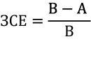
де А – кількість живих плодів;
В – кількість жовтих тіл вагітності.
2. Передімплантаційна смертність визначалась за формулою:
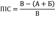
де А – кількість живих плодів;
Б – кількість загиблих (резорбованих) плодів;
В – кількість жовтих тіл вагітності.
3. Постімплантаційна смертність визначалась за формулою:
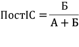
де А – кількість живих плодів;
Б – кількість загиблих (резорбованих) плодів.
4. Кількість плодів на 1 самку (середній показник у групах).
5. Середні значення вагових показників ембріонів (г).
Статистичну обробку даних здійснювали для порівняння показників, що характеризують ембріотоксичний ефект за різних умов експозиції. Оцінювання відмінностей між незалежними групами проводили із застосуванням t-критерію Стьюдента. Статистична обробка даних проводилась із застосуванням Microsoft Exel R версія 4.3.1 (https//www.r-project.org/) [16]. Аналіз виконували відповідно до сучасних стандартів прозорості та відтворюваності біостатистичних досліджень.
РЕЗУЛЬТАТИ ТА ЇХ ОБГОВОРЕННЯ
У нашому експерименті всі самиці вижили, впродовж дослідження залишались активні, добре споживали їжу та воду. Забір ембріонального матеріалу, підрахування кількості жовтих тіл вагітності в яєчниках проводився на 13-ту та 20-ту добу ембріогенезу. Хронічна інтоксикація хлоридом кадмію в дозі 3 мг/кг вже на 13-ту добу експерименту спровокувала порушення топографії ембріонів у рогах матки, а саме: ми визначали асиметричну імплантацію у 8,43% самиць. Для норми в поліестральних багатоплідних ссавців характерний рівномірний розподіл зародків в обох рогах матки. Фізіологічні інтервали між зонами імплантації зазвичай зумовлені поодинокими резорбціями ембріональних та плацентарних структур, що зрідка фіксується і в контрольній групі. Проміжки в маткових трубах між місцями прикріплення вказують на випадки резорбції плаценти чи плода, які поодиноко трапляються і в контролі. Проте в групі ізольованої дії кадмієвої солі поширеність часткової або повної топографічної асиметрії була значно вищою, а при ізольованому введенні хлориду кадмію частота випадків повної або часткової асиметрії розподілу ембріонів була суттєво вищою. Цей факт визначався нами і на 20-ту добу дослідження. Таким чином, хронічне введення хлориду кадмію призвело до того, що на 13-ту та 20-ту добу в самиць щурів спостерігалася девіація нормального розміщення ембріонів у вигляді асиметричної імплантації.
Як показало обчислення кількісних показників, середня кількість живих ембріонів у контрольній групі на 13-ту добу становила 12,50±0,28, тоді як у групі ізольованого впливу хлоридом кадмію цей показник знизився до 8,88±0,24 без достовірного зниження середньої кількості жовтих тіл у яєчниках в обох групах (рис. 1).
{slider title="Рис. 1. Кількість живих ембріонів на 1 самицю на 13-ту добу гестації в експериментальних групах (M±m) ↓" class="tbl-articles my-figure" open="false"}
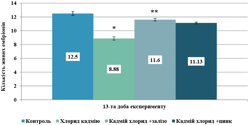
Статистична значущість за t-критерієм Стьюдента: * – p<0,05 порівняно з контролем; ** – p<0,05 порівняно з групою хлорид кадмію. {/sliders}
Аналіз та порівняння отриманих числових обрахувань продемонстрували, що пренатальний ізольований щоденний вплив хлориду кадмію в досліджуваній дозі знижує показник кількості живих ембріонів на 13-ту добу на 28,9%. Таким чином, вже на 13-ій добі експерименту виявлено достовірне зниження показника порівняно з контролем (p<0,05) Суттєве зниження кількості живих ембріонів порівняно з контролем підтверджує той факт, що кадмій є сильною ембріотоксичною отрутою, яка долає плацентарний бар’єр та здатна викликати високу ембріональну смертність.
У групах комбінованого застосування кадмію з металосукцинатами спостерігався чіткий модифікуючий ефект. У групі комбінації кадмію із сукцинатом заліза відмічалась тенденція до збільшення кількості живих ембріонів порівняно з групою ізольованого впливу – 11,6±0,19, що свідчило про зростання кількості ембріонів більше ніж на 25% відносно показника групи кадмію, хоча й залишалося на 11% нижче за контроль. Недостовірно нижчий показник протекторного впливу порівняно з групою комбінації кадмію із сукцинатом заліза зафіксовано в четвертій групі комбінованого надходження кадмію із сукцинатом цинку, де досліджуваний показник становив 11,13±0,13. У цій групі кількість живих ембріонів також наближалася до фізіологічної норми, що вказує на антитоксичну ефективність сукцинату цинку як антагоніста кадмію при їх одночасному надходженні в організм в експерименті на щурах.
На 20-ту добу ембріогенезу плоди вилучали оперативно разом з плацентою для подальшого морфологічного аналізу й верифікації їхньої відповідності стадіям розвитку за критеріями Гамбургера-Гамільтона (НН). На 20-ту добу гестації середня кількість живих ембріонів у групі ізольованого введення хлориду кадмію була найнижчою серед усіх експериментальних груп і становила 8,75±0,27, тоді як у контрольній групі цей показник становив 12,63±0,19 (достовірне зниження показника порівняно з контролем (p<0,05)). У групах комбінованого введення середня кількість живих ембріонів була вищою за групу ізольованого введення хлориду кадмію, але не мали достовірної різниці при їх порівнянні між собою: 11,00±0,20 у групі комбінації хлориду кадмію із сукцинатом заліза та 11,38±0,28 у групі одночасного введення хлориду кадмію та сукцинату цинку. Тобто, при комбінованому введенні кадмію із сукцинатами цинку або заліза спостерігається збереження показників середніх значень кількості ембріонів, що свідчить про антагоністичний характер дії сукцинатів на кадмієву інтоксикацію в підгострому експерименті на щурах при їх одночасному надходженні в досліджуваних дозах (рис. 2).
{slider title="Рис. 2. Кількість живих ембріонів на 1 самицю на 20-ту добу гестації в експериментальних групах (M±m) ↓" class="tbl-articles my-figure" open="false"}
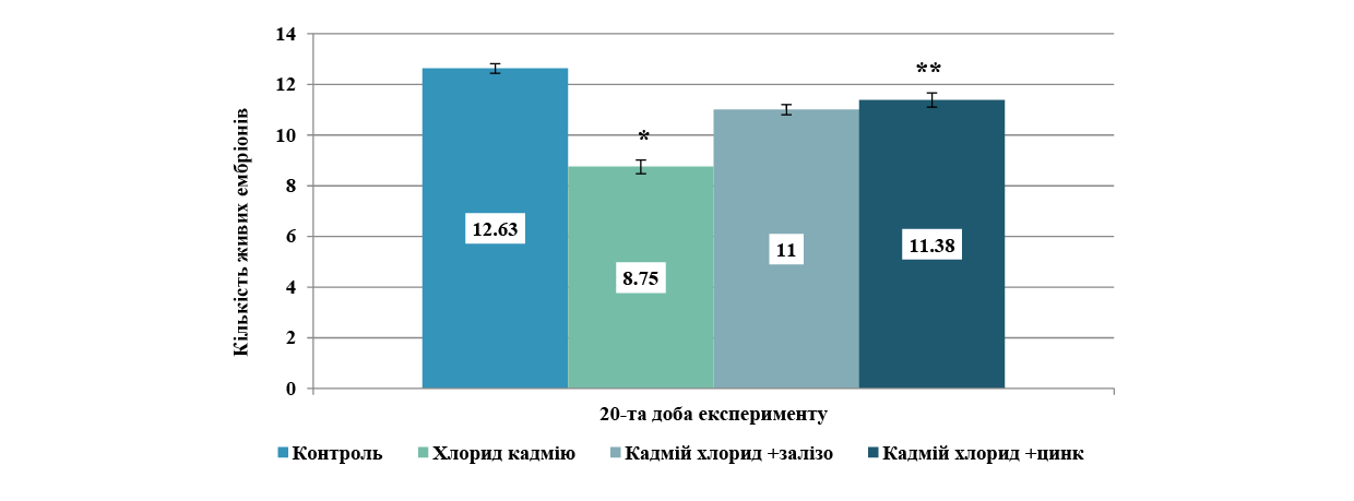
Статистична значущість за t-критерієм Стьюдента: * – p<0,05 порівняно з контролем; ** – p<0,05 порівняно з групою хлорид кадмію. {/sliders}
Таким чином, завершальний етап пренатального розвитку плодів дозволив кількісно оцінити ступінь ембріотоксичної дії хлориду кадмію та протекторну ефективність металосукцинатів упродовж усього гестаційного періоду. У групі ізольованого впливу хлоридом кадмію спостерігається, як і на попередньому терміні дослідження, критичне зниження показника кількості ембріонів у посліді – на 30,7% відносно контролю. Це підтверджує стійку антенатальну токсичність кадмію, яка зберігається протягом усього періоду гестації.
Поєднання токсиканту із сукцинатом заліза забезпечило часткове відновлення виживаності плодів, а саме: показник зріс на 23,9% порівняно з групою ізольованого введення кадмію. Застосування сукцинату цинку продемонструвало найвищий захисний ефект ембріотоксичності кадмію. Показник зріс на 30,1% відносно групи кадмієвої інтоксикації і максимально наблизився до рівня контрольної групи (відставання від норми становить лише 11,6%). Отримані результати доводять, що сукцинати цинку та заліза проявляють модифікуючий вплив на показники ембріотоксичності хлориду кадмію при їх комбінованому надходженні в підгострому експерименті на щурах.
Одним з важливих критеріїв ембріотоксичності є вагові показники ембріонів. У нашому дослідженні на 20-ту добу ембріогенезу в контрольній групі середня вага ембріона становила 2,89±0,06 г, що відповідає фізіологічному еталону, а ізольоване введення хлориду кадмію призвело до вірогідного зниження маси тіла плодів до 2,65±0,07 г, що на 8,30% менше порівняно з контрольними значеннями. Сумісне застосування кадмію із сукцинатом заліза продемонструвало позитивну динаміку: маса ембріонів зберігалася до 2,77± 0,05 г, перевищуючи показник ізольованої інтоксикації на 4,53% (рис. 3).
{slider title="Рис. 3. Вагові показники ембріонів (г) на 20-ту добу гестації в експериментальних групах ↓" class="tbl-articles my-figure" open="false"}
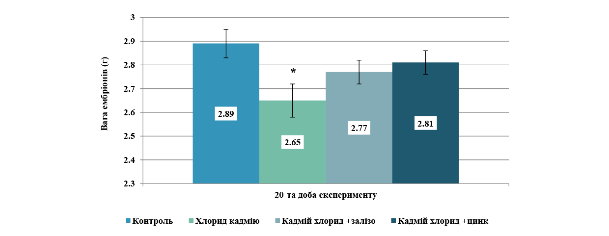
Статистична значущість за t-критерієм Стьюдента: * - p<0,05 порівняно з контролем. {/sliders}
Найбільш виражений ростостимулюючий та антитоксичний ефект зафіксовано в групі поєднання хлориду кадмію із сукцинатом цинку. Середня маса плодів тут досягла 2,81±0,05 г, що на 6,04% вище за рівень ізольованого впливу і практично нівелює затримку розвитку, відстаючи від фізіологічної норми контролю лише на 2,77%.
Таким чином, хлорид кадмію викликає синдром затримки пренатального розвитку плодів, що проявляється зменшенням їхньої кількості та маси. Сукцинат цинку при одночасному надходженні з кадмієм в організм дослідних тварин демонструє вищу протективну здатність щодо збереження кількості та соматичного росту плодів, ніж сукцинат заліза, майже повністю відновлюючи масові параметри до рівня контролю.
Аналіз та порівняння кількісних та вагових даних по ембріонам доводить, що на 13-ту добу ембріогенезу ізольоване введення хлориду кадмію призводить до високопрезентативного зниження кількості живих ембріонів відносно контролю (p<0,001). Введення металосукцинатів на тлі інтоксикації забезпечує статистично значущий протективний ефект: у групі сукцинату заліза показник вірогідно зростає порівняно з ізольованим впливом важкого металу (p<0,055), аналогічна високозначуща тенденція до нормалізації реєструється і за умов застосування сукцинату цинку (p<0,01). Наприкінці гестаційного періоду (20-та доба ембріогенезу) ізольована дія хлориду кадмію призводила до глибокого та статистично значущого пригнічення виживаності плодів порівняно з контрольною групою – кількість живих ембріонів зменшилась до 8,75±0,27 проти 12,63±0,19 у контролі (p<0,05). Застосування досліджуваних металосукцинатів забезпечило стійку протекцію ембріонального матеріалу від ксенобіотичного пресингу. У групі сукцинату заліза показник живих ембріонів вірогідно зріс до 11,0±0,20 порівняно з ізольованою інтоксикацією (p<0,05). Найбільш виражений та статистично високорепрезентативний захисний ефект зафіксовано за умов введення сукцинату цинку, де кількість живих плодів досягла 11,38±0,28, що демонструє максимальний рівень значущості відновлення показника відносно групи інтоксикації (p<0,05).
Окрім кількісних та вагових показників ембріонів, нами для визначення ступеня ембріотоксичності досліджуваних речовин обчислювались відносні показники ембріональної смертності, які враховують не лише кількість ембріонів, а й кількість резорбцій, кількість жовтих тіл вагітності та дозволяють зіставити їх між собою. Найбільш значущим та доказовим показником ембріотоксичності речовини є показник загальної ембріональної смертності (ЗЕС). Обрахування цього показника на 13-ту добу ембріогенезу продемонструвало, що зниження кількості ембріонів у групі ізольованого введення хлориду кадмію відбувалось за рахунок високої ембріональної смертності – 0,26±0,03, тобто показник збільшувався у 8,6 раза (p<0,05) порівняно з контролем (0,03±0,01). Водночас у групах комбінованого введення показники ЗЕС були достовірно нижчі. У групі впливу кадмію разом із сукцинатом заліза цей показник становив 0,06±0,02, що було в 4,3 раза нижче за групу ізольованого впливу (p<0,05), а при комбінації з цинком – 0,09±0,02, що у 2,8 раза нижче (p<0,05), ніж у групі ізольованого впливу хлоридом кадмію (рис. 4).
{slider title="Рис. 4. Показник загальної ембріональної смертності на 13-ту добу гестації в експериментальних групах ↓" class="tbl-articles my-figure" open="false"}
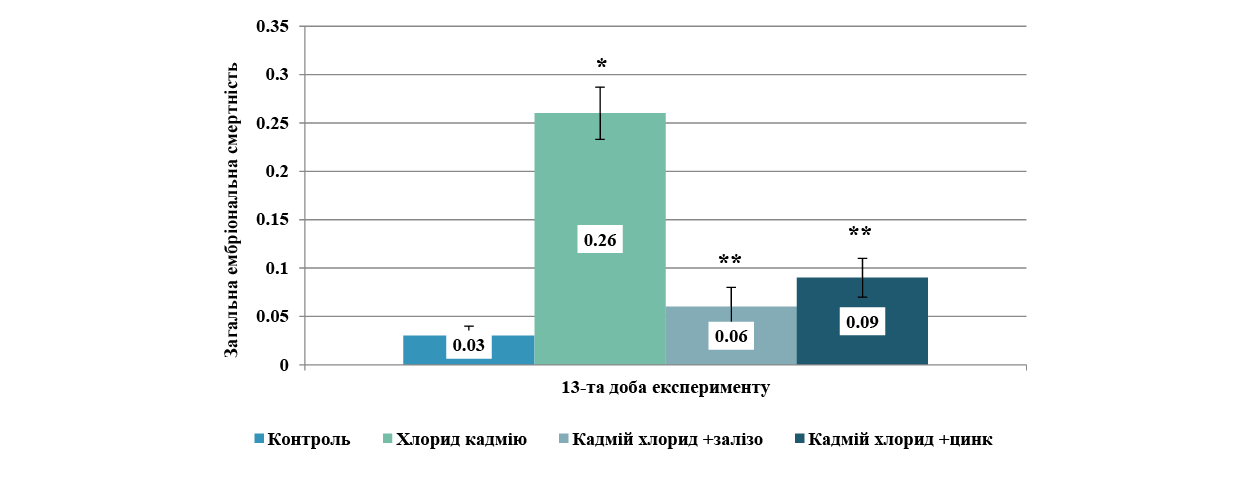
Статистична значущість за t-критерієм Стьюдента: * – p<0,05 порівняно з контролем; ** – p<0,05 порівняно з групою хлорид кадмію.{/sliders}
Таким чином, експозиція хлоридом кадмію під час вагітності має виражену ембріотоксичність уже на 13-ту добу ембріогенезу щурів, що проявляється стрімким зростанням загальної ембріональної смертності та активізацією процесу резорбції ембріонів. Сукцинати заліза та цинку виявляють протективні властивості: комбіноване застосування цих сполук разом із ксенобіотиком ефективно нівелює його негативний ембріотоксичний вплив. Одночасне надходження з хлоридом кадмію сукцинату заліза знижує показник ЗЕС порівняно з групою ізольованого впливу кадмію, повертаючи цей показник у бік до контрольних значень.
На 20-й добі експерименту показник ЗЕС збільшувався при ізольованому впливі кадмієм до 0,32±0,02, що перевищувало контрольні показники в 10,7 раза й було максимальним результатом серед усіх груп. У групах комбінованого введення із сукцинатом цинку показник ЗЕС становив 0,10±0,2, а із сукцинатом заліза дорівнював 0,13±0,03, такі дані були достовірно нижчими за групу ізольованого впливу. У групі кадмієвої інтоксикації наприкінці вагітності самиць щурів наростали темпи ембріональної смертності порівняно з попереднім терміном дослідження, а показник ЗЕС збільшився в 1,2 раза.
Привертала увагу статистично незначуща відмінність між показниками ЗЕС у групах комбінованого введення хлориду кадмію в поєднанні із сукцинатом заліза та хлориду кадмію в поєднанні із сукцинатом цинку на 13-ту добу експерименту, водночас на 20-ту добу експерименту різниця між середніми показниками ЗЕС також не є достовірною, хоча до контрольних показників наближається саме група комбінації із сукцинатом цинку.
Аналіз таких даних вказує, що на ранніх стадіях ембріогенезу сукцинат заліза має більш значущий антагоністичний ефект по відношенню до ембріотоксичності кадмію порівняно із цинком, водночас на 20-ту добу експерименту цинк та залізо мають приблизно однакову ефективність, проте показник ЗЕС у групі комбінації із цинком найнижчий серед експериментальних груп.
Сукцинати заліза та цинку виявляють виражений захисний ефект щодо ембріотоксичності кадмію вагітних щурів на 13-ту та 20-ту добу гестації при їх одночасному потраплянні в організм. Такі результати перегукуються з даними експериментальних досліджень, у яких проводили пошук біоантагоністів токсичності важким металам серед органічних солей біометалів – цитратів. У дослідженнях також аналізувались результати комбінованого впливу солей важких металів та цитратів мікроелементів (заліза, церію, германію, селену та ін.) і визначали зниження рівня загальної ембріональної смертності та збільшення кількості живих плодів. Експериментально доведено модифікуючий вплив цитратів мікроелементів на ембріотоксичні властивості кадмію [8, 12, 13]. Дослідники експериментально довели, що нутрієнтні втручання (зокрема залізо) можуть бути перспективним способом зменшення рівня інтоксикації кадмієм. Робота підкреслює важливість балансу мікроелементів у харчуванні та їхнього впливу на токсикологічні процеси [8]. Дослідниками-морфологами Дніпровського державного медичного університету проводилось також попередні дослідження з виявлення потенційних біоантагоністичних характеристик сукцинатів на тлі кадмієвої інтоксикації вагітних самиць, проте доза кадмію була менша. Вже в цих експериментах комбіноване введення хлориду кадмію із сукцинатами цинку або міді сприяло зниженню всіх показників ембріотоксичності хлориду кадмію та покращенню репродуктивних показників. Досліджувались також показники доімплантаційної, післяімплантаційної та загальної ембріональної смертності [14, 15], але в цих дослідженнях не вивчали захисні біопротекторні властивості сукцинату заліза, а доза кадмію була менша на третину.
ВИСНОВКИ
- Пренатальний ізольований вплив хлориду кадмію в дозі 3мг/кг на вагітних самиць викликає стійкий і статистично значущий ембріотоксичний ефект упродовж усього періоду гестації, що проявляється як достовірним зниженням порівняно з контролем (p<0,05) кількості живих плодів (8,75±0,27), тоді як у контрольній групі цей показник становив 12,63±0,19, так і затримкою їхнього соматичного росту.
- Щоденне введення хлориду кадмію в досліджуваній дозі викликає глибокі ембріотоксичні зміни, які маніфестують уже на ранніх етапах. На 13-ту добу ембріогенезу фіксується стрімке зростання загальної ембріональної смертності за рахунок активізації процесів резорбції та високопрезентативне зниження кількості живих ембріонів відносно контролю. Наприкінці гестаційного періоду (20-та доба) токсичний пресинг кадмію досягає максимуму: кількість живих плодів критично зменшується, а показник загальної ембріональної смертності зростає і в 10,7 раза перевищує рівень контрольної групи.
- На ранніх стадіях ембріогенезу сукцинат цинку і сукцинат заліза виявляють виражений та статистично значущий протективний ефект, ефективно нівелюючи ембріолетальний вплив кадмію при їх одночасному надходженні в організм. При цьому на 13-ту добу сукцинат заліза демонструє дещо потужніший первинний антагоністичний потенціал порівняно із сукцинатом цинку, забезпечуючи вірогідне зростання кількості живих ембріонів та ефективне зниження показника загальної ембріональної смертності в бік до контрольних значень. Різниця між показниками загальної ембріональної смертності в групах заліза та цинку на цьому терміні є статистично незначущою.
- Наприкінці ембріонального розвитку (20-та доба) вектор максимальної захисної ефективності зміщується в бік сукцинату цинку. Хоча обидва препарати вірогідно знижують загальну ембріональну смертність (ЗЕС) порівняно з ізольованою токсикацією, а різниця між їхніми середніми показниками залишається недостовірною, саме за умов застосування сукцинату цинку зафіксовано найнижчий рівень загальної ембріональної смертності серед усіх експериментальних груп і найбільше наближення до норми. Це підтверджується максимальним рівнем статистичної значущості відновлення кількості живих плодів.
- Сукцинат заліза та сукцинат цинку є потенційними високоефективними засобами корекції кадмієвої ембріотоксичності в експерименті на щурах. Їхня дія має пролонгований характер і має відмінності, залежні від стадії ембріогенезу: залізо виявляє сильніший захисний ефект на етапі раннього органогенезу, тоді як цинк забезпечує максимальну виживаність плодів другої половини вагітності.
Внески авторів:
Нефьодова О.О. – концептуалізація, ведення, нагляд, методологія, адміністрування проєкту;
Шевченко І.В. – проведення дослідження, курація даних, формальний аналіз, написання: рецензування та редагування.
Фінансування. Дослідження не має зовнішніх джерел фінансування.
Конфлікт інтересів. Автори заявляють про відсутність конфлікту інтересів.
REFERENCES
- Kolosova II, Shatorna VF. Toxicity of cadmium salts on indicators of embryogenesis of rats. Regulatory Mechanismsin 2022;13(4):330-8. doi: https://doi.org/10.15421/022243
- Ma R, Wang X, Zhuang H, Chen Y, Zhu P, Shao S, et al. Joint impacts of cadmium exposure and anxiety on in vitro fertilization outcomes: a couple-based cohort study. Environ2026;301:124506. doi: https://doi.org/10.1016/j.envres.2026.124506
- Shatorna VF, Lomyha LL. Changes in embryotoxicity indicators of cadmium chloride in a chronic experiment in rats. Bulletin of problems biology and medicine. 2023;2(169):397-401. doi: https://doi.org/10.29254/2077-4214-2023-2-169-397-401
- Ebokaiwe AP, Ezedom T, Denis-Eboh U, Odeghe OB, Obimma JN, Olasehinde O. Redox Perturbation in cadmium-mediated hypothalamic, pituitary, and testicular impairment and protective effects of zinc in rats is accompanied by modulation of tryptophan catabolism enzyme/protein. Mol Neurobiol. 2026 Jan 25;63(1):394. doi: https://doi.org/10.1007/s12035-026-05689-z
- Yu HT, Zhen J, Leng JY, Cai L, Ji HL, Keller BB. Zinc as a countermeasure for cadmium toxicity. Acta Pharmacol2021;42(3):340-6. doi: https://doi.org/10.1038/s41401-020-0396-4
- Mikolić A, Schönwald N, Piasek M. Cadmium, iron and zinc interaction and hematological parameters in rat dams and their offspring. J Trace Elem Med Biol. 2016 Dec;38:108-16. doi: https://doi.org/10.1016/j.jtemb.2016.08.008
- Rudenko KM. [Reduction of the degree of embryotoxicity of cadmium chloride under combined administration with copper succinates in an experiment on rats]. Visnyk Problem Biolohiyi i Medytsyny. 2020;4(158):69-73. Ukrainian.
doi: https://doi.org/10.29254/2077-4214-2020-4-158-69-73 - Brzóska MM, Rogalska J. Protective effect of zinc supplementation against cadmium-induced oxidative stress and the RANK/RANKL/OPG system imbalance in the bone tissue of rats. Toxicol Appl Pharmacol. 2013 Oct 1;272(1):208-20. doi: https://doi.org/10.1016/j.taap.2013.05.016
- Alkharashi NAO, Periasamy VS, Athinarayanan J, Alshatwi AA. Cadmium triggers mitochondrial oxidative stress in human peripheral blood lymphocytes and monocytes: Analysis using in vitro and system toxicology approaches. J Trace Elem Med Biol. 2017;42:117-28. doi: https://doi.org/10.1016/j.jtemb.2017.04.014
- Prenatal Development Toxicity Study. OECD Guideline for the Testing of Chemicals No. 414. Paris:OECDPublishing; 2018. doi: https://doi.org/10.1787/9789264070820-en
- Nordberg GF, Fowler BA, Nordberg M, editors. Handbook on the Toxicology of Metals. 4th ed. Amsterdam: Academic Press; 2015.
- Lee JK, Yu Y, DeLellis RR, Ebea P, Zhu S, Theophilus R, et al. Establishing dietary iron requirements for mouse and rat models of hereditary hemochromatosis. FASEB J. 2022;36(Suppl 1):R2602. doi: https://doi.org/10.1096/fasebj.2022.36.S1.R2602
- Genchi G, Sinicropi MS, Lauria G, Carocci A, Catalano A. The effects of cadmium toxicity. Int J Environ Res Public Health. 2020;17(11):3782. doi: https://doi.org/10.3390/ijerph17113782
- Nefodova OO, Bilyshko DV. [Experimental determination of the effect of cadmium chloride when administered alone and in combination with selenium citrate on the indicators of rat embryogenesis]. Visnyk Problem Biolohii i Medytsyny. 2018;3(145):301-5. Ukrainian.
doi: https://doi.org/10.29254/2077-4214-2018-3-145-301-305 - Costa MCV, Kmecick M, Kolodziejski SC, Vieira da Costa CR, Vargas JE, Ortolani-Machado CF. Morphological and cellular effects of lead, cadmium, and perfluorooctanoic acid on Gallus gallus embryonic development: insights into cephalic region vulnerabilities. Environ Pollut.2025;380:126543. doi: https://doi.org/10.1016/j.envpol.2025.126543
- Hruzieva TS, Lekhan VM, Ohniev VA, Haliienko LI, Kriachkova LV, Palamar BI, et al. Biostatistics. Vinnytsia: Nova knyha; 2020. 384 p.
Ключові слова: порушення зору, бойові офтальмологічні травми, двосторонні ураження, вікова вразливість, сторонні тіла
Abstract
Gunshot and explosive injuries to the visual organs are one of the most severe and socially significant forms of ophthalmological pathology, especially in the context of armed conflicts, terrorist attacks, and man-made disasters. The anatomical complexity of the eye, its vulnerability to high-energy impact, and the vital importance of visual function determine the extreme severity of the consequences of such injuries. These injuries not only reduce the quality of life of the victims but also produce a substantial load on healthcare and social support systems due to the high incidence of disabling outcomes. The study aimed to investigate the clinical and functional characteristics of gunshot and explosive injuries of the visual organs with subsequent analysis of treatment outcomes in a specialised trauma centre. A retrospective study was conducted at the University Trauma Clinic in Tirana, Albania, between January 1995 and December 2006. The sample included 172 patients (240 eyes) admitted within the first 24 hours after injury. The methodology included the analysis of medical records and ophthalmological examination data, including visometry using Snellen tables, biomicroscopy using a slit lamp, ophthalmoscopy, and orbital radiography. In cases of suspected foreign bodies in the posterior segment, computed tomography was utilised. Statistical data processing was performed using IBM SPSS Statistics v.22.0, utilising Student's t-test for quantitative indicators and Pearson's χ² test for categorical variables. Epidemiological analysis revealed peaks in morbidity in 1997 (25%) and 1999 (22%), corresponding to periods of political crisis and armed conflict. The victims were predominantly male (92%) with an average age of 23.9±14.1 years. Notably, 54% of victims were minors, and 71% resided in rural areas. Explosions caused the largest proportion of cases (49%), followed by gunshot wounds (31%), indicating high-energy injury mechanisms. Clinical evaluation showed that severe eye injuries predominated (76%), often accompanied by combined injuries to other organs (75%). Foreign bodies were detected in 75% of eyes, and bilateral injuries occurred in 39% of patients. Despite treatment, severe anatomical outcomes (enucleation, atrophy) were recorded in 57% of patients, and irreversible loss of vision was recorded in 39% of eyes. The high level of disability identified highlights the need for systemic preventive measures and the readiness of ophthalmology services to receive seriously injured people on a massive scale. The practical significance of the data suggests that healthcare authorities must plan medical evacuation, train personnel, and create specialised ophthalmic trauma centres to manage complex, combined injuries effectively. Special attention must be paid to vulnerable groups, particularly children and rural residents, to prevent injuries during periods of social instability.
Реферат
Вогнепальні та вибухові травми очей. Барді Н. Поранення органів зору від вогнепальної зброї та вибухових пристроїв є однією з найтяжчих і соціально значущих форм офтальмологічної патології, особливо в умовах збройних конфліктів, терористичних актів та техногенних катастроф. Анатомічна складність ока, його вразливість до високоенергетичного впливу та життєво важливе значення зорової функції визначають надзвичайну тяжкість наслідків таких травм. Ці травми не тільки знижують якість життя потерпілих, але й створюють значне навантаження на системи охорони здоров'я та соціального забезпечення через високу частоту інвалідності. Мета дослідження полягала у вивченні клінічних та функціональних особливостей вогнепальних та вибухових травм органів зору з подальшим аналізом результатів лікування у спеціалізованому травматологічному центрі. Ретроспективне дослідження було проведено в Університетській травматологічній клініці в Тирані, Албанія, у період із січня 1995 року до грудня 2006 року. До вибірки увійшли 172 пацієнти (240 очей), госпіталізовані протягом перших 24 годин після травми. Методологія включала аналіз медичних карт та даних офтальмологічного обстеження, включаючи візометрію за допомогою таблиць Снеллена, біомікроскопію за допомогою щілинної лампи, офтальмоскопію та рентгенографію орбіти. У випадках підозри на наявність сторонніх тіл у задньому сегменті використовувалася комп'ютерна томографія. Статистична обробка даних проводилася за допомогою IBM SPSS Statistics v.22.0 з використанням t-критерію Стьюдента для кількісних показників та χ²-критерію Пірсона для категоріальних змінних. Епідеміологічний аналіз виявив піки захворюваності в 1997 (25%) та 1999 (22%) роках, що відповідало періодам політичної кризи та збройного конфлікту. Жертвами були переважно чоловіки (92%) із середнім віком 23,9±14,1 року. Примітно, що 54% жертв були неповнолітніми, а 71% проживали в сільській місцевості. Найбільшу частку випадків становили вибухи (49%), за ними йшли вогнепальні поранення (31%), що вказує на механізми травмування з високою енергією. Клінічна оцінка показала, що переважали тяжкі травми очей (76%), які часто супроводжувалися комбінованими травмами інших органів (75%). Чужорідні тіла були виявлені в 75% очей, а двосторонні травми траплялися в 39% пацієнтів. Незважаючи на лікування, тяжкі анатомічні наслідки (енуклеація, атрофія) були зафіксовані в 57% пацієнтів, а незворотна втрата зору – у 39% очей. Високий рівень інвалідності, виявлений у дослідженні, підкреслює необхідність проведення системних профілактичних заходів та готовності офтальмологічних служб до прийому великої кількості людей з тяжкими травмами. Практичне значення даних свідчить про те, що органи охорони здоров'я повинні планувати медичну евакуацію, навчати персонал та створювати спеціалізовані офтальмологічні травматологічні центри для ефективного лікування складних комбінованих травм. Особливу увагу слід приділяти вразливим групам населення, зокрема дітям та мешканцям сільської місцевості, з метою запобігання травмам у періоди соціальної нестабільності.
Gunshot and explosive injuries to the visual organs are one of the most severe and socially significant forms of ophthalmological pathology, especially in the context of armed conflicts, terrorist attacks and man-made disasters. The anatomical complexity of the eye, its vulnerability to high-energy impact and the vital importance of visual function determines the extreme severity of the consequences of such injuries. Between 1995 and 2006, the problem was significantly exacerbated by the growing number of localized armed conflicts, instability in border and rural areas, and the proliferation of improvised explosive devices. The high incidence of disabling outcomes, including complete loss of vision and eyeballs, not only reduces the quality of life of the victims but also produces a substantial load on healthcare and social support systems. In this context, the study of the clinical features and outcomes of ophthalmic injuries caused by firearms and explosives is of relevance to improve approaches to medical care, rehabilitation and prevention.
A substantial clinical problem is the high incidence of bilateral eye injuries in blast injuries, which significantly worsens the prognosis of restoration of visual function and reintegration of the victim into society. N. Bardhi [1] emphasised that the main pathogenetic mechanism of bilateral lesions is diffuse energy transfer through the bone structures of the facial skull, accompanied by multiple foreign bodies in the orbital tissues. Despite the proposed algorithms for diagnosis and treatment, approaches to emergency care remain fragmented, and the rate of outcomes with complete vision loss remains high, which indicates the need for a comprehensive review of protocols for the management of such patients. A significant obstacle to effective ophthalmic care for combat injuries is the high incidence of combined injuries, which makes early surgical intervention difficult. G. Warjri et al. [2] emphasised that ophthalmological care in patients with multiple organ trauma often falls by the wayside, leading to delayed treatment of ocular injuries. Despite the introduction of multidisciplinary teams, there is a lack of coordination between ophthalmologists and surgeons of other specialties, which reduces the effectiveness of rehabilitation measures.
High incidence of underage drinking, especially in rural areas where access to specialized medical care is limited, is notable. D. Zekaj et al. [3] highlighted that the lack of safety education programmes and affordable personal protective equipment for children and adolescents makes them particularly vulnerable. Despite the steps taken to prevent injuries among young people, the statistics of severe eye injuries in this age group grow, indicating the need for targeted prevention initiatives and training in basic safety skills in hazardous environments. The problem of preserving functional vision even with partial anatomical restoration of the eye remains unresolved. V. Kumar et al. [4] demonstrated that restoration of the eyeball anatomy does not guarantee the return of visual function, especially in cases of damage to the optic nerve and central retinal structures. Despite the development of microsurgery and reconstructive technologies, the rates of visual rehabilitation after blast injuries remain extremely low, which requires the search for new biotechnological solutions and methods of neurorehabilitation.
The absence of specialised ophthalmic trauma centres in most regions of Albania significantly reduces the efficiency and effectiveness of care. G. Nallbani and M. Krasniqi [5] addressed the shortage of staff, insufficient equipment and low readiness of institutions to receive mass casualties. Even if medical evacuation is available, the lack of standardised patient routes leads to delays in care and, consequently, to worsening outcomes. This highlights the need for strategic planning of resource provision and development of a network of specialised institutions. The high degree of disability after ophthalmic injuries is also associated with psychological consequences, which are often ignored in the treatment process. Y. Zhang et al. [6] noted that vision loss is accompanied by the development of post-traumatic stress disorders, depression and social isolation, especially in young patients. At the same time, comprehensive psychological support is practically not integrated into standard rehabilitation protocols, which limits the effectiveness of returning victims to active life.
There is no systematic database reflecting the structure, causes, and outcomes of ophthalmic injuries in the context of hostilities. D. Prat et al. [7] emphasized that the fragmentation of statistical sources does not provide an accurate assessment of the scale of the problem and makes it difficult to forecast and plan resources. In the context of growing instability and the probability of mass admission of severely injured people, this situation is unacceptable, which requires the development of a centralised information system and registers of ophthalmic injuries. The mechanisms for providing emergency ophthalmological care in the first hours after an injury are also insufficiently developed. A. Mohamed et al. [8] highlighted that the effectiveness of treatment largely depends on the time of transport and the availability of standard operating protocols. However, in most cases, victims are delivered late, and the initial examination is conducted by untrained specialists. These factors significantly limit the potential for preserving vision and restoring ocular structures, which requires regulatory regulation and improved training of medical staff.
The combination of these problems suggests that the existing system of ophthalmological care for gunshot and blast injuries requires significant modernisation at both the organisational and clinical levels. The study aimed to investigate the clinical and functional characteristics of gunshot and explosive injuries of the visual organs with subsequent analysis of treatment outcomes in a specialised trauma centre. The objectives of the study included a retrospective analysis of medical records, identification of the main factors influencing the severity of injuries, and evaluation of the effectiveness of the care provided.
MATERIALS AND METHODS OF RESEARCH
The study was conducted at the Trauma University Hospital (formerly Central Military University Hospital) [9] in Tirana, Albania, between January 1995 and December 2006. The study is a retrospective design based on the analysis of medical records and diagnostic materials of patients admitted with gunshot and blast injuries of the eye. The sample included all patients admitted to the hospital within the first 24 hours after the injury, with a documented injury to the eye or orbit due to gunshot or explosive exposure and a complete medical record. Cases with isolated soft tissue injuries of the eyelids without involvement of the eyeball, patients with incomplete documentation and those who died before admission were excluded. Thus, the study included 172 patients (158 men and 14 women) with a total of 240 injured eyes. The average age of the victims was 23.9±14.1 years. For each case, socio-demographic characteristics (age, gender, occupation, place of residence), circumstances of injury, type of injury (bullet, shell and mine fragments, explosives), severity of eye injury and the presence of combined injuries to other organs and systems were recorded. The latter were classified into five main groups: maxillofacial area, brain, limbs, thorax and abdomen. This study was considered by the members of the Research Ethics Council of the University Hospital of Trauma. Based on the vote of the Commission's members, a decision was made: All procedures performed in the study were in accordance with the Declaration of Helsinki [10] and the rules of Good Clinical Laboratory Practice (GCLP) [11]. The scientific research can be implemented in its current form, No. 258101.
The ophthalmological examination included visometry using Snellen tables [12] (Germany), biomicroscopy using a Haag-Streit BM 900 slit lamp (Switzerland), ophthalmoscopy (Welch Allyn 3.5V, USA), orbital radiography using a Siemens Multix Pro (Germany), and, if necessary, ultrasound A- and B-scan (Tomey UD-6000, Japan). Intraocular pressure was measured by Goldmann applanation tonometry in eyes with preserved globe integrity. In patients with suspected or confirmed open-globe injuries, tonometry was not performed before primary wound closure because external pressure could aggravate the injury; intraocular pressure was assessed after restoration of globe integrity when clinically indicated. In case of suspected foreign bodies in the posterior segment of the eye, computed tomography (GE HiSpeed CT/i, USA) was used. The injuries were classified into three degrees: mild (contusions, superficial injuries), moderate (penetrating wounds without massive destruction of the eyeball), and severe (destruction of the eyeball, massive injuries with large foreign bodies). Bilateral injuries were addressed separately. The treatment included both conservative methods (antibiotic therapy, anti-inflammatory drugs, antihypertensive therapy, and local wound treatment) and surgical interventions. In patients with clinical signs of purulent complications, material from the ocular wound and, when clinically obtainable, aqueous humor or vitreous samples collected during surgery were submitted for microbiological culture and identification of the causative microorganism. Antibiotic susceptibility testing was performed for the isolated microorganisms, and antibacterial therapy was subsequently adjusted according to the obtained results. Depending on the clinical situation, primary surgical treatment of wounds, removal of foreign bodies, corneal and scleral reconstructive surgery, orbital reconstruction, and, in cases of irreversible damage, enucleation of the eye were performed.
The outcomes were assessed comprehensively. The dynamics of visual acuity on admission and discharge were measured, and anatomical results (preservation of the eyeball, development of atrophy or subatrophy), and the incidence of complications (endophthalmitis, panophthalmitis, secondary glaucoma) were analyzed. Visual outcomes were classified into four levels: preserved vision (≥0.3), moderate reduction (0.1-0.29), severe reduction (0.01-0.09) and functional blindness (≤0.01 or enucleation). Statistical data processing was performed using IBM SPSS Statistics v.22.0 [13] (USA). Student's t-test was used for quantitative indicators, and Pearson's χ² test for categorical variables. Multivariate logistic regression was used to identify factors associated with unfavorable outcomes (eye loss, functional blindness). The level of statistical significance was assumed to be p<0.05. Based on the retrospective analysis, practical recommendations were formulated to optimize the care of victims of gunshot and blast eye injuries. Timeliness of interventions, the sequence of diagnostic and therapeutic measures, and the factors associated with the most adverse visual outcomes were emphasized.
Statistical data processing was performed using IBM SPSS Statistics v.22.0 [13] (USA) [License No. 3022941]. The normality of the distribution of quantitative data was assessed using the Kolmogorov-Smirnov test. Quantitative data following a normal distribution are presented as Mean ± Standard Deviation (SD), while categorical variables are presented as absolute numbers (n) and percentages (%). Since the quantitative data met the assumption of normality, Student's t-test [14] was used for comparative analysis. Pearson's χ² test [15] was utilized for categorical variables. Multivariate logistic regression [16] was used to identify factors associated with unfavorable outcomes (eye loss, functional blindness). The level of statistical significance was assumed to be p<0.05. Based on the retrospective analysis, practical recommendations were formulated to optimize the care of victims of gunshot and blast eye injuries. Timeliness of interventions, the sequence of diagnostic and therapeutic measures, and the factors associated with the most adverse visual outcomes were emphasised.
RESULTS AND DISCUSSION
The issue of annual dynamics of eye injuries caused by gunshot and explosive impact was a key element of the epidemiological assessment of the war and post-war period. The chronological distribution of cases was used to identify peaks in morbidity associated with specific political and social events, including internal conflicts, armed clashes and massive weapons proliferation (Table 1). The analysis by year reflected the duration of the impact of crises on the civilian population and the emergency eye care system. In times of instability, such data was used to plan the allocation of resources, staff training and rapid response of clinics. In addition, temporal patterns in the admission of patients with severe eye injuries formed the basis for subsequent prevention measures and legal regulation in the field of civilian security.
{slider title="Table 1. Distribution of eye injuries by year (1995-2006) ↓" class="tbl-articles my-figure" open="false"}
|
Years |
Number of cases (damaged persons) |
Percentage |
|
1995 |
1 |
0.5 |
|
1996 |
6 |
3.5 |
|
1997 |
43 |
25.0 |
|
1998 |
14 |
8.1 |
|
1999 |
37 |
21.5 |
|
2000 |
19 |
11.0 |
|
2001 |
15 |
8.7 |
|
2002 |
15 |
8.7 |
|
2003 |
6 |
3.5 |
|
2004 |
8 |
4.7 |
|
2005 |
7 |
4.1 |
|
2006 |
1 |
0.5 |
|
Sum |
172 |
100 |
{/sliders}
The annual analysis demonstrated a pronounced irregularity in the frequency of eye injuries caused by gunshot and explosive impact. The highest number of cases was recorded in 1997 – 43 patients (25% of the total number over 12 years). This sharp increase corresponded to the political crisis in Albania, which was accompanied by massive arms seizures and spontaneous violence. The second peak occurred in 1999, with 37 cases (22%), of which a significant number came from Kosovo amid the armed conflict. In total, 80 incidents (47%) occurred in these two years, which is almost half of all recorded cases. In the remaining years, the number of victims ranged from one to nineteen, showing sporadic but significant spikes in injuries.
The study of the gender of victims of gunshot and blast eye injuries established socio-behavioural and demographic patterns typical of conflict periods (Table 2). In the context of military or near-military activity, gender differences in morbidity reflected not only biological but also socio-cultural factors such as participation in hostilities, access to weapons, lifestyle characteristics, and risks associated with certain professions. Notably, in the context of this study, the gender imbalance was not only statistically significant, but also preventive: it indicated risk groups requiring targeted intervention and special attention in educational and restrictive programmes.
{slider title="Table 2. Distribution of cases by gender ↓" class="tbl-articles my-figure" open="false"}
|
Gender (sex) |
Number of cases (persons) |
% |
|
Male (M) |
158 |
92 |
|
Female (F) |
14 |
8 |
|
M + F |
172 |
100 |
Notes: χ²(1)=120.6; p<0.001. {/sliders}
The gender distribution of the victims showed a pronounced predominance of men: 158 (92%) of the 172 patients were men, and only 14 (8%) were women. This imbalance was consistent with the nature of the armed incidents and the typical social role of men in situations of instability. Men, especially younger men, were significantly more likely to be involved in or witness confrontations, to have access to weapons, and to be exposed to the risks associated with hostilities, illicit arms trafficking and domestic conflicts involving explosives. Women, on the other hand, were more likely to be outside the epicentre of active hostilities, which explains the extremely low percentage of casualties. This indicator also highlighted the disproportionate involvement of men in the events that led to injuries and was used as a substantial indicator of social vulnerability. The data suggested that in conditions of increased militarisation of society or social destabilization, men were the main risk group for severe eye injuries, which should be considered when developing targeted prevention and rehabilitation strategies.
The study of the age characteristics of victims of gunshot and blast injuries to the eye identified the most vulnerable age groups and to identify demographic shifts typical of periods of armed instability (Table 3). The average age of patients and gender disaggregation were central for research of the structure of the affected population, their level of activity, involvement in armed conflict or its consequences. Age analysis was key in terms of assessing the severity of injuries, prognosis of recovery of visual functions and the possibility of further rehabilitation. Data in this category was also central in planning medical care and preventive measures aimed to protect the most at-risk age groups.
{slider title="Table 3. Average age of victims ↓" class="tbl-articles my-figure" open="false"}
|
Number of cases (persons-patients) |
Average age (years) |
Gender |
|
|
male |
female |
||
|
172 |
23.94±14.11 |
158 |
14 |
Notes: data are presented as Mean±Standard Deviation (SD); t≈–1.30; p=0.21 (not significant). {/sliders}
The average age of patients with eye injuries caused by gunshot or explosive impacts was 23.94±14.11 years, indicating a predominance of young people among the victims. Men represented the absolute majority in this sample, which was also reflected in their dominance by age. This age structure corresponded to the social reality of the period, when young men were more likely to be involved in armed conflicts or to become accidental victims.
An analysis of the distribution of victims by age group provided a more detailed assessment of the extent to which different generational categories were involved in the armed conflict (Table 4). This breakdown was substantial for the identification of the most at-risk groups, especially among children and adolescents, where the consequences of trauma had not only medical but also serious social impact. Determining age-related vulnerability was also of practical importance in developing primary prevention programs, planning educational campaigns, and organising eye care and long-term rehabilitation. In the context of the study period, which included years of political crisis and military conflicts, the age distribution reflected the overall dynamics of violent events and the extent of civilian casualties.
{slider title="Table 4. Distribution of victims by age group ↓" class="tbl-articles my-figure" open="false"}
|
Age -group |
Number of cases (persons) |
% |
Note |
|
0-10 years |
19 |
11 |
54 |
|
11-20 years |
74 |
43 |
|
|
21-30 years |
35 |
20 |
46 |
|
>30 years |
44 |
26 |
|
|
Sum |
172 |
100 |
100 |
{/sliders}
The age structure data revealed the highest concentration of ocular injuries in young people: 74 patients (43%) belonged to the age group of 11-20 years, and 19 cases (11%) were children under 10 years of age. Thus, more than half of the victims (54%) were minors. The next largest group was 21-30 years of age (20%), while the proportion of patients over 30 years of age was only 26%. These results reflected the high degree of involvement of children and adolescents in traumatic events that occurred in a context of social instability and uncontrolled proliferation of weapons. It is noteworthy that a significant number of victims were among the younger age groups, indicating violations of the public security system and insufficient preventive measures. Such a young age structure also indicated a high risk of permanent disability with severe consequences for decades to come. Given the shortage of specialized assistance in the post-conflict period, these findings highlighted the importance of developing and implementing multisectoral strategies to protect children and adolescents in times of crisis.
The professional and social status of the victims assessed the structure of involvement of different categories of the population in situations involving gunshot and blast injuries to the organ of vision (Table 5). This information was substantial for identification of vulnerable social groups that were at the highest risk in the context of civil conflict, armed clashes and an unstable social environment. The comparative analysis of the professional composition of the victims also made it possible to establish indirect links between the type of activity, age, place of residence and the nature of the injuries sustained. In practical terms, this information served as a basis for targeting prevention resources and developing educational programmes aimed for certain social groups at increased risk of injury.
Children and schoolchildren accounted for the largest share of the victims – 70 people (41%). The second largest group consisted of people without official employment, pensioners and the temporarily unemployed, with 66 cases (38%). This structure reflected on the one hand, the low social protection and high degree of vulnerability of these categories, and, on the other hand, their potential involvement in informal or illegal activities, including the use of weapons. Military personnel accounted for only 21% of the total number of victims (36 people), indicating the predominantly civilian nature of most of the cases reported.
{slider title="Table 5. Distribution of victims by professional status ↓" class="tbl-articles my-figure" open="false"}
|
Profession and occupation |
Number of cases (persons) |
% |
|
Children -pupils |
70 |
41 |
|
Military persons |
36 |
21 |
|
Out of work, others |
66 |
38 |
|
Sum |
172 |
100 |
{/sliders}
The analysis of the victims’ place of residence was a substantial aspect of the study of the epidemiology of ocular injuries caused by gunshot and blast exposure (Table 6). Comparison of urban and rural populations in terms of injury rates identified territorial risk areas and determined the impact of infrastructural, economic and cultural factors on the prevalence of severe eye injuries. The spatial distribution could also indicate the availability of weapons, population density and level of medical care in different regions. This data was of strategic importance in the development of preventive measures, including targeted information and crisis response programmes adapted to the specifics of urban and rural environments.
{slider title="Table 6. Distribution of victims by residence ↓" class="tbl-articles my-figure" open="false"}
|
Residence (social origin) |
No of cases |
% |
Odds Ratio (OR) |
95% CI |
p-value |
|
Country-village |
122 |
71 |
2.66 |
1.36-5.17 |
0.005 |
|
City |
50 |
29 |
1.00 |
1.00-1.00 |
1.000 |
|
Sum |
172 |
100 |
2.66 |
1.36-5.17 |
0.005 |
{/sliders}
Of the 172 victims, the majority (122 people, or 71%) lived in rural areas, while only 50 patients (29%) were urban residents. This ratio reflected both the territorial structure of Albania's population at that time and the specific nature of access to weapons and explosives. In rural areas, especially during the crisis years, there was a wider distribution of firearms stored outside official control. In addition, inadequate medical infrastructure and safety education also contributed to the increase in the number of serious injuries. Despite higher population density, urban areas had a lower proportion of victims, which may have been due to better public safety regulations and more rapid access to medical care. These data highlighted the need to strengthen preventive measures in rural areas, including restricting unauthorised access to weapons and conducting educational campaigns to prevent domestic and non-combat injuries among the rural population.
The study of the immediate causes of eye injuries associated with gunshot and blast exposure made it possible to classify the damaging factors and assess their relative contribution to the structure of injuries (Table 7). The etiological classification was substantial in the research of the mechanism of injury, its potential severity and the probability of concomitant injuries. In addition, the data on the types of weapons and explosives that caused eye injuries reflected both the nature of the armed conflict and the level of uncontrolled proliferation of dangerous objects among the civilian population. This information was directly relevant to the development of legal and prevention strategies aimed to limit access to the most traumatic munitions.
The analysis showed that the most common cause of eye injuries was explosions of various origins, accounting for 49% of cases (84 victims). This category included injuries caused by TNT, dynamite and other detonating substances used both in the military and in the home. Injuries caused by firearms bullets, fragments and cartridges were the second most frequent (31%, 53 cases), reflecting the effects of both hostilities and careless handling of weapons. In third place were injuries caused by grenades, mines and bombs (20%, 35 cases), characterized by an extremely high degree of tissue destruction and frequent bilateral lesions. This structure confirmed that the most dangerous for the organ of vision were explosive impacts with significant kinetic and thermal energy, leading to multiple injuries to both the eyeball and the orbital region. Moreover, most of the incidents occurred outside of organised hostilities, indicating the civilian nature of the injuries and the critical situation with explosives control in society.
{slider title="Table 7. Distribution of cases by type of injury factor ↓" class="tbl-articles my-figure" open="false"}
|
Cause of injury |
Number of cases (persons) |
Sum |
% |
Odds Ratio (OR) |
95% CI |
p-value |
|
|
Bullets and shells |
Bullets (pieces of them) |
48 |
53 |
31 |
3.69 |
1.79-7.58 |
0.001 |
|
Shells |
5 |
1.00 |
1.00-1.00 |
1.000 |
|||
|
Explosions |
TNT, dynamite |
26 |
84 |
49 |
4.86 |
2.01-11.75 |
0.001 |
|
Other explosions |
58 |
3.69 |
1.79-7.58 |
0.001 |
|||
|
Bombs, Grenades and Mines |
Bombs, Grenades |
20 |
35 |
20 |
3.69 |
1.79-7.58 |
0.001 |
|
Mines |
15 |
1.00 |
1.00-1.00 |
1.000 |
|||
|
Sum |
172 |
172 |
100 |
4.86 |
2.01-11.75 |
0.001 |
|
{/sliders}
The analysis of combined injuries to the organ of vision with injuries to other body systems assessed the overall severity of the victims' condition and the complexity of managing such patients in a clinic setting (Table 8). Combined injuries significantly increased the risk of adverse outcomes, prolonged treatment time and required the participation of multidisciplinary teams. The presence of multiple injuries, especially in the context of blast or gunshot exposure, reflected not only the mechanisms of injury but also the level of overall lethality of the weapon used. These data were of practical importance for both prognostic assessment and planning of medical evacuation, surgical care and subsequent rehabilitation in conditions of limited resources.
{slider title="Table 8. Distribution of cases by the presence of combined injuries ↓" class="tbl-articles my-figure" open="false"}
|
Total number of cases (persons)with injuries to the eyes |
Number of cases (persons)with eye injuries and other organs |
% |
|
172 |
129 |
75 |
Notes: χ²(1)=43.0; p<0.001. {/sliders}
Of the 172 victims, 129 (75%) had combined injuries to the eye and other body systems. Only 43 cases (25%) were isolated eye injuries, indicating the high-energy nature of most injuries, especially those resulting from explosions, where the damage was systemic. Injuries to the skull, brain, maxillofacial area, chest and abdomen were common. The high proportion of combined injuries required ophthalmologists to work with neurosurgeons, maxillofacial surgeons, traumatologists and anaesthetists. In addition, the presence of multiple injuries significantly complicated both the treatment tactics and prognosis to save the patient's vision and life. These data pointed to the need to organize specialized multidisciplinary teams in clinics that receive victims of war and post-war trauma, as well as the importance of early comprehensive diagnosis on admission. A correlation between the severity of ophthalmic lesions and the prevalence of concomitant injuries was noted, especially in blast injuries, which emphasizes the systemic damage to the body that was typical for this period.
Assessment of the severity of ocular injuries was a key parameter in stratifying patients into clinical categories and the prognosis of visual function recovery (Table 9). Classification of injuries into mild, moderate and severe made it possible not only to standardise treatment approaches but also to compare results between different groups based on the initial level of damage. This gradation was crucial for the rapid triage of victims during mass admissions, as well as for long-term planning of surgical interventions and rehabilitation. In addition, the severity of the injury was directly correlated with the need for radical intervention, the incidence of complications and the likelihood of permanent disability.
{slider title="Table 9. Distribution of victims by severity of eye injuries ↓" class="tbl-articles my-figure" open="false"}
|
Number of damage cases (persons) |
Degree (Step) of eye injuries |
Sum |
||
|
light {I} |
medium {II} |
hard {III} |
||
|
172 |
22 |
19 |
131 |
172 |
{/sliders}
Most patients had severe eye injuries, 131 people, which accounted for 76% of the total number of victims. Medium-severity injuries were recorded in 19 patients (11%), and mild injuries in only 22 (13%). These data underlined the highly destructive nature of the injuries, mainly caused by blast exposure and proximity to the epicentre of the injury. Severe injuries were usually accompanied by severe destruction of the eyeball, foreign bodies, haemophthalmos and optic nerve damage. Injuries of moderate severity were more often characterized by penetrating wounds without massive structural damage. Minor injuries, such as contusions or superficial lesions, were much less common and probably did not always lead to hospitalization, which could also distort the overall statistics towards more severe cases. Thus, the study sample primarily reflected the most severe segment of ophthalmic injuries requiring inpatient and often surgical treatment. These data also confirmed that the clinic where the study was conducted received the most severe patients in the context of the military and post-crisis strain on the healthcare system.
Comparison of the severity of ocular injuries with the presence of concomitant injuries to other organs revealed correlations between the anatomical distribution of the lesion and the clinical severity of the condition (Table 10). This cross-assessment was indicative of the formation of clinical and tactical teams and the identification of priorities in the provision of assistance. The high severity of ophthalmic injuries was often accompanied by multiorgan injuries, which required a comprehensive approach to treatment. Establishment of these links can be used not only to predict the outcome, but also to clarify the nature of the injury mechanism, especially in the context of blast injuries.
{slider title="Table 10. Extent of eye injuries depending on the presence of combined injuries ↓" class="tbl-articles my-figure" open="false"}
|
Eye injury degree (Step) |
Number of cases (injured person) |
% |
Combined injuries |
Chi-Square Value |
df |
p-value |
|
|
No. |
% |
||||||
|
Light {I} |
22 |
13 |
18 |
14 |
0.903 |
2 |
0.637 |
|
Medium {II} |
19 |
11 |
15 |
12 |
0.903 |
2 |
0.637 |
|
Hard {III} |
131 |
76 |
96 |
74 |
0.903 |
2 |
0.637 |
|
{I+II+III} |
172 |
100 |
129 |
100 |
0.903 |
2 |
0.637 |
{/sliders}
Of the 131 patients with severe eye injuries, 96 (74%) had simultaneous injuries to other organs, confirming the systemic nature of the lesion due to high-energy mechanisms. In the moderate severity group, 15 out of 19 patients (79%) had combined injuries, and in the mild injury category, 18 out of 22 (82%) had combined injuries. Thus, regardless of the degree of ophthalmic damage, the presence of combined injuries remained significant in all clinical groups. However, the absolute numbers and the proportion of severe cases among the total number of victims emphasized that severe injuries were most closely associated with multiple injuries. These results concluded that eye injuries sustained in conditions of gunshot and especially explosive exposure were almost always accompanied by widespread damage to the body. This required not only specialized ophthalmological care, but also the constant readiness of multidisciplinary teams to provide comprehensive medical support. This systematic nature of injuries was not only clinically but also organisationally relevant, affecting the allocation of resources, the stages of treatment and medical triage.
The analysis of the location of ocular injuries on the side of the lesion, whether unilateral or bilateral, was substantial for assessment of the anatomical severity of the injury and prognosis of visual function recovery (Table 11). Injury to one eye often resulted in functional vision, while bilateral injuries were associated with a higher risk of total blindness, disability and severe psychosocial consequences. The frequency of bilateral injuries also reflected the specifics of the impact mechanism, where blast injuries had the greatest potential for symmetrical damage to the face and eyes. This data was used to develop emergency care standards, plan prosthetic care and psychological support for patients.
{slider title="Table 11. Distribution of victims by the nature of eye damage (one or both eyes) ↓" class="tbl-articles my-figure" open="false"}
|
Eyes Injuries |
Number of cases(persons) |
% |
|
In one eye (R or L) |
104 |
61 |
|
Both eyes (R +L) |
68 |
39 |
|
Sum |
172 |
100 |
Notes: χ²(1)=7.54; p=0.006. {/sliders}
Of the 172 patients, 104 (61%) were diagnosed with unilateral eye injuries, while 68 (39%) had bilateral injuries. Thus, bilateral damage was observed in almost every third case. These data emphasized the high intensity of the traumatic factor, especially in patients with blast injuries, where the radial spread of energy and the presence of multiple fragments contributed to the damage to both orbits. At the same time, patients with unilateral injuries had prospects for maintaining their ability to work and autonomy in everyday life, which could not be said about victims with bilateral injuries requiring long-term rehabilitation support. The high proportion of cases with bilateral injuries, especially in peacetime, indicated a deep systemic security crisis and uncontrolled access to powerful traumatic means. This further strengthened the requirements for reforming the system of public control over the circulation of weapons and explosives, as well as the development of ophthalmological services capable of providing care to patients with complete visual deficits.
A detailed analysis of the number of affected eyes, depending on the nature of the injury, unilateral or bilateral, was used to assess the distribution of the burden on ophthalmological services and to clarify the extent of damage in the clinical sample (Table 12). In contrast to the analysis by the patient, consideration of the eyes as separate anatomical units gave a more accurate picture of the extent of the injury, the degree of involvement of the visual organs and the prognosis for the restoration of visual function. These data were substantial for both clinical assessment and statistical modelling of outcomes in mass trauma, especially in the context of crises.
Among the 240 injured eyes, 104 (44%) were in patients with unilateral injuries, with 54 eyes being right and 50 being left. The remaining 136 eyes (56%) were injured in patients with bilateral injuries, with an equal proportion of 68 right and left eyes. These results confirmed the high frequency of symmetrical lesions characteristic of explosive trauma mechanisms, especially at close range or in confined spaces. The predominance of bilateral lesions in terms of the number of eyes compared to the number of patients indicated a severe degree of damage in such injuries and a potential threat of complete vision loss. In addition, the even distribution between the right and left eyes confirmed the absence of selectivity in the mechanism of injury, which is typical for non-selective damaging factors, such as fragments from explosive devices. From a practical point of view, this meant that a significant number of patients required symmetrical restoration or prosthetics, as well as comprehensive psychological support in cases of complete or almost complete loss of vision.
{slider title="Table 12. Distribution of injured eyes by side and nature of the lesion ↓" class="tbl-articles my-figure" open="false"}
|
Injuries |
Number of cases (persons) |
% |
Cases of injured eyes |
|||
|
R |
L |
Sum |
% |
|||
|
In one eye (R or L) |
104 |
61% |
54 |
50 |
104 |
44% |
|
Both eyes (R + L) |
68 |
39% |
68 |
68 |
136 |
56% |
|
Sum |
172 |
100% |
122 |
118 |
240 |
100% |
{/sliders}
The presence of foreign bodies in the eyes after trauma was a substantial indicator of the mechanism of injury and determined the specifics of treatment tactics and the outcome of ophthalmological interventions (Table 13). Intraocular and orbital foreign bodies posed a substantial threat to both anatomical integrity and functional prognosis, increasing the risk of infectious complications, retinal detachment and permanent visual loss. Their frequency served not only as a marker of injury severity but also as a central diagnostic criterion for triaging patients requiring emergency surgery. These data were also significant for the organizational structure of the trauma service, requiring the availability of high-tech imaging diagnostics and microsurgical equipment.
{slider title="Table 13. Presence of foreign bodies in the eyes of victims ↓" class="tbl-articles my-figure" open="false"}
|
Nominations of cases damaged by Gunshots and Explosions |
No. of cases (persons & eyes) |
No. of cases (with foreign ocular bodies) |
% |
|
Damaged persons (patients) |
172 |
129 |
75 |
|
Injured eyes |
240 |
179 |
75 |
Notes: OR=6.1, 95% CI 2.8-13.1; p<0.001. {/sliders}
Among the 172 victims, foreign bodies were found in the eyes of 129 people (75%), and a total of 179 eyes out of 240 (also 75%) were affected. This meant that in many cases, foreign bodies were present in both eyes of the same patient. Such a high frequency indicated the predominance of an explosive mechanism of injury, in which fragments of metal, stone or other materials penetrated the tissues of the eye and orbit at high speed. The presence of foreign bodies required urgent imaging studies using X-ray, ultrasound and, if necessary, computed tomography. In addition, given the limited time and resources available, their presence determined the need for priority surgical intervention. Clinical experience showed that the longer foreign bodies remained in the structures of the eye, the higher the risk of developing endophthalmitis, choroidoretinal inflammation, and secondary glaucoma. This indicator also emphasised the extremely traumatic nature of the weapons used and the complexity of managing patients, who often required several stages of surgical removal of fragments, followed by restoration of the integrity of the eyeball or prosthetics.
Assessment of the effectiveness of surgical removal of foreign bodies from the eye was key to analysing the quality of care provided and the level of technical equipment of the clinic (Table 14). The ability to remove the foreign body at an early stage significantly reduced the risk of infectious complications and increased the probability of preserving residual vision. At the same time, the presence of residual fragments in the eye or orbit indicated either the severity of anatomical damage that prevented complete removal or limited imaging and microsurgery capabilities in the acute period. These data are also substantial for the research of the long-term prognosis and the incidence of late complications.
Among 129 patients with registered foreign bodies, surgical removal was performed in 78 patients (60%). Accordingly, in 51 patients (40%), the fragments remained in the eye tissue. A similar proportion was observed at the eye level: out of 179 affected eyes, only 104 (58%) had the bodies removed, while 75 (42%) remained. This indicated a high proportion of cases where removal was either technically impossible, or the risk of intervention exceeded the potential benefit. Such situations often arose when foreign bodies were in the posterior segment of the eye, close to the optic nerve or surrounded by destroyed structures, where surgery could lead to complete anatomical loss. In addition, the retention of foreign bodies in the eye implied the need for long-term follow-up due to the risk of late inflammation or a reactive response. The high percentage of non-explanted fragments also raised the issue of the need to upgrade surgical equipment and implement reoperation protocols aimed for secondary removal of the bodies after the patient's condition stabilised. The data emphasised the complexity of the early surgical stage in ophthalmic injuries in the context of blast trauma.
{slider title="Table 14. Removal and preservation of foreign bodies in victims ↓" class="tbl-articles my-figure" open="false"}
|
Nomination of cases |
Number of cases with ocular foreign bodies |
Exited or moved |
% |
Remained yet |
% |
χ² |
p-value |
|
Damaged persons |
129 |
78 |
60 |
51 |
40 |
5.65 |
0.017 |
|
Injured eyes |
179 |
104 |
58 |
75 |
42 |
4.70 |
0.030 |
{/sliders}
Determination of the severity of ocular injuries with a focus on cases resulting in anatomical or functional loss of the organ of vision identified the most severe clinical subgroup (Table 15). This category included patients with destruction of the eyeball, enucleation, atrophy of the eyeball and complete blindness. This stratification was substantial for assessment of the extent of disability, planning prosthetics and rehabilitation, and analyzing the effectiveness of the treatment measures applied. In addition, bilateral cases of severe injuries were of pronounced psychosocial significance, as they resulted in complete loss of vision and complete dependence on others.
{slider title="Table 15. Incidence of severe and extremely severe eye injuries (in one or both eyes) ↓" class="tbl-articles my-figure" open="false"}
|
Number of damaged or injured persons |
Very Hard Injuries, in one eye (R or L) |
Very Hard Injuries in both eyes (R+L) |
Total sum |
% |
||||||||
|
descruction enucleation (anophthalmia) |
atrophia, subatrophia, b.auli |
vizusi |
sum |
% |
blinded in both eyes |
dead |
sum |
% |
||||
|
172 |
32 |
16 |
30 |
78 |
45% |
16 |
4 |
20 |
12% |
98 |
57% |
|
{/sliders}
Of the 172 victims, 78 (45%) had extremely severe damage to one eye, while 20 (12%) had similar damage in both eyes. In total, this amounted to 98 people (57%) of the entire sample. These injuries included destruction of the eyeball, its surgical removal (enucleation), severe atrophy or subatrophy, and persistent functional blindness. These data indicated that more than half of the patients who had suffered gunshot or blast injuries to the eyes were left with permanent damage incompatible with the preservation of visual function. The fact that 20 patients were completely blind due to bilateral lesions was indicative. This circumstance required a comprehensive approach to subsequent rehabilitation, including prosthetics, psychological support and restoration of basic life skills.
The re-systematization of data on extremely severe ocular injuries with clarification of anatomical outcomes and distribution by side of the lesion contributed to the research of the extent of disability in the study cohort (Table 16). The presentation of such data was necessary to substantiate clinical and social rehabilitation decisions, including those related to referral of patients for prosthetics, disability registration, social support and specialized care. In addition, the detailing of the types of anatomical damage improved the accuracy of recording of cases of anopthalmia, atrophy and total blindness, which is relevant for long-term medical and epidemiological monitoring.
{slider title="Table 16. Number of all casualties (injured) with very severe eye injuries (VSI) ↓" class="tbl-articles my-figure" open="false"}
|
Number of damaged (injured) persons |
Very hard injuries in one eye(R^L) |
Very hard injuries in both eyes(R+L) |
Total sum |
% |
||||
|
descruction enucleation (anophthalmia) |
atrophia, subatrophia, b.auli |
vizusi |
blinded in both eyes |
dead |
||||
|
172 |
32 |
16 |
30 |
16 |
4 |
98 |
57% |
|
{/sliders}
Among 172 patients, 98 (57%) had extremely severe anatomical consequences of trauma. This included 32 people who had their eyeballs destroyed and then removed (enucleation), 16 who had anophthalmia due to destruction, and 30 who had severe eyeball atrophy with complete loss of function. Sixteen patients with complete bilateral blindness were identified separately, four of whom also had concomitant fatal outcomes or irreversible CNS damage. These data demonstrated that in most cases the outcome was irreversible and accompanied by a significant reduction in quality of life. Such a high concentration of severe anatomical outcomes in a single clinical sample reflected not only the high traumatic nature of the damaging factors used, but also the fact that the study included mainly patients with the most severe forms of damage. This category of patients required the most comprehensive approach to further management, including prosthetics, spatial orientation training and psychological adaptation, as well as systematic state support.
The study of the severity of eye injuries on admission was key to determining the treatment tactics and prognosis (Table 17). At the initial examination stage, doctors had to distinguish between mild, moderate and severe injuries, as this determined both the extent of surgical intervention and the likelihood of preserving vision. Given the massive number of casualties typical of military conflicts and explosive situations, this classification can be used for prompt allocation of resources and prioritisation of treatment. In addition, the statistical distribution of cases by severity was used to assess the real burden on the ophthalmic traumatology system and identify which forms of injuries prevailed in clinical practice.
{slider title="Table 17. Number of cases (eye injuries), by G&E, depending on the degree of eye damage, on admission ↓" class="tbl-articles my-figure" open="false"}
|
No. |
Degree of eye injuries |
Evaluation of eye injuries |
Injured eyes |
Sum |
% |
|
|
OD (R) |
OS (L) |
|||||
|
1 |
I |
Light (L) |
12 |
11 |
23 |
10% |
|
2 |
II |
Medium (M) |
11 |
13 |
24 |
10% |
|
3 |
III |
Hard (H) |
99 |
94 |
193 |
80% |
|
4 |
(I+II+III) |
(L+M+H) |
122 |
118 |
240 |
100% |
{/sliders}
On admission, the 240 ocular injuries were distributed as follows: mild injuries were observed in 23 eyes (10%), moderate injuries in 24 eyes (10%), and most severe injuries were in 193 eyes (80%). Thus, every fifth patient was admitted with a relatively mild injury, while 4 out of 5 cases were associated with devastating damage to the eyeball. This emphasized the specificity of military and explosive injuries, where the probability of severe trauma was extremely high. The prevalence of severe injuries was explained by both the power of the traumatic factor and the proximity of the victims to the epicentre of the explosion or the line of fire. Clinically, these patients required immediate surgical interventions, often in conditions of time and resource constraints. Such a high proportion of severe injuries also meant that ophthalmological care in these conditions almost always dealt with critical cases, where the prognosis was unfavorable from the outset. Later, the dynamics of these patients' outcomes were compared with those at discharge, which assessed the effectiveness of the treatment measures applied.
The analysis of the dynamics of visual functions at the time of discharge was of value for assessing the effectiveness of the treatment provided (Table 18). In contrast to the data on admission, where severe damage was significantly prevalent, the results after conservative and surgical interventions traced positive changes. Comparison of these indicators gave an idea of how realistic it was to improve the condition of the eyeball at different degrees of severity. Notably, even in the case of an unfavourable outcome, the treatment was substantial in preventing complications and preserving the anatomical integrity of the organ of vision. Thus, the dynamics of the indicators reflected not only the medical but also the social significance of ophthalmological care provided in the context of war trauma.
{slider title="Table 18. Number of cases (Eye injuries), by G&E, according to the degree of eye injury, at discharge ↓" class="tbl-articles my-figure" open="false"}
|
No. |
Degree of eye injuries |
Evaluation of eye injuries |
Injured eyes |
Sum |
% |
|
|
OD (R) |
OS (L) |
|||||
|
1 |
I |
Light (L) |
40 |
43 |
83 |
34% |
|
2 |
II |
Medium (M) |
8 |
13 |
21 |
9% |
|
3 |
III |
Hard (H) |
73 |
63 |
136 |
57% |
|
4 |
(I+II+III) |
(L+M+H) |
121 |
119 |
240 |
100% |
{/sliders}
At the time of discharge, the distribution of eyes by severity had changed significantly. Mild injuries were already observed in 83 eyes (34%), which represented a significant increase compared to the baseline data. Moderate injuries accounted for 21 eyes (9%), which was slightly lower than at admission. Severe injuries decreased to 136 eyes (57%) but remained dominant. This shift indicated that some of the eyes initially classified as severe had moved into the milder or moderate category after treatment. Clinically, this was due to successful surgical treatment of wounds, removal of foreign bodies and restorative interventions that preserved the structure and function of the eye. Despite this, more than half of the eyes remained severely injured, reflecting the limited therapeutic options available for destructive injuries. Nevertheless, positive dynamics were recorded, confirming the effectiveness of timely comprehensive treatment in reducing the number of severe outcomes.
Comparative analysis of the frequency of severe and extremely severe eye injuries identified a general trend and determined the most devastating injuries among all victims (Table 19). This indicator was of fundamental importance, as severe and extremely severe injuries were the main causes of disability and loss of vision among the victims. The high proportion of these injuries indicated the specifics of combat and explosive injuries, where the mechanical energy of a projectile or shrapnel caused damage far beyond mild or moderate injuries. Thus, the table demonstrated the real clinical severity of trauma cases that came to the clinic.
{slider title="Table 19. Injured eyes with severe and very severe injuries in proportion to the total number of (injured) eyes ↓" class="tbl-articles my-figure" open="false"}
|
Total number of cases (eyes), injured |
Number of cases (eyes), |
Number of cases (eyes), |
Total |
% |
|
240 |
52 |
94 |
146 |
61 |
{/sliders}
Of the 240 eyes affected, 146 (61%) were classified as severe to extremely severe. Among them, 52 eyes (36%) were classified as severe, while 94 eyes (64%) were considered extremely severe. Thus, every six out of ten affected eyes had devastating damage, and most of it was irreversible. These data underlined that ophthalmologist were dealing mainly with the most severe category of injuries, where the prognosis for restoring vision was extremely limited. Clinically, this meant that a significant proportion of patients required radical interventions, including enucleation or treatment of complications such as subatrophy or endophthalmitis. Such a high proportion of extremely severe injuries also indicated that the conditions in which the injuries occurred were associated with exposure to powerful traumatic factors of explosions and close-range gunfire.
Determination of the clinical diagnosis and residual visual acuity in patients with severe and extremely severe injuries was a key step in assessing the outcomes of treatment (Table 20). This information was not only medically but also socially relevant, as it reflected the level of disability of the victims and the possibility of their rehabilitation. In the context of military ophthalmic traumatology, it was necessary not only to preserve the anatomical integrity of the eye but also to assess the functional outcome, expressed in the preservation of at least minimal visual function. The data presented divided patients based on clinical outcomes from enucleation to various degrees of residual vision, which gave a holistic view of the consequences of severe and extremely severe injuries.
{slider title="Table 20. Clinical diagnosis and visual acuity (vizus) of injured eyes in relation to severe and very severe injuries ↓" class="tbl-articles my-figure" open="false"}
|
Clinic diagnosis and visual acuity |
Number of cases (eyes) with hard and very hard injuries |
|||||
|
OD |
OS |
sum |
final |
% |
note |
|
|
Enucleation, excision (destruction of the boculi), Anophthalm |
17 |
17 |
34 |
94 |
64 |
VH injuries |
|
Atrophia,Subatrophia b.oculi |
8 |
12 |
20 |
|||
|
Visual acuity vizus-zero Ø |
23 |
17 |
40 |
|||
|
Visual acuity vizus-0.01-0.2 |
27 |
25 |
52 |
52 |
36 |
H injuries |
|
Sum |
75 |
71 |
146 |
146 |
100 |
|
{/sliders}
Among the 146 eyes with severe and extremely severe injuries, a significant proportion (34 eyes) resulted in anatomical loss by enucleation or destruction of the eyeball, which corresponded to extremely severe injuries. In 20 eyes, atrophy or subatrophy developed, which also led to irreversible loss of visual function. In 40 cases, zero visual acuity (vizus Ø) was recorded, while 52 eyes retained residual vision within 0.010.2. Thus, about 64% of the eyes were classified as extremely severe damage, and 36% as severe. These results demonstrated that even with active surgical and conservative therapy, there was a high probability of functional blindness. Clinically, this meant that most patients were left without visual capabilities, while only a small proportion had a chance of minimal visual recovery.
The assessment of the outcomes of very severe eye injuries determined the actual degree of disability of the victims (Table 21). This analysis was of fundamental importance, as it was the “very severe injuries” category that reflected the most severe consequences of war-related eye injuries. In contrast to the previous tables, which addressed both severe and extremely severe injuries, the focus was exclusively on eyes with irreversible changes that led to complete loss of visual function. These data represented the final picture of outcomes for patients whose injuries could not be effectively treated even with the use of the full range of surgical and conservative methods.
Of the total 240 injured eyes, 94 (39%) were found to have extremely severe injuries. Among them, 34 eyes resulted in anatomical loss due to enucleation or destruction of the eyeball, 20 cases resulted in atrophy or subatrophy, and 40 eyes had zero visual acuity. Thus, almost four out of ten eyes had an outcome of functional or anatomical blindness. These figures demonstrated that extremely severe injuries accounted for a significant proportion of all cases and determined the overall level of disability among the victims. Clinically, this meant that even with timely treatment and active surgical interventions, it was impossible to preserve visual function in a significant proportion of patients. Such a high proportion of irreversible outcomes underscored the devastating nature of war trauma and pointed to the need to develop strategies for prevention and improve the protection of military personnel and civilians in combat.
{slider title="Table 21. Number of cases (injured eyes with very severe injuries (VSI) – outcome ↓" class="tbl-articles my-figure" open="false"}
|
Total number of cases (eyes)injured |
Number of cases (eyes) with Very Hard Injuries |
Sum |
% |
||
|
anophthalm |
atrophia b.oculi |
visual acuity (zero)ø |
|||
|
240 |
34 |
20 |
40 |
94 |
39 |
{/sliders}
The study demonstrated a clear dependence of the frequency of eye injuries on socio-political instability and armed conflict. The most pronounced peaks in morbidity occurred in 1997 and 1999, which correlated with large-scale crises and internal clashes. Similar patterns are described in the studies by M. Laubscher et al. [17] and R. Abaza et al. [18], highlighting a correlation between the intensity of armed incidents and the growth of ophthalmic injuries. However, in contrast to these studies, the present study focuses on the long-term effect of instability, covering a wider timeframe, which provides improved objectivity in the conclusions. The predominance of males among the victims (92%) underlines the gender specificity of participation in armed events. A. Straszewski et al. [19] achieved similar results, highlighting the increased involvement of men in hostilities. At the same time, J. Jabara et al. [20] offer a different interpretation, insisting on the equal exposure of women and men in urban conflict. However, the data do not support this conclusion, as the percentage of women among the victims was extremely low, which makes the hypothesis of J. Jabara et al. [20] controversial in the context of the region and period under study.
The average age of the victims (23.94 years) and the high proportion of young people in the overall structure of injuries confirm the social vulnerability of the younger generation in the context of political and social instability. The age indicator indicates that most patients were in the most active phase of their lives, both socially and professionally, which increases the significance of the consequences of their injuries. These results are confirmed by A. Hussein et al. [21] and S. Maqungo et al. [22], noting that people under the age of 30 are the main risk group in conflicts. According to these authors, young people, especially men, are more likely to be involved in armed episodes either by virtue of their status as participants or as direct witnesses and victims. These authors attribute this to their high mobility, social instability, and lack of awareness of potential risks. In addition, young people often lack sustainable forms of employment and support, which makes them more susceptible to the impact of an aggressive environment. These observations are fully consistent with the patterns identified in the study and highlight the need to develop age-appropriate prevention and social adaptation programs.
Children and adolescents (under 20 years of age) accounted for more than half of all victims, a demographic structure that indicates not only the physical vulnerability of this age group, but also significant gaps in the system of public and institutional protection. The involvement of minors in situations involving gunshot and explosive injuries may be due to their direct presence in combat zones and the lack of effective measures to restrict access to weapons in civilian environments. H. Qi and K. Li [23] emphasised the importance of age stratification in assessing the consequences of trauma, but do not include minors in a separate category, which limits the value of their analysis, especially in the context of conflict and post-conflict areas. In contrast, D. Bieler et al. [24] identified children as a critically vulnerable group, highlighting the need to include special mechanisms of protection, prevention and long-term support, which is confirmed by the results of the study. This proportion of child and adolescent injuries requires the development of targeted educational programmes, strengthening of interagency control and the creation of sustainable channels of interaction between healthcare, education and social services to effectively identify and support minors at risk. In terms of occupational status, the largest proportion was children and schoolchildren (41%), as well as people without formal employment (38%). These data demonstrate the insufficient protection of the most vulnerable social categories. Similar findings are presented by A. Żyluk et al. [25] and H. Ghoraba et al. [26], emphasizing the correlation between low employment and the risk of injury in conflict.
The distribution of cases by place of residence showed a predominance of rural residents (71%). This can be explained by both the level of access to weapons and the limited medical infrastructure. S. Eksert et al. [27] identified similar trends in post-conflict regions, confirming the importance of the geographical factor. At the same time, K. Likhari [28] highlighted that there are no differences between urban and rural areas, which is not consistent with the results of the present analysis and probably reflects different urbanisation conditions in the study sample. The analysis of the causes of injuries revealed the dominance of blast injuries (49%). S. Stewart et al. [29] emphasise that explosive devices cause the most severe ophthalmological consequences, which is consistent with the results presented. On the contrary, M. Kalaycı et al. [30] believe that firearms are the leading cause of severe injuries, but this hypothesis was not confirmed in the present study. The high proportion of combined injuries (75%) indicates the systemic nature of the traumatic impact. Similar data were previously reported by J. Menezes et al. [31], describing multiorgan damage as a frequent occurrence in blast injuries. The results obtained complement their findings, emphasizing the need for a multidisciplinary approach in the treatment of victims [32].
Most injuries (76%) were classified as severe, requiring high clinical preparedness. J.S. Daniels et al. [33] noted similar severity values in armed conflict zones. H. Baker et al. [34] noted that symmetrical lesions are characteristic of shrapnel wounds, which is confirmed by the data presented here. Thus, the results of the study contributed to the research on the specifics of the damaging factors in mass traumatism.
The presence of foreign bodies in 75% of eyes emphasizes the destructive nature of the injury and indicates a high-energy mechanism of injury typical of blast exposure. The presence of metal, glass or other fragments in the structures of the eye and orbit significantly increases the risk of secondary complications, including infectious processes, endophthalmitis, chronic inflammation and mechanical damage to the internal structures of the eye. Similar data are presented by J. Scherer et al. [35], describing a high incidence of shrapnel wounds involving foreign bodies, emphasising their key role in the formation of late ophthalmic complications and disability. However, the efficiency of their removal remains insufficient (60%), which indicates technical limitations, especially when the fragments are in the posterior segment of the eye or near the optic nerve. This points to the need for technical re-equipment and staff training, as noted by M. Bakhiet et al. [36] in a study on surgical ophthalmic traumatology, emphasizing the importance of using high-precision equipment, improved imaging techniques and a multi-stage approach to the management of patients with intraocular and orbital foreign bodies.
The final category of patients with very severe injuries, accounting for 57% of the sample, demonstrates the extent of disability and a high probability of complete loss of visual function. In these patients, enucleation, anatomical destruction of the eyeball, severe atrophy, and persistent blindness, including bilateral lesions, were recorded. This clinical picture indicates a significant deterioration in the quality of life, long-term disability and the need for comprehensive rehabilitation. These data correlate with the analysis of A. Baig et al. [37] emphasise the high level of blindness and anopthalmia in post-conflict regions, linking them to the use of unguided high-explosive munitions. Therefore, the present study not only confirms but also extends these findings, demonstrating the systemic inability of early response infrastructures to provide timely intervention. This fact highlights the need for a systematic response to mass ophthalmic trauma, including the development of specialised ophthalmology centres, training of multidisciplinary teams and the creation of effective triage and follow-up protocols.
Taken together, these findings highlight the critical importance of implementing multisectoral prevention and rehabilitation programs in the context of armed instability. The high proportion of severe and extremely severe eye injuries, the identified age, gender and social characteristics, and the limited treatment options in post-crisis settings require proactive preparation and coordination with other civilian security institutions.
CONCLUSIONS
- The study confirmed a strong correlation between socio-political instability and eye injury spikes, with peak morbidity occurring during the crises of 1997 (25%) and 1999 (22%).
- Victims were predominantly male (92%) and young (average age 23.9 years), with minors (under 20) accounting for 54% of cases. Schoolchildren (41%) and the unemployed (38%) represented the most vulnerable groups.
- Rural residence was a significant risk factor, accounting for 71% of all recorded cases.
- From a medical point of view, the clinical picture of the injuries was extremely severe: 76% of patients had grade III injuries, and 75% had combined injuries of other systems. The incidence of bilateral eye injuries was 39%, and in 57% of cases, the injuries resulted in extremely severe anatomical consequences.
- Irreversible damage was diagnosed in 39% of the eyes, including anophthalmia, atrophy and complete blindness. Despite the treatment provided, the proportion of severe outcomes remained dominant, reflecting the limited capabilities of medicine in high-energy trauma mechanisms.
- The study is limited by its retrospective design and restriction to inpatients, potentially undercounting milder cases. Future efforts should focus on long-term psychosocial analysis, social adaptation of the blind, and the development of rehabilitation models for young victims.
Funding. There is no funding for the study.
Conflict of interests. The author declares no conflict of interest.
REFERENCES
- Bardhi N. Ophthalmological injuries caused by gunshot wounds and shell fragments. OphthaTherapy. 2025;12(1):88-95. doi: https://doi.org/10.24292/01.OT.020525
- Warjri G, Beniwal A, Vashistha V, Sharma S, Dhiman R, Saxena R. Bilateral blindness with a single bullet injury. Indian J Ophthalmol Case Rep. 2022;2(4):959-61. doi: https://doi.org/10.4103/ijo.ijo_380_22
- Zekaj D, Kaçaj M, Roshi E. Estimated burden of selected eye diseases in Albania during the period 1990-2021. Healthc Low-Resour Settings. 2025;13(2):13619. doi: https://doi.org/10.4081/hls.2025.13619
- Kumar V, Surve A, Banerjee M. Sclopetaria with retinal folds. BMJ Case Rep. 2020;13(12):e238016. doi: https://doi.org/10.1136/bcr-2020-238016
- Nallbani G, Krasniqi M. Evaluation of ocular injuries among athletes in Albania. Sport Mont J. 2023;21(1):31-55. doi: https://doi.org/10.26773/smj.230205
- Zhang Y, Kang X, Wu Q, Zheng Z, Ying J, Zhang M. Explosive eye injuries: characteristics, traumatic mechanisms, and prognostic factors for poor visual outcomes. MilMed 2023;10:2. doi: https://doi.org/10.1186/s40779-022-00438-4
- Prat D, Braun M, Givon A, Goldman S, Katorza E, Shapira S. How do gunshot and explosive injuries to the lower extremities differ in severity and treatment? A comparative study from the Israel-Gaza conflict. Clin Orthop Relat Res. 2025 Nov 1;483(11):2037-43. doi: https://doi.org/10.1097/CORR.0000000000003498
- Mohamed A, Ibrahim H, Taşkoparan H, Ibrahim Y. Epidemiological characteristics and comparative outcome of blast versus gunshot injuries of the extremities in Somalia. J Orthop Surg Res. 2023;18:44. doi: https://doi.org/10.1186/s13018-023-03527-9
- Trauma University Hospital Tirana [Internet]. Tirana: Ministry of Health and Social Protection [cited 2025 Oct 3]. Available from: https://shendetesia.gov.al/
- World Medical Association. World Medical Association Declaration of Helsinki: ethical principles for medical research involving human subjects. JAMA. 2013;310(20):2191-4. doi: https://doi.org/10.1001/jama.2013.281053
- World Health Organization. Good clinical laboratory practice (GCLP) [Internet]. 2009 [cited 2025 Oct 3]. Available from: https://iris.who.int/server/api/core/bitstreams/be7adb3b-63e7-426c-b8d9-86406bc76b05/content
- Snellen Eye Chart. HVES [Internet]. [cited 2025 Oct 3]. Available from: https://www.hves.com/wp-content/uploads/snellen-chart.pdf
- IBM SPSS Statistics for Windows, Version 22.0 [Internet]. Armonk, NY: IBM Corp; 2013 [cited 2025 Oct 3]. Available from: https://www.ibm.com/support/pages/release-notes-ibm-spss-statistics-22
- Mishra P, Singh U, Pandey CM, Mishra P, Pandey G. Application of student's t-test, analysis of variance, and covariance. Annals Card Anaes. 2019;22(4):407-11. doi: https://doi.org/10.4103/aca.ACA_94_19
- Zhou Z, Ku HC, Huang Z, Xing G, Xing C. Differentiating the Cochran‐Armitage trend test and Pearson's χ2 test: Location and dispersion. Annals Hum Gen.2017;81(5):184-9. doi: https://doi.org/10.1111/ahg.12202
- O'brien SM, Dunson DB. Bayesian multivariate logistic regression. Biometrics. 2004;60(3):739-46. doi: https://doi.org/10.1111/j.0006-341X.2004.00224.x
- Laubscher M, Ferreira N, Birkholtz F, Graham S, Maqungo S, Held M. Civilian gunshot injuries in orthopaedics: a narrative review of ballistics, current concepts, and the South African experience. Eur J Orthop Surg Traumatol. 2021;31:923-30. doi: https://doi.org/10.1007/s00590-021-02934-0
- Abaza R, Lukens-Bull K, Bayouth L, Smotherman C, Tepas J, Crandall M. Gunshot wound incidence as a persistent, tragic symptom of area deprivation. Surgery. 2020;168(4):671-5. doi: https://doi.org/10.1016/j.surg.2020.05.016
- Straszewski A, Schultz K, Dickherber J, Dahm J, Wolf J, Strelzow J. Gunshot-related upper extremity nerve injuries at a level 1 trauma center. J Hand Surg Am. 2022;47(1):88.e1-88.e6. doi: https://doi.org/10.1016/j.jhsa.2021.03.020
- Jabara J, Gannon N, Vallier H, Nguyen M. Management of civilian low-velocity gunshot injuries to an extremity. J Bone Joint Surg Am. 2021;103(11):1026-37. doi: https://doi.org/10.2106/JBJS.20.01544
- Hussein A, Ali A, Ahmed S, Mohamud M, Ahmed M, Kizilay M. Our experience with blast and gunshot induced traumatic vascular injuries at Somalia’s major vascular referral center. Sci Rep. 2024;14:13004. doi: https://doi.org/10.1038/s41598-024-63686-5
- Maqungo S, Kauta N, Held M, Mazibuko T, Keel M, Laubscher M, et al. Gunshot injuries to the lower extremities: issues, controversies and algorithm of management. Injury.2020;51(7):1426-31. doi: https://doi.org/10.1016/j.injury.2020.05.024
- Qi H, Li K. Civilian gunshot wounds to the head: a case report, clinical management, and literature review. ChinNeurosurg 2021;7:12. doi: https://doi.org/10.1186/s41016-020-00227-9
- Bieler D, Franke A, Kollig E, Güsgen C, Mauser M, Friemert B, et al. Terrorist attacks: common injuries and initial surgical management. Eur J Trauma Emerg Surg. 2020;46:683-94. doi: https://doi.org/10.1007/s0068-020-01342-z
- Żyluk A, Puchalski P, Szlosser Z. Gunshot injury to the upper limb with an unusual bullet trajectory. Handchir Mikrochir Plast Chir. 2020;52(1):35-7. doi: https://doi.org/10.1055/a-1075-2356
- Ghoraba H, Leila M, Ghoraba H, Abdelfattah H, Elgemai E. Endophthalmitis secondary to occult intraocular eyelashes following perforating gunshot injury: a case report. Int Med Case Rep J. 2022;15:657-60. doi: https://doi.org/10.2147/IMCRJ.S387929
- Eksert S, Aşık M, Kaya M. Assessment of firearm injuries undergoing advanced airway management: Role II hospital experience. Ulus Travma Acil Cerrahi Derg. 2020;26(2):301-5. doi: https://doi.org/10.14744/tjtes.2020.59956
- Likhari K. Bullet in the orbital apex: explosive situation! Indian J Ophthalmol Case Rep. 2023;3(3):856-8. doi: https://doi.org/10.4103/ijo.ijo_7_23
- Stewart S, Tunstall C, Stevenson T. Gunshot wounds in civilian practice: a review of epidemiology, pathophysiology and management. Orthop Trauma. 2023;37(4):216-21. doi: https://doi.org/10.1016/j.mporth.2023.05.002
- Kalaycı M, Er S, Tahtabaşı M. Bomb explosion: ocular effects of primary, secondary and tertiary mechanisms. Clin2020;14:1145-51. doi: https://doi.org/10.2147/OPTH.S253438
- Menezes J, Batra K, Zhitny V. A nationwide analysis of gunshot wounds of the head and neck: morbidity, mortality, and cost. J Craniofac Surg. 2023;34(6):1655-60. doi: https://doi.org/10.1097/SCS.0000000000009268
- Bakhritdinova F, Narzikulova Kumri Islamovna KI, Yusupov ShA, Egamberdieva ME. Estimation of tolerance of a domestic non-steroid anti-inflammatory drug in the postoperative period of extraction of cataracts. BullMedBiol 2021;3(2):103-6. doi: https://doi.org/10.11603/bmbr.2706-6290.2021.2.12343
- Daniels JS, Albakry I, Braimah RO, Samara MI, Albalasi RA, Al-Rayshan SMA. Management of maxillofacial gunshot injuries with emphasis on damage control surgery during the Yemen civil war: review of 173 victims from a level 1 trauma hospital in Najran, Kingdom of Saudi Arabia. Craniomaxillofac Trauma Reconstr. 2022;15(1):58-65. doi: https://doi.org/10.1177/19433875211012211
- Baker H, Straszewski A, Dahm J, Dickherber J, Krishnan P, Dillman D, et al. Gunshot-related lower extremity nerve injuries. Eur J Orthop Surg Traumatol. 2023;33:851-6. doi: https://doi.org/10.1007/s00590-022-03220-3
- Scherer J, Jensen K, Suda A, Lefering R, Kollig E, Pape H, et al. Gunshot injuries in Central Europe: epidemiology and outcome in Germany, Switzerland and Austria – an analysis based on the TraumaRegister DGU. Injury.2024;55(10):111734. doi: https://doi.org/10.1016/j.injury.2024.111734
- Bakhiet M, Koko M, Kabbashi A, Gismalla M. Gunshot injuries: patterns, presentations, and outcomes of civilian hospital experiences in a developing country setting. Res Sq [Preprint]. 2020 Jul 15:1-15. doi: https://doi.org/10.21203/rs.3.rs-42232/v1
- Baig A, Momin S, Ho P, Anwar F, Shallard G, Davies D, et al. Cranial gunshot wounds: comparative analysis of management and outcomes in a United Kingdom major trauma centre and the United States. Br J Neurosurg. 2026 Apr;40(2):228-34. doi: https://doi.org/10.1080/02688697.2025.2503770
Ключові слова: бронхіальна астма, тривожність, автономна дисфункція, автономна нервова система, варіабельність серцевого ритму
Abstract
This analytical cross-sectional study investigated the complex relationship between anxiety severity, autonomic nervous system (ANS) status, heart rate variability (HRV), and clinical control in patients with bronchial asthma. The study enrolled 60 asthmatic patients aged 35-65 years (30 men and 30 women) undergoing inpatient treatment at the Allergology Center in Dnipro, Ukraine, during 2024-2025. Participants were symmetrically allocated into four age- and sex-matched factorial cells (15 patients each) based on their asthma control status (controlled versus uncontrolled) and the presence or absence of clinically significant anxiety assessed via the Hospital Anxiety and Depression Scale (HADS). Pulmonary function was evaluated by spirometry (Spirolab III®, MIR) to determine forced expiratory volume in 1 second (FEV1). Autonomic status was quantified using 5-minute electrocardiographic recordings captured with a Polar H10 chest sensor and processed via Kubios HRV Scientific software to extract key time-domain (MHR, SDNN, RMSSD) and frequency-domain (LF, HF, LF norm, HF norm, LF/HF ratio) parameters. Factorial analysis using a rank-based Aligned Rank Transformation (ART) ANOVA revealed strong, independent main effects of asthma control on pulmonary function and major spectral HRV measures, alongside a profound independent main effect of anxiety on total heart rate variability indexed by SDNN (F=80.20, p<0.001). Patients with comorbid anxiety demonstrated a significant shift toward sympathetic dominance driven primarily by the severe suppression of parasympathetic activity. In patients with controlled asthma and anxiety, the LF/HF ratio reached a peak of 1.96 (1.39-2.55), while RMSSD and SDNN decreased to 10.60 (8.10-13.20) ms and 23.50 (17.90-29.30) ms, respectively. Conversely, uncontrolled asthma in the absence of anxiety was characterized by a distinct hyper-vagal state, exhibiting the highest RMSSD value of 45.60 (34.80-56.90) ms and the lowest LF/HF ratio of 0.64 (0.49-0.79). Crucially, significant interaction effects were detected for RMSSD and LF norm, whereas the interaction for HF norm was not statistically significant, indicating that the association of anxiety with these outcomes was not uniform across asthma-control strata. Secondary analyses revealed a strong inverse correlation between anxiety severity and total HRV (Spearman's ρ= -0.882 for the entire cohort) and a powerful synergistic relationship between psychological distress and autonomic imbalance regarding overall asthma controllability (multiple correlation coefficient =0.914). In conclusion, clinically significant anxiety in bronchial asthma induces a profound autonomic dysregulation that disrupts the predictable physiological links between the lungs and the cardiorespiratory axis, leading to a consistent decline in clinical asthma control. Short-term HRV assessment using validated wearable sensors represents a practical, non-invasive digital tool for routine clinical settings, and systematic psychological screening should be integrated into comprehensive asthma management strategies to optimize therapeutic outcomes.
Реферат
Тривожність та автономна дисфункція в пацієнтів з астмою. Височина І.Л., Березуцький І.В. Це аналітичне поперечне дослідження присвячене вивченню комплексного взаємозв'язку між рівнем тривожності, станом автономної нервової системи (АНС), варіабельністю серцевого ритму (ВСР) та клінічним контролем у пацієнтів з бронхіальною астмою. У дослідження було включено 60 пацієнтів з бронхіальною астмою віком 35-65 років (30 чоловіків і 30 жінок), які проходили стаціонарне лікування в Алергологічному центрі м. Дніпро, Україна, протягом 2024-2025 років. Учасники були симетрично розподілені на чотири зіставлені за віком та статтю факторіальні групи (по 15 пацієнтів у кожній) відповідно до рівня контролю астми (контрольована або неконтрольована) та наявності чи відсутності клінічно значущої тривожності, яку оцінювали за Госпітальною шкалою тривоги та депресії (HADS). Функцію зовнішнього дихання оцінювали методом спірометрії (Spirolab III®, MIR) з визначенням об'єму форсованого видиху за 1-шу секунду (FEV₁). Статус автономної нервової системи оцінювали за даними 5-хвилинної електрокардіографії, зареєстрованої за допомогою нагрудного датчика Polar H10 з подальшим аналізом у програмі Kubios HRV Scientific для визначення ключових часових (MHR, SDNN, RMSSD) та спектральних (LF, HF, LF norm, HF norm, співвідношення LF/HF) показників. Факторіальний аналіз із застосуванням непараметричного методу ART-ANOVA виявив сильні незалежні головні ефекти контролю астми на функцію легень та основні спектральні параметри ВСР, а також глибокий незалежний головний ефект тривожності на загальну варіабельність серцевого ритму, індексовану показником SDNN (F=80,20, p<0,001). У пацієнтів із коморбідною тривожністю виявлено значний зсув вегетативного балансу в бік симпатикотонії, зумовлений насамперед вираженим пригніченням парасимпатичної активності. У групі контрольованої астми з тривожністю співвідношення LF/HF досягало пікового значення 1,96 (1,39-2,55), тоді як показники RMSSD та SDNN суттєво знижувалися до 10,60 (8,10-13,20) мс і 23,50 (17,90-29,30) мс відповідно. Навпаки, неконтрольована астма за відсутності тривожності характеризувалася вираженим гіпервагусним станом з найвищим значенням RMSSD 45,60 (34,80-56,90) мс та найнижчим коефіцієнтом LF/HF 0,64 (0,49-0,79). Важливо, що для RMSSD та LF norm було виявлено статистично значущі ефекти взаємодії факторів, тоді як взаємодія для HF norm не була статистично значущою, що доводить здатність тривожності активно пригнічувати вихідний парасимпатичний тонус незалежно від рівня контролю захворювання. Вторинний аналіз продемонстрував тісний обернений кореляційний зв'язок між тяжкістю тривоги та загальною ВСР (коефіцієнт Спірмена ρ= -0,882 для всієї когорти), а також потужну синергетичну взаємодію між психологічним стресом й автономним дисбалансом щодо загального контролю перебігу астми (коефіцієнт множинної кореляції становив 0,914). На завершення, клінічно значуща тривожність при бронхіальній астмі індукує глибоку автономну дисрегуляцію, яка порушує передбачувані фізіологічні зв’язки легень з кардіореспіраторною віссю, призводячи до стійкого погіршення клінічного контролю астми. Короткочасне оцінювання ВСР за допомогою валідованих портативних сенсорів є практичним неінвазивним цифровим інструментом для повсякденної клінічної практики, а систематичний психологічний скринінг має бути інтегрований у комплексні стратегії ведення пацієнтів з астмою для оптимізації результатів лікування.
Asthma is frequently accompanied by emotional disorders, primarily anxiety. Recent data indicate that this comorbidity occurs in 60-70% of cases and consistently worsens the clinical course of asthma [1]. Research has shown that the relationship between anxiety and asthma is bidirectional: anxiety can lead to the development of bronchospastic syndrome, while asthma can trigger anxiety symptoms [2]. Additionally, an imbalance in the autonomic nervous system (ANS) is known to play a significant role in the development of both conditions. The dysfunction of the ANS in anxiety disorders has been extensively studied, with most research showing increased activity in the sympathetic nervous system (SNS) and reduced activity in the parasympathetic nervous system (PNS) [3]. However, regarding ANS dysfunction in asthma, researchers do not have a unanimous perspective. Some studies indicate increased PNS activity in patients with uncontrolled asthma (UA), while others suggest a predominance of SNS activity [5]. The pathogenesis of ANS dysfunction in the context of asthma and anxiety has only been partially studied [6, 7, 8, 9]. Understanding how the ANS contributes to the interaction between asthma and anxiety could lead to the development of more effective treatment strategies for these patients.
The aim of this study is to investigate the ANS status in patients with asthma and anxiety.
MATERIALS AND METHODS OF RESEARCH
This study utilized an analytical cross-sectional design and was conducted at Dnipro State Medical University, Ukraine. We selected 60 asthmatic patients aged between 35 and 65 years, 30 men and 30 women, undergoing inpatient treatment in the allergological center in Dnipro. Participants were stratified according to asthma control and clinically significant anxiety status, resulting in a balanced allocation across four factorial cells of 15 patients each. All 60 patients received the same treatment regimen, which included a combination of inhaled corticosteroids (ICS) and long-acting beta-agonists (LABAs). The four factorial cells were clinically defined as follows: the first group included patients with CA who were at risk for clinical stress and anxiety (CASA); the second group consisted of patients with controlled asthma without risk of clinical stress and anxiety (CA); the third group was made up of patients with UA at risk for clinical stress and anxiety (UASA); and the fourth group comprised patients with UA who did not have risk factors for clinical stress and anxiety (UA) [10].
The HADS (Hospital Anxiety and Depression Scale) questionnaire was given individually to each patient. This allowed for the assessment of anxiety using the following criteria: 0-7 points – normal; 8-10 points – subclinical anxiety (mild impairment); 11 points and above – clinically significant anxiety [11]. Lung function was assessed using a computerized spirograph (Spirolab III® MIR). Forced Expiratory Volume in 1 second (FEV1) was determined.
ANS status was assessed using Heart Rate Variability (HRV). The ANS supplies nerves to both the heart and lungs. It regulates the heart rate and the force of contraction in the heart, as well as bronchial smooth muscle constriction and mucus hypersecretion in the lungs. There is a connection between these two components. Therefore, HRV is recognized as a reliable method for diagnosing autonomic dysfunction in both the myocardium and respiratory systems. Non-invasive assessments of the ANS are conducted using HRV based on established measurement standards, physiological interpretations, and clinical uses outlined in the guidelines from the working group of the European and American Society of Cardiology and Electrophysiology [12]. In 2024, the Psychophysiological Research Societies Committee released an updated report on Heart Rate and Heart Rate Variability. This report provides guidelines for methodology, interpretation, and reporting in HRV research. It supports the use of mobile HRV recording devices and encourages the application of short-term HRV measurements [13]. Recent studies have demonstrated the validity and reliability of short-term heart rate variability (HRV) parameters in assessing the status of ANS [14, 15, 16].
HRV parameters were derived from 5-min electrocardiogram recordings by the Polar H10 sensor chest strap device (Polar Electro Oy, Kempele, Finland) in a seated position after ensuring clean ECG signals, absence of movement artifacts and comfortable breathing. After extraction of the 1-channel ECG data and RR data (exported from the Elite HRV app) import into Kubios HRV Scientific Software version 3.5.0 (Biosignal Analysis and Medical Imaging Group, Department of Physics, University of Kuopio, Finland) was conducted. For the HRV analysis, we used both the time domain and frequency domain methods: heart rate (MHR), the square root of the variance of RR intervals (SDNN), the square root of the mean squared differences of successive RR intervals (RMSSD); very low frequency (VLF), low frequency (LF), high frequency (HF), normalized low frequency (LF Norm), normalized high frequency (HF Norm), and the LF/HF ratio.
The study procedure was explained to the patients. We only included research participants who were willing to provide written Patient Informed Consent for the Study (Code: 22253/2024). The patients were selected based on their clinical history. We excluded patients with other diseases like hypertension, diabetes, thyroid disorders, and ischemic heart disease. Smokers, alcoholics and pregnant women also were excluded from our study. All patients underwent clinical examination and investigations during the period from 09.00 AM to 12.00 PM to avoid confounding effects of circadian rhythm on HRV. On the day of the HRV study, patients did not receive inhalation therapy. Statistical evaluation was performed using the Microsoft Office Excel and IBM SPSS Statistics (version 27.0.1.0) [17].
Continuous variables were summarized as median (Q1-Q3). The two categorical factors were asthma control (controlled vs uncontrolled) and clinically significant anxiety (present vs absent). Because the outcomes were analyzed without assuming normality, a rank-based aligned-rank transformation (ART) procedure was applied to the full 2×2 factorial structure. The ART approach permits separate testing of the main effect of asthma control, the main effect of anxiety, and the asthma-control × anxiety interaction while retaining the factorial design. No Student's t-test was used, because the analysis did not assume normally distributed continuous outcomes.
For each factorial effect, F statistics with 1 and 56 degrees of freedom, two-sided p values, and partial eta-squared (partial η²) were reported. P values for factorial outcomes were unadjusted for multiplicity and were interpreted at α=0.05. Spearman rank correlations were used as secondary analyses to quantify associations involving HADS-A and selected HRV measures. The sign of each coefficient was interpreted according to the direction of the association; negative coefficients indicate inverse relationships and positive coefficients indicate direct relationships.
The study protocol was approved by the Biomedical Ethics Committee of Dnipro State Medical University (Extract from the Minutes No. 33, dated 17 December 2025). The study was conducted in accordance with the ethical principles of the Declaration of Helsinki (World Medical Association, 2013 revision). Written informed consent was obtained from every participant before enrollment. Clinical and imaging data were analysed in anonymised form to protect confidentiality.
RESULTS AND DISCUSSION
A total of 60 patients with bronchial asthma were included in the study. According to asthma control and anxiety status, participants were allocated into four age- and sex-matched factorial cells comprising 15 patients in each factorial cell.
Descriptive characteristics
The four factorial cells were balanced. Table 1 presents the baseline clinical, pulmonary, heart-rate, and HRV variables represented as median and interquartile range (Q1-Q3). HADS-A values were substantially higher in the clinically significant anxiety groups, whereas ACQ-7 and FEV₁ differentiated controlled from uncontrolled asthma as expected from the factorial classification. ACQ-7 and HADS-A are shown descriptively because they were used to define the corresponding experimental factors.
Factorial Analysis
The Aligned Rank Transformation (ART) analysis of the pulmonary and HRV outcomes is shown in Table 2. The principal findings revealed strong main effects of asthma control on pulmonary function and several key HRV measures, together with a strong main effect of anxiety on SDNN (F=80.20, p<0.001). Significant interactions were detected for RMSSD and LF norm, whereas the interaction for HF norm was not statistically significant, indicating that the association of anxiety with these outcomes was not uniform across asthma-control strata.
{slider title="Table 1. Clinical, pulmonary, and HRV characteristics by factorial cell ↓" class="tbl-articles my-figure" open="false"}
|
Variable |
CA + anxiety (CASA) |
CA + no anxiety (CA) |
UA + anxiety (UASA) |
UA + no anxiety (UA) |
|
ACQ-7 |
0.65 (0.43–0.86) |
0.55 (0.50–0.61) |
4.82 (4.18–5.47) |
3.78 (3.34–4.22) |
|
HADS-A |
15.50 (12.80–18.40) |
4.30 (3.80–4.90) |
16.10 (12.90–19.50) |
5.20 (4.40–6.00) |
|
FEV1 (%) |
76.60 (72.40–81.20) |
81.80 (76.60–87.30) |
64.20 (59.70–68.80) |
68.20 (64.30–72.10) |
|
MHR (bpm) |
87.60 (81.20–94.30) |
80.10 (75.30–85.00) |
71.20 (66.40–76.00) |
61.40 (57.80–65.00) |
|
SDNN (ms) |
23.50 (17.90–29.30) |
46.50 (35.80–56.70) |
21.50 (16.20–26.80) |
44.20 (36.40–52.30) |
|
RMSSD (ms) |
10.60 (8.10–13.20) |
16.80 (13.40–20.10) |
25.40 (20.50–30.20) |
45.60 (34.80–56.90) |
|
LF (ms²) |
838.50 (646.20–1034.10) |
762.70 (588.30–939.80) |
435.60 (324.50–546.80) |
321.00 (196.40–444.20) |
|
HF (ms²) |
421.50 (295.30–546.60) |
386.50 (270.80–503.70) |
698.70 (556.80–840.50) |
835.60 (634.70–1037.20) |
|
LF norm (n.u.) |
40.60 (30.20–51.40) |
50.40 (38.60–62.10) |
43.80 (31.40–56.20) |
33.20 (22.70–43.60) |
|
HF norm (n.u.) |
21.40 (15.80–27.20) |
32.50 (24.90–40.30) |
38.20 (27.50–48.90) |
58.80 (46.50–71.40) |
|
LF/HF ratio |
1.96 (1.39–2.55) |
1.52 (1.09–1.95) |
1.04 (0.74–1.32) |
0.64 (0.49–0.79) |
Notes: CA, controlled asthma; UA, uncontrolled asthma; HADS-A, Hospital Anxiety and Depression Scale Anxiety subscale; FEV1, forced expiratory volume in 1 second; MHR, mean heart rate; SDNN, standard deviation of normal-to-normal intervals; RMSSD, root mean square of successive differences; LF, low-frequency power; HF, high-frequency power; n.u., normalized units. {/sliders}
Autonomic and correlation patterns
Several distinct autonomic patterns across the factorial cells become evident from the consolidated data:
Anxiety and total HRV suppression: A notable finding across the factorial cells is the low SDNN in patients with clinically significant anxiety (CASA: 23.50 ms; UASA: 21.50 ms), reflecting a restricted range of beat-to-beat variation and an unadaptable autonomic nervous system (ANS). Statistical analysis revealed a strong inverse correlation between SDNN and HADS-A scores: Spearman's rank correlation coefficient for all 60 patients combined reached -0.882.
Parasympathetic regulation and vagal shifts: RMSSD and HF parameters-both specific indices of parasympathetic nervous system (PNS) tone-demonstrated unique behavior across asthma strata. Contrary to simplified models, PNS tone was higher in patients with uncontrolled asthma (UA branches) compared to those with controlled disease (CA branches), reflecting a pattern of increased vagal modulation in uncontrolled asthma. However, within both strata, the presence of anxiety was associated with reduced vagal activity. For instance, HF norm decreased from 32.50 n.u. (CA) to 21.40 n.u. (CASA), and from 58.80 n.u. (UA) to 38.20 n.u. (UASA).
Autonomic balance and disease control: The LF/HF ratio confirmed a marked predominance of sympathetic activity in patients with controlled asthma, which was further amplified by concurrent anxiety (LF/HF =1.96 in CASA). In contrast, the uncontrolled asthma group without anxiety showed a clear parasympathetic shift (LF/HF =0.64). Vagal suppression induced by anxiety shifted the autonomic balance toward a relative sympathetic increase in both groups, showing a strong direct correlation with HADS-A scores (Spearman's ρ=0.769 in Group 1; Spearman's ρ=0.875 in Group 3).
Interaction of control and autonomic status: Statistical evaluation exploring the relationship between asthma control (overall ACQ-7 score) and ANS status (LF/HF ratio) revealed a strong correlation. In the anxiety factorial cells (1 and 3), Spearman's ρ coefficient was 0.775, while in the non-anxiety factorial cells (2 and 4), it was even higher at 0.894, suggesting that anxiety shifts or confounds the direct relationship between asthma clinical control and autonomic regulation.
{slider title="Table 2. Aligned rank transformation (ART) ANOVA results for factorial effects ↓" class="tbl-articles my-figure" open="false"}
|
Outcome |
Effect |
F-value |
p-value |
Partial η² |
|
FEV1 |
Asthma control |
85.63 |
<0.001 |
0.605 |
|
MHR |
Asthma control |
74.10 |
<0.001 |
0.570 |
|
SDNN |
Asthma control |
0.29 |
0.595 |
0.005 |
|
RMSSD |
Asthma control |
218.27 |
<0.001 |
0.796 |
|
LF norm |
Asthma control |
13.74 |
<0.001 |
0.197 |
|
HF norm |
Asthma control |
63.06 |
<0.001 |
0.530 |
|
LF/HF |
Asthma control |
54.86 |
<0.001 |
0.495 |
{/sliders}
Given the complexity of the relationships among asthma controllability, anxiety, and ANS parameters, a single correlation does not fully capture the nature of their interaction. Therefore, a multiple correlation coefficient was calculated to describe the combined relationship. The high multiple correlation coefficient of 0.914 indicates a strong joint association of anxiety severity (HADS-A) and autonomic balance (LF/HF ratio) with asthma controllability.
The principal finding of this study is the presence of a profound autonomic nervous system (ANS) imbalance in patients with bronchial asthma, which is compounded and structurally altered by the presence of clinically significant anxiety. By utilizing a rigorous Aligned Rank Transformation (ART) factorial design, we were able to isolate the independent main effects of asthma control and anxiety, as well as their significant interactions, on specific time- and frequency-domain heart rate variability (HRV) parameters.
A central finding across all asthma factorial cells is the severe suppression of total heart rate variability, indexed by low SDNN values, in patients with concomitant anxiety (CASA: 23.50 ms; UASA: 21.50 ms). Factorial ANOVA confirmed a highly significant main effect of anxiety on SDNN (F=80.20, p<0.001), while asthma control showed no significant independent impact on this parameter (F=0.29, p=0.595). This pattern is further supported by the secondary correlation analysis, which demonstrated a remarkably strong inverse relationship between total HRV and anxiety severity, both within the anxious cohort (Spearman's ρ= -0.856) and across the entire study cohort of 60 patients (Spearman's ρ= -0.882). These data align with compelling evidence from previous psychophysiological research indicating that low SDNN serves as a reliable and robust digital biomarker for clinical anxiety disorders, reflecting an unadaptable, rigid ANS unable to flexibly respond to environmental and internal stressors [3, 14].
However, evaluating ANS status solely through total variability provides an oversimplified view of the pathogenic mechanisms at play. Our analysis of parasympathetic nervous system (PNS) modulations via RMSSD and normalized high-frequency power (HF norm) revealed a far more complex, bi-directional dynamic. In contrast to traditional paradigms suggesting uniform autonomic suppression in chronic diseases, our data demonstrated a strong independent main effect of asthma control on both RMSSD (F=218.27, p<0.001) and HF norm (F=63.06, p<0.001). Patients with uncontrolled asthma without anxiety (the UA group) exhibited a marked predominance of parasympathetic modulation, characterized by the highest RMSSD (45.60 ms) and HF norm (58.80 n.u.) values, alongside the lowest mean heart rate (61.40 bpm). Physiologically, this hyper-vagal state represents the classic autonomic hallmark of uncontrolled airway inflammation, driving bronchoconstriction, airway hyperresponsiveness, and mucus hypersecretion, which is consistent with several respiratory and functional studies [4, 19, 20].
The critical clinical insight, however, emerges from the significant interaction effects detected for RMSSD (F=16.84, p<0.001) and LF norm (F=9.74, p=0.003). These statistics indicate that the impact of anxiety on autonomic outcomes is not uniform across different asthma severity strata. Within both controlled and uncontrolled asthma branches, the presence of clinically significant anxiety actively dampens and suppresses the vagal component [3, 9, 10]. This is explicitly visible in the drop in HF norm from 32.50 n.u. (CA) to 21.40 n.u. (CASA), and from 58.80 n.u. (UA) to 38.20 n.u. (UASA).
This finding challenges simplified clinical assumptions regarding autonomic interactions [6, 7, 8, 9]. Theoretically, a shift toward sympathetic dominance (such as the highest LF/HF ratio of 1.96 observed in the CASA group) should promote bronchodilation via β1adrenergic pathways. However, in the clinical reality of comorbid anxiety, this protective mechanism is absent. Instead, anxiety-induced sympathetic activation coupled with simultaneous parasympathetic withdrawal appears to cause a profound autonomic dysregulation or "arrhythmia" of the respiratory-cardiac axis. This mechanism is reflected in the ACQ-7 values: the score increased from 0.55 in the CA group to 0.65 in CASA, and from 3.78 in the UA group to 4.82 in UASA. This underscores that while anxiety increases SNS activity, it consistently leads to a simultaneous decline in asthma controllability, emphasizing the bidirectional relationship between the two conditions [1, 2].
The tight physiological coupling between autonomic balance and asthma control is further highlighted by the strong inverse correlation between the ACQ-7 overall score and the LF/HF ratio, which was particularly prominent in the non-anxiety groups (Spearman's ρ= -0.894) compared to the anxiety groups (ρ= -0.775). This discrepancy suggests that while autonomic tone directly mirrors asthma control under stable psychological conditions, the presence of clinical anxiety acts as a profound confounding and disruptive factor that distorts the predictable physiological relationship between the lungs and the ANS.
Ultimately, because chronic asthma controllability, psychological distress, and autonomic tone are inherently interwoven, a simple double-factor analysis cannot fully capture the clinical picture. A multiple rank correlation analysis showed a strong combined association of HADS-A and the LF/HF ratio with ACQ-7 (multiple correlation coefficient, R=0.914), indicating that anxiety severity (HADS-A) and autonomic imbalance (the LF/HF ratio) are jointly and strongly associated with overall asthma controllability [5, 19, 20].Consequently, optimizing therapeutic outcomes in bronchial asthma requires a holistic approach that moves beyond basic bronchodilation to actively screen for and manage comorbid anxiety, thereby stabilizing the underlying autonomic equilibrium.
Limitations. The present study has several limitations. First, it was a single-center study with a relatively small sample size and no control group, which may have influenced the results. Additionally, the respiratory rate, as it is known to impact HRV analysis, was not recorded in this study. Efforts were made to reduce any potential effects, but this remains a limitation. Furthermore, although the non-parametric Aligned Rank Transformation (ART) ANOVA design is highly robust for non-normal data, it evaluates significance based on rank-based transformations rather than the original physical metrics (raw milliseconds or percentages). Furthermore, although the non-parametric Aligned Rank Transformation (ART) ANOVA design is highly robust for non-normal data, it evaluates significance based on rank-based transformations rather than the original physical metrics (raw milliseconds or percentages). This approach establishes the statistical presence of factorial interactions but inherently limits direct parametric modeling of raw effect sizes. On the positive side, the strength of this study was the formation of age- and sex-matched groups of asthma patients, categorized according to their level of asthma control and anxiety. When analyzing the HRV assessment results, we attempted to minimize the influence of inhalation therapy: patients refrained from using ICS, LABA or LAMA for several hours before the study. However, patients with UA were allowed to use salbutamol. The existing data on the effects of inhaled LABA and LAMA on HRV are contradictory. For instance, Sarah Elhage et al. concluded that LABA and LAMA do not significantly affect HRV in asthma patients [21]. In contrast, Hajar Ali et al. reported that the administration of salbutamol within two hours prior to HRV testing or the use of LABA within two weeks prior resulted in decreased PNS activity and increased SNS activity in asthma patients [20]. Furthermore, Ching-Hui Tsou et al. found prolonged SNS activation following β2-agonist inhalation [22]. In their review of recent studies, Lorena Soto-Retes et al. concluded that, currently, there is no reliable evidence regarding the impact of LABA and LAMA on the ANS in asthma patients [7].
CONCLUSIONS
- Patients with bronchial asthma and comorbid, clinically significant anxiety exhibit severe autonomic dysfunction characterized by marked sympathetic hyperactivity, profound withdrawal of parasympathetic modulation, and a critical suppression of total heart rate variability. This rigid autonomic profile indicates a diminished regulatory capacity and impaired systemic adaptability to psychophysiological stress.
- A state of uncontrolled asthma is fundamentally associated with a hyper-vagal shift and elevated parasympathetic activity, reflecting the pathophysiological mechanisms of airway inflammation and bronchial hyperresponsiveness. However, the chronic suppression of overall heart rate variability (low SDNN) induced by concurrent anxiety strongly correlates with poorer clinical control of the disease, establishing a distinct relationship between autonomic dysregulation and clinical severity.
- Clinically significant anxiety actively disrupts the predictable physiological equilibrium between the respiratory system and the autonomic nervous system, contributing to a profound autonomic imbalance across all asthma factorial cells. Psychological distress does not merely act as an isolated comorbidity but serves as a disruptive factor that impairs autonomic stability, directly exacerbating the clinical course and controllability of bronchial asthma.
- Routine screening for anxiety and emotional distress in patients with bronchial asthma is highly recommended in clinical practice to facilitate the early identification of individuals at an increased risk of poor disease control, thereby allowing for the timely implementation of targeted, individualized management strategies.
- Short-term heart rate variability assessment using standard electrocardiography or validated wearable sensors represents a practical, non-invasive, and cost-effective digital method for evaluating autonomic function. Incorporating short-term HRV monitoring into clinical protocols may serve as a valuable additional tool for the comprehensive and objective assessment of asthma patients in routine clinical settings.
Acknowledgement. This research is not currently receiving funding from any source. We would like to thank the authors who contributed to this work
Contributors:
Vysochyna I.L. – conceptualization, methodology, formal analysis;
Berezutskyi I.V. – resources, data curation, investigation, software.
Funding.Thisresearchreceivednoexternalfunding.
Conflict of interests. The authors declare no conflict of interest.
REFERENCES
- Kotsiou OS, Barkas GI, Kokkinidou K, Siachpazidou DI, Gourgoulianis KI, Daniil Z. Psychopathology in asthma patients: analysis of demographic and clinical associations. J2025;62(2):290-302. doi: https://doi.org/10.1080/02770903.2024.2400601
- Rogulj M, Lušić Kalcina L. How anxiety affects asthma? Liječ Vjesn. 2024;146(Suppl 1):71-76. doi: https://doi.org/10.26800/LV-146-supl1-12
- Tomasi J, Zai CC, Pouget JG, Tiwari AK, Kennedy JL. Heart rate variability: Evaluating a potential biomarker of anxiety disorders. Psychophysiology. 2024;61(2):e14481. doi: https://doi.org/10.1111/psyp.14481
- Pushparaj T, Sathiyaseelan M. Association between spectral analysis of heart rate variability and pulmonary function tests in bronchial asthma patients. Niger J Physiol Sci. 2022;37(2):195-8. doi: https://doi.org/10.54548/njps.v37i2.5
- Schiwe D, Vendrusculo FM, Becker NA, Donadio MVF. Impact of asthma on heart rate variability in children and adolescents: Systematic review and meta-analysis. Pediatr2023;58(5):1310-21. doi: https://doi.org/10.1002/ppul.26340
- Frost A, Wright RJ. Depression and asthma: need to consider autonomic imbalance in asthma management. J Allergy Clin Immunol Pract. 2023;11(2):529-31. doi: https://doi.org/10.1016/j.jaip.2023.01.001
- Soto-Retes L, Milagro J, Crespo-Lessmann A, Curto E, Medina ÉFM, Bailón R, et al. Parasympathetic nervous system: A key role in control and mood disorders in patients with asthma. Ann Allergy Asthma Immunol. 2024;133(4):430-6. doi: https://doi.org/10.1016/j.anai.2024.07.022
- Tashchuk VK, Rusnak IT, Amelina TM, Hinhuliak OM, Korbut BA. [Heart rate variability: clinical opportunities and control]. Clinical and Experimental Pathology. 2025;24(1):93-98. Ukrainian. doi: https://doi.org/10.24061/1727-4338.xxiv.1.91.2025.14
- Vovkanych L, Fedkiv M. Heart rate variability: modern approaches and prospects of use in sports, medicine and biology. Studia Biologica. 2025;11(1):58-64. doi: https://doi.org/10.33989/2025.11.1.336858
- McDowell R, Heaney L, Brown T, Bunting B, Burhan H, Chaudhuri R, et al. UK Severe Asthma Registry. An examination of factorial invariance of the Asthma Control Questionnaire among adults with severe asthma. PloS2023;18(12):e0295493. doi: https://doi.org/10.1371/journal.pone.0295493
- Zigmond AS, Snaith RP. The Hospital Anxiety and Depression Scale. Acta Psychiatr Scand. 1983;67(6):361-70. doi: https://doi.org/10.1111/j.1600-0447.1983.tb09716.x
- Heart rate variability: standards of measurement, physiological interpretation and clinical use. Task Force of the European Society of Cardiology and the North American Society of Pacing and Electrophysiology. Circulation. 1996;93(5):1043-65. PMID: 8598068
- Quigley KS, Gianaros PJ, Norman GJ, Jennings JR, Berntson GG, de Geus EJC. Publication guidelines for human heart rate and heart rate variability studies in psychophysiology – Part 1: Physiological underpinnings and foundations of measurement. Psychophysiology. 2024;61(9):e14604. doi: https://doi.org/10.1111/psyp.14604
- Schaffarczyk M, Rogers B, Reer R, Gronwald T. Validity of the Polar H10 Sensor for Heart Rate Variability Analysis during Resting State and Incremental Exercise in Recreational Men and Women. Sensors (Basel). 2022;22(17):6536. doi: https://doi.org/10.3390/s22176536
- Walecka I, Gąsior JS, Wieniawski P, Werner B. Elite HRV smartphone application using Polar H10 is valid for short-term heart rate variability analysis in pediatric cardiac patients. Kardiol Pol. 2024;82(12):1275-7. doi: https://doi.org/10.33963/v.phj.103321
- Besson C, Baggish AL, Monteventi P, Schmitt L, Stucky F, Gremeaux V. Assessing the clinical reliability of short-term heart rate variability: insights from controlled dual-environment and dual-position measurements. Sci Rep.2025;15(1):5611. doi: https://doi.org/10.1038/s41598-025-89892-3
- Wassertheil-Smoller S, Smoller J. Biostatistics and Epidemiology. Springer International Publishing: New York; 2024. 232 p. doi: https://doi.org/10.1007/978-3-031-53043-2
- Liu W, Wang S, Gu H, Li R. Heart rate variability, a potential assessment tool for identifying anxiety, depression, and sleep disorders in elderly individuals. Front2025;16:1485183. doi: https://doi.org/10.3389/fpsyt.2025.1485183
- Sinha M, Behera AK, Sinha R, Parganiha A, Pande B, Sharma R, et al. Circadian rhythmicity of heart rate variability and its impact on cardiac autonomic modulation in asthma. Chronobiol Int. 2021;38(11):1631-9. doi: https://doi.org/10.1080/07420528.2021.1938595
- Ali H, Brooks C, Tzeng YC, Crane J, Beasley R, Gibson P, et al. Heart rate variability as a marker of autonomic nervous system activity in young people with eosinophilic and non-eosinophilic asthma. J Asthma. 2023;60(3):534-42. doi: https://doi.org/10.1080/02770903.2022.2070763
- Elhage S, Laurent L, Diallo K, Bouhaddi M, Ritter O, Clairet AL, et al. Effects of long-acting bronchodilators on cardiac autonomic control in COPD. Respir Med2022;82:100968. doi: https://doi.org/10.1016/j.resmer.2022.100968
- Tsou CH, Pon LS, Liang JZ, Chan YH, Chen KJ, Cheng FS, et al. Response of heart rate variability and cardiorespiratory phase synchronization to routine bronchodilator test in patients with asthma. Chin J Physiol. 2021;64(4):177-85. doi: https://doi.org/10.4103/cjp.cjp_19_21
Ключові слова: тиреоїдит Хашимото, автоантитіла, соціально-економічні фактори, спосіб життя, здоров’я
Abstract
Hashimoto’s thyroiditis (HT) is a prevalent autoimmune disorder in which clinical expression is defined not only by immunological mechanisms but also by lifestyle and socioeconomic determinants. These factors may be particularly influential in newly diagnosed, untreated patients, where autoimmune activity reflects the natural course of the disease. Understanding how behavioral and social variables relate to subjective health and autoantibody levels may support early, non-pharmacological interventions.Aim: to assess the relationship between health behaviors, socioeconomic indicators, subjective health, and autoantibody levels in newly diagnosed, untreated HT patients.A case-control study was conducted among 105 newly diagnosed HT patients and 25 healthy controls. Sociodemographic characteristics, lifestyle behaviors, and dietary patterns were assessed using the validated KomPAN questionnaire. Thyroid function (thyroid-stimulating hormone,free thyroxine (TSH, fT4)) and serum anti-TPO(thyroid peroxidase) and anti-TG (thyroglobulin) concentrations were measured. Correlation analyses were performed to assess relationships between questionnaire-derived numerical scores of lifestyle and socioeconomic variables and autoimmune markers.HT patients exhibited significantly higher TSH and autoantibody levels and lower fT4 compared with controls. Diet quality, physical activity, and financial status showed significant correlations with anti-TPO antibody concentrations. Socioeconomic indicators demonstrated significant inverse associations with anti-TG antibody levels: financial status (r= -0.232), education (r= -0.182), and employment (r= -0.260) were inversely correlated with anti-TG levels (all p<0.01). Patients more frequently rated their health as “worse than peers,” reflecting the psychosocial burden of early disease stages.Lifestyle behaviors were closely linked to subjective health, while socioeconomic vulnerability was associated with higher anti-TG antibody levels in untreated HT patients. These findings suggest that financial insecurity, lower level of education, and unstable employment may be related to increased autoimmune reactivity, potentially through mechanisms involving chronic stress and limited access to healthcare. Early lifestyle optimization and multidisciplinary support may improve well-being even before hormone therapy is initiated. Larger prospective studies are needed to confirm these associations and clarify the pathways through which behavioral and social determinants influence autoimmune activity.
Реферат
Вплив способу життя та соціально-економічних детермінантів на автоімунну активність у пацієнтів з уперше діагностованим тиреоїдитом Хашимото. Левтерова Б.А., Томов Д.Г. Тиреоїдит Хашимото (ТХ) є поширеним автоімунним захворюванням, клінічний перебіг якого визначається не лише імунологічними механізмами, але й факторами способу життя та соціально-економічними детермінантами. Ці фактори можуть мати особливе значення для пацієнтів з новим діагнозом, які ще не отримували лікування, оскільки автоімунна активність у них відображає природний перебіг захворювання. Розуміння взаємозв’язків між поведінковими та соціальними чинниками, суб’єктивним здоров’ям і рівнями автоантитіл може сприяти ранньому впровадженню немедикаментозних втручань. Мета – оцінити взаємозв’язок між поведінковими факторами здоров’я, соціально-економічними показниками, суб’єктивним здоров’ям та рівнями автоантитіл у пацієнтів з новодіагностованим нелікованим тиреоїдитом Хашимото (ТХ). Проведено дослідження типу «випадок-контроль» за участю 105 пацієнтів з новодіагностованим ТХ та 25 здорових осіб контрольної групи. Соціодемографічні характеристики, поведінкові звички та харчові патерни оцінювали за допомогою валідованого опитувальника KomPAN. Визначали функцію щитоподібної залози (тиреотропний гормон, вільний тироксин (ТТГ, fT4)) та концентрації сироваткових anti-TPO (антитіла до тиреоїдної пероксидази) й anti-TG (антитіла до тиреоглобуліну) антитіл. Для оцінювання взаємозв’язків між отриманими за допомогою опитувальника числовими показниками способу життя та соціально-економічними змінними й маркерами автоімунної активності було проведено кореляційний аналіз. Пацієнти з ТХ мали достовірно вищі рівні ТТГ та автоантитіл і нижчі значення fT4 порівняно з контрольною групою. Якість харчування, фізична активність та фінансовий стан продемонстрували значущі кореляції з концентраціями anti-TPO антитіл. Соціально-економічні показники продемонстрували значущі зворотні асоціації з рівнями anti-TG антитіл: фінансовий стан (r= -0,232), рівень освіти (r= -0,182) та зайнятість (r= -0,260) були зворотно пов’язані з рівнями anti-TG (усі p<0,01). Пацієнти частіше оцінювали свій стан здоров’я як «гірший, ніж у ровесників», що відображає психосоціальне навантаження на ранніх етапах захворювання. Поведінкові фактори здоров’я були тісно пов’язані із суб’єктивним здоров’ям, тоді як соціально-економічна вразливість була пов’язана з вищими рівнями anti-TG антитіл у нелікованих пацієнтів з ТХ. Отримані результати свідчать, що фінансова нестабільність, нижчий рівень освіти та нестійка зайнятість можуть бути пов’язані з підвищеною автоімунною реактивністю, потенційно через механізми, пов’язані з хронічним стресом та обмеженим доступом до медичної допомоги. Раннє покращення способу життя та мультидисциплінарна підтримка можуть сприяти покращенню самопочуття ще до початку гормональної терапії. Необхідні масштабні проспективні дослідження для підтвердження цих асоціацій та уточнення шляхів, через які поведінкові й соціальні детермінанти впливають на автоімунну активність.
Hashimoto’s thyroiditis (HT) is a chronic autoimmune disorder of the thyroid gland and represents the most common form of autoimmune thyroid disease. It predominantly affects women of reproductive age and is characterized by lymphocytic infiltration and progressive destruction of the thyroid parenchyma [1]. A key diagnostic feature is the presence of autoantibodies against thyroid peroxidase (anti‑TPO) and thyroglobulin (anti‑TG), which correlate with the severity of the autoimmune process [1, 2]. Although the pathogenesis of HT remains incompletely understood, it is well established that the disease results from a complex interplay between genetic predisposition, environmental influences, and epigenetic mechanisms [3, 4]. Numerous studies support the concept of a multifactorial etiology, in which the combination of inherited factors and environmental exposures determines the clinical manifestation and severity of the disease [4].
Podolska et al. report that “lifestyle factors, including diet, physical activity, and stress levels, may influence both symptoms and autoimmune activity” [5]. Nutritional status is of particular importance, as deficiencies in micronutrients such as selenium and vitamin D, as well as excessive iodine intake, may contribute to modulation of autoimmune responses [6]. Additionally, Ruggeri et al. demonstrated that dietary habits influence oxidative stress markers in patients with Hashimoto’s thyroiditis, while the impact on antibody levels may vary between individuals [2].
Physical activity has also been shown to play a significant role in immune regulation. Gleeson et al. found that moderate exercise enhances immune function and reduces inflammatory markers [7]. In the context of HT, regular physical activity may alleviate fatigue and improve quality of life, contributing to better overall well‑being [5].
Socioeconomic factors – such as education, income, and employment – exert substantial influence on access to healthcare, dietary quality, and the ability to manage stress. Braveman et al. emphasize that “the social determinants of health are critical for chronic diseases, including autoimmune conditions” [8]. Lower socioeconomic status is often associated with limited access to preventive care and treatment, higher levels of psychosocial stress, and poorer dietary patterns, all of which may intensify autoimmune activity [9]. Education is a key determinant, as higher health literacy facilitates understanding of the disease and adherence to therapeutic recommendations [10].
Although socioeconomic status does not appear to substantially influence the incidence of Hashimoto’s thyroiditis, increasing evidence suggests that it may affect disease severity, access to care, and clinical outcomes. Large population studies do not show significant differences in incidence across socioeconomic groups [11]. However, global prevalence is higher in low‑ and middle‑income regions, likely due to reduced access to healthcare and preventive services. Women are affected approximately four times more often than men, regardless of socioeconomic background [4, 11]. Income and employment influence disease outcomes through two main pathways: lower income is associated with reduced access to nutrient‑rich foods (such as those containing selenium and vitamin D) and to regular medical check‑ups [12]. Furthermore, chronic stress arising from socioeconomic insecurity may alter immune responses through changes in hormonal regulation and cytokine profiles [13, 14].
The aim of the present study is to examine the relationship between health behaviors, socioeconomic characteristics, and autoantibody levels in newly diagnosed patients with Hashimoto’s thyroiditis.
Our hypothesis is that lifestyle factors and socioeconomic conditions are associated with subjective health and autoimmune activity in patients with newly diagnosed Hashimoto’s thyroiditis.
MATERIALS AND METHODS OF RESEARCH
A case-control observational study was conducted using a structured questionnaire to assess dietary habits through a direct anonymous survey. The study included 130 participants: 105 newly diagnosed patients with Hashimoto’s thyroiditis and 25 healthy controls. All patients were untreated at the time of enrolment, thereby eliminating the potential influence of hormone therapy on immune activity. This approach is consistent with the recommendations of Vargas-Uricoechea et al. (2023), who emphasize that antibody assessment in untreated individuals provides a more accurate reflection of autoimmune activity [15]. Inclusion criteria: age 18-65 years; newly diagnosed Hashimoto’s thyroiditis confirmed by serological and/or ultrasonographic criteria; and no prior initiation of thyroid hormone therapy. Exclusion criteria included: presence of other autoimmune diseases; previous treatment with levothyroxine or immunosuppressive agents; pregnancy or lactation; and severe chronic comorbidities (e.g., cardiovascular, renal, hepatic disorders).
An original Bulgarian-language self-administered questionnaire consisting of 58 items was developed specifically for this study. Selected items and scoring components from the validated KomPAN questionnaire (Dietary Habits, Nutrition, and Beliefs Questionnaire, version 2.2) were incorporated to assess key lifestyle domains, including dietary habits and physical activity [16, 17]. The complete KomPAN questionnaire was not administered; only those sections relevant to the predefined objectives and hypothesis of the present study were selected. Additional questions were developed by the authors to assess socioeconomic characteristics, health-related behaviors, and subjective health status. The final instrument therefore represented a combination of validated KomPAN-derived items and study-specific questions. An internal validation process, including expert assessment of content and face validity, was performed to ensure conceptual clarity and relevance of the questionnaire for the study population.
The study was conducted in accordance with the principles of the Declaration of Helsinki. Written informed consent was obtained from all participants prior to enrolment. The research protocol was approved by the Research Ethics Committee of the Medical University of Plovdiv (No. 4/04.05.2023).
Hashimoto’s thyroiditis was diagnosed based on a combination of serological and ultrasonographic criteria. Patients were included when autoimmune thyroiditis was confirmed by the presence of elevated thyroid autoantibodies (anti-TPO and/or anti-TG) and/or characteristic ultrasonographic features, including thyroid heterogeneity, hypoechogenicity, and micronodularity. Patients with seronegative Hashimoto’s thyroiditis, defined as the absence of elevated anti-TPO and anti-TG antibodies but the presence of characteristic ultrasonographic findings consistent with autoimmune thyroiditis, were also included. Ultrasound evaluation was performed by an experienced endocrinologist according to standard diagnostic criteria for autoimmune thyroiditis. Serum concentrations of anti-TPO and anti-TG antibodies were measured in all participants using a chemiluminescent immunoassay on a Mindray analyzer (Mindray Bio-Medical Electronics Co., Ltd., Shenzhen, China). Additional laboratory parameters were analyzed using enzyme-linked immunosorbent assay (ELISA) [18]. Autoantibody concentrations were evaluated as markers of autoimmune activity and were not used as the sole criterion for diagnosis.
Coding of questionnaire variables: responses derived from the KomPAN-based items and study-specific questions were not analyzed as individual nominal or categorical variables. Instead, composite scores were calculated from multiple questionnaire items to represent predefined constructs, including diet quality, physical activity, financial status, education, and employment status. These composite scores were treated as numerical indices reflecting the relative level of each construct rather than as isolated categories. Higher scores represented higher levels of the assessed domains (e.g., better diet quality, greater physical activity, or more favorable socioeconomic status). The scoring procedure followed the structure of the questionnaire domains, with ordered numerical values assigned to the respective categories (diet quality: 1-4; physical activity: 1-3; financial status: 1-3). Prior to correlation analysis, the relationships between the derived indices and antibody concentrations were examined for linearity. The transformed questionnaire scores demonstrated approximately linear relationships with antibody concentrations, which allowed the application of Pearson’s correlation coefficient for exploratory assessment of linear associations between the derived indices and autoimmune markers. The selection of questionnaire-derived variables was predefined according to the study hypothesis and focused on lifestyle and socioeconomic domains considered potentially relevant to autoimmune activity and subjective health outcomes.
Statistical analysis was performed using IBM SPSS Statistics for Windows, Version 23.0 (licensed institutional version). The distribution of continuous variables, including age, BMI, thyroid-stimulating hormone (TSH), free thyroxine (fT4), anti-TPO, and anti-TG antibody concentrations, was assessed using the Shapiro-Wilk test. As these variables demonstrated non-normal distribution, continuous data were presented as median values with minimum–maximum ranges. Comparisons between the two independent groups (patients with Hashimoto’s thyroiditis and healthy controls) were performed using the Mann-Whitney U-test. Categorical variables were summarized as frequencies and percentages. Differences between groups were assessed using Pearson’s chi-square test when the assumptions for its application were met. Fisher’s exact test was used when the expected cell counts were less than 5.
Associations between questionnaire-derived numerical indices (diet quality, physical activity, financial status, education, and employment status) and antibody concentrations (anti-TPO and anti-TG) were evaluated using Pearson’s correlation coefficient. These questionnaire-derived indices were obtained through predefined scoring procedures and were analyzed as numerical measures reflecting the respective lifestyle and socioeconomic domains. The use of Pearson’s correlation was based on the assessment of linear relationships between the derived indices and antibody concentrations. Separate correlation analyses were performed for anti-TPO and anti-TG antibody levels. A p-value <0.05 was considered statistically significant [19].
RESULTS AND DISCUSSION
The socio-demographic, lifestyle, dietary and socioeconomic characteristics of the study participants are presented in Table 1. Among patients with Hashimoto’s thyroiditis, 78.1% were women, which is consistent with epidemiological data indicating that the disease affects women 5-10 times more frequently than men [11]. The median age was 38 years in the patient group and 31 years in the control group. BMI was higher in the HT group (median 27.4 kg/m²) compared with controls (median 23.9 kg/m²), although this difference did not reach statistical significance (p=0.079). Serum anti-TPO and anti-TG concentrations were significantly higher in patients compared with controls (both p<0.001), with a wider distribution of values among patients. Differences in physical activity, dietary habits, education, and financial situation did not reach statistical significance, whereas self-reported health differed markedly between groups (p=0.001).
Education level did not differ significantly between patients and controls (p=0.549). A total of 63.8% of participants had higher education, which is above the national average. This aligns with the findings of Vuletić et al., who reported that individuals with autoimmune diseases are more likely to seek medical care and participate in research when they have a higher educational level [20].
Financial situation also showed no statistically significant differences (p=0.272), although patients were more concentrated in the “average” category. This is consistent with the observations of Braveman et al., who emphasized the association between chronic diseases and social vulnerability [8].
Regarding diet, 66.9% rated their dietary habits as “good,” although patients more frequently reported “poor” diet quality. Ruggeri et al. found that patients with Hashimoto’s thyroiditis often exhibit deficiencies in selenium, vitamin D, and antioxidants, particularly when dietary patterns are unbalanced [2].
In terms of physical activity, 55.4% reported moderate leisure-time activity, with patients being less frequently represented in the “high activity” category, although the difference was not statistically significant. Gleeson et al. demonstrated that moderate activity improves immune function, whereas excessive activity may enhance inflammatory processes [7].
Average sleep duration ranged between 6-8 hours, with no significant differences between groups. Screen time averaged 3-5 hours per day, with patients being slightly less digitally active. Chronic stress has been associated with immune dysregulation, which may represent a relevant pathway linking psychosocial factors and autoimmune activity [21].
Recent evidence indicates that environmental and behavioral factors may influence the development and progression of autoimmune thyroiditis. Correlation analysis revealed statistically significant associations between lifestyle factors and anti-TPO antibody levels in patients with Hashimoto’s thyroiditis (Table 2). Dietary quality (r=0.287, p<0.01), physical activity (r=0.296, p<0.01), and financial status (r=–0.224, p<0.01) were significantly associated with anti-TPO antibody concentrations. These findings suggest that lifestyle and socioeconomic factors may be related to autoimmune activity [22].
{slider title="Table 1. Socio-demographic, lifestyle, dietary and socioeconomic characteristics of study participants (n=130) ↓" class="tbl-articles my-figure" open="false"}
|
Variable |
Patients (n=105) |
Controls (n=25) |
p‑value |
|
Sex |
0.008** |
||
|
– Female |
82 (78.1%) |
13 (52.0%) |
|
|
– Male |
23 (21.9%) |
12 (48.0%) |
|
|
Age (years, median [Min–Max]) |
38 [19–65] |
31 [18–55] |
0.028* |
|
BMI (kg/m², median [Min–Max]) |
27.4 [18.2–45.1] |
23.9 [17.5–33.2] |
0.079* |
|
anti‑TPO (IU/mL, median [Min–Max]) |
0.30 [0.10–92.50] |
0.20 [0.10–0.50] |
<0.001* |
|
anti‑TG (IU/mL, median [Min–Max]) |
0.20 [0.10–35.40] |
0.10 [0.10–0.40] |
<0.001* |
|
Physical activity |
0.081** |
||
|
– Low |
31 (29.5%) |
4 (16.0%) |
|
|
– Moderate |
59 (56.2%) |
13 (52.0%) |
|
|
– High |
15 (14.3%) |
8 (32.0%) |
|
|
Work activity level |
0.326*** |
||
|
– Low |
40 (38.1%) |
13 (52.0%) |
|
|
– Moderate |
53 (50.5%) |
11 (44.0%) |
|
|
– High |
12 (11.4%) |
1 (4.0%) |
|
|
Eating habits |
0.290*** |
||
|
– Poor |
31 (29.5%) |
4 (16.0%) |
|
|
– Good |
69 (65.7%) |
18 (72.0%) |
|
|
– Very good |
5 (4.8%) |
3 (12.0%) |
|
|
Education |
0.549*** |
||
|
– Primary/Secondary |
38 (36.2%) |
9 (36.0%) |
|
|
– College/Bachelor |
26 (24.8%) |
6 (24.0%) |
|
|
– Master’s degree |
41 (39.0%) |
10 (40.0%) |
|
|
Financial situation |
0.272*** |
||
|
– Below average |
8 (6.2%) |
0 |
|
|
– Average |
80 (76.2%) |
19 (76.0%) |
|
|
– Above average |
17 (16.2%) |
6 (24.0%) |
|
|
Self-reported health |
0.001*** |
||
|
Worse than peers |
20 (19.0%) |
0 (0%) |
|
|
Same as peers |
59 (56.2%) |
10 (40.0%) |
|
|
Healthier than peers |
26 (24.8%) |
15 (60.0%) |
Notes: p-value <0.05; * – Mann-Whitney U-test; ** – Pearson’s chi-square test; *** – Fisher’s exact test (used when expected cell counts were <5). {/sliders}
The inverse association between financial status and anti-TPO antibody levels suggests that socioeconomic conditions may be associated with variations in autoimmune activity. Previous studies support this pattern: Ruggeri et al. reported that antioxidant-rich diets may improve oxidative balance, although their impact on antibody levels varies individually [2], while Gleeson et al. demonstrated that moderate physical activity exerts anti-inflammatory effects and enhances immune function [7]. Taken together, these findings indicate that lifestyle behaviors (diet and physical activity) and financial status are significantly associated with autoimmune activity in patients with Hashimoto’s thyroiditis.
{slider title="Table 2. Correlations between lifestyle factors and anti-TPO antibody levels in patients with Hashimoto’s thyroiditis (n=105) ↓" class="tbl-articles my-figure" open="false"}
|
Variable |
Pearson R |
p‑value |
|
Diet |
0.287 |
<0.01 |
|
Physical activity |
0.296 |
<0.01 |
|
Financial status |
-0.224 |
<0.01 |
{/sliders}
As shown in Table 3, the correlation analysis identified significant inverse relationships between socioeconomic indicators and autoantibody concentrations in patients with Hashimoto’s thyroiditis.
{slider title="Table 3. Correlations between anti-TG antibody levels and socioeconomic determinants in patients with Hashimoto’s thyroiditis (n=105) ↓" class="tbl-articles my-figure" open="false"}
|
Variable |
Pearson R |
p‑value |
|
Financial status |
-0.232 |
<0.01 |
|
Education |
-0.182 |
<0.01 |
|
Employment status |
-0.260 |
<0.01 |
{/sliders}
Financial status (r= -0.232, p<0.01), education (r= -0.182, p<0.01), and employment status (r= -0.260, p<0.01) were inversely associated with anti-TG antibody levels, indicating that lower socioeconomic indicators were associated with higher anti-TG antibody levels. These findings highlight the relevance of socioeconomic determinants as modulators of immune dysregulation in Hashimoto’s thyroiditis. Similar observations were reported by Braveman et al., who identified social determinants as key contributors to chronic disease burden [8]. The negative association between educational level and antibody titres may reflect differences in health literacy and engagement in preventive behaviors, consistent with the findings of Vuletić et al., who noted higher healthcare participation among individuals with higher education [20].
Our findings are consistent with those of Chen et al., who demonstrated that socioeconomic disparities significantly influence healthcare access and clinical outcomes in patients with thyroid disorders [12]. Similarly, the systematic review by Hu et al. reported a higher prevalence of Hashimoto’s thyroiditis in resource-limited regions, suggesting that socioeconomic inequalities may contribute to disease burden [11].
Subjective health assessment also differed markedly between patients and controls. A total of 53.1% of participants rated their health as “the same as their peers,” while 19% of patients perceived their health as “worse than others,” compared with none of the controls. Conversely, controls more frequently described themselves as “healthier than their peers” (60% vs. 24.8% among patients). These observations align with the findings of Podolska et al., who reported that subjective health in patients with Hashimoto’s thyroiditis is often lower than objective clinical indicators, particularly in early disease stages [5]. Danailova et al. similarly noted that healthier lifestyle behaviors are associated with better subjective health [6]. Additional studies have shown that dietary improvements combined with increased physical activity may enhance perceived health status in individuals with autoimmune thyroid disorders [23, 24, 25].
CONCLUSION
- Lifestyle behaviors were associated with subjective health and showed significant correlations with anti-thyroid peroxidase antibody levels in patients with newly diagnosed Hashimoto’s thyroiditis.
- Socioeconomic factors – especially financial status, education, and employment, showed significant associations with anti-thyroglobulin antibody concentrations, suggesting that social disadvantage may may be associated with increased autoimmune reactivity, potentially reflecting mechanisms related to chronic stress and healthcare accessibility.
- The results emphasize the importance of assessing dietary habits and socioeconomic context in newly diagnosed patients with Hashimoto’s thyroiditis.
- A multidisciplinary approach involving medical, nutritional, and psychosocial expertise is essential for optimal disease management.
- Early adoption of healthy dietary patterns and moderate physical activity may improve subjective well-being and support the overall management of autoimmune thyroiditis before hormone therapy is initiated.
Limitations. This study has several limitations. First, the cross-sectional case–control design allows identification of associations but does not permit causal inference. Reverse causality cannot be excluded; it remains unclear whether lower socioeconomic status contributes to higher autoantibody levels or whether the clinical manifestations of Hashimoto’s thyroiditis reduce work capacity and subsequently worsen financial status. Second, the statistically significant difference in sex distribution between patients and controls may represent an important confounding factor influencing both lifestyle behaviors and immunological parameters. Third, several variables, including diet, physical activity, and subjective health, were self-reported, which may introduce recall and reporting bias. The relatively small sample size also limits more detailed stratification and adjustment for potential confounders. Future research should include larger and more diverse cohorts and apply multivariable regression models adjusting for age, sex, BMI, TSH, and fT4, as well as prospective designs that enable monitoring of autoantibody dynamics and the long‑term impact of socioeconomic factors.
Contributors:
Levterova B.A. – conceptualization, methodology, validation, investigation, formal analysis, resources, data curation, writing – original draft, writing – review & editing;
Tomov D.G. – conceptualization, methodology, investigation, resources, writing – review & editing, visualization, supervision, funding acquisition, project administration.
Funding. Project No. HO-06/2023 was carried out with the financial support of the Medical University – Plovdiv.
Conflict of interests. The authors declare no conflict of interest.
REFERENCES
- Ragusa F, Fallahi P, Elia G, Gonnella D, Paparo SR, Giusti C, et al. Hashimotos' thyroiditis: Epidemiology, pathogenesis, clinic and therapy. Best Pract Res Clin Endocrinol Metab. 2019 Dec;33(6):101367. doi: https://doi.org/10.1016/j.beem.2019.101367
- Ruggeri RM, Giovinazzo S, Barbalace MC, Cristani M, Alibrandi A, Vicchio TM, et al. Influence of Dietary Habits on Oxidative Stress Markers in Hashimoto's Thyroiditis. Thyroid. 2021 Jan;31(1):96-105. doi: 10.1089/thy.2020.0299. Epub 2020 Nov 12.
Erratum in: Thyroid. 2021 Apr;31(4):709. doi: https://doi.org/10.1089/thy.2020.0299.correx - Xiao S, Hu Y, Wang X, Yu H. Epigenetic Mechanisms in Autoimmune Thyroid Diseases: Bridging Research and Clinical Applications. Int J Mol Sci. 2025;26(24):11823. doi: https://doi.org/10.3390/ijms262411823
- Patel J, Griffing GT. Hashimoto Thyroiditis: Practice Essentials, Background, Etiology. Medscape [Internet]. 2024 [cited 2026 Mar 24]. Available from: https://emedicine.medscape.com/article/120937-overview
- Podolska A, Woźniacka A, Boba B, Chojnacka B, Wątor J, Krzeszowiak J, et al. The impact of lifestyle on the course of Hashimoto’s disease: the role of diet, physical activity, and stress – a literature review. Quality Sport. 2025;43:61288. doi: https://doi.org/10.12775/QS.2025.43.61288
- Danailova Y, Velikova T, Nikolaev G, Mitova Z, Shinkov A, Gagov H, et al. Nutritional Management of Thyroiditis of Hashimoto. Int J Mol Sci. 2022 May 5;23(9):5144. doi: https://doi.org/10.3390/ijms23095144
- Gleeson M, Bishop NC, Stensel DJ, Lindley MR, Mastana SS, Nimmo MA. The anti-inflammatory effects of exercise: mechanisms and implications for the prevention and treatment of disease. Nat Rev Immunol. 2011;11(9):607-15. doi: https://doi.org/10.1038/nri3041
- Braveman P, Egerter S, Williams DR. The social determinants of health: coming of age. Annu Rev Public Health. 2011;32:381-98. doi: https://doi.org/10.1146/annurev-publhealth-031210-101218
- Marmot M. Health equity in England: the Marmot review 10 years on. BMJ. 2020 Feb 24;368:m693. doi: https://doi.org/10.1136/bmj.m693
- Nutbeam D, McGill B, Premkumar P. Improving health literacy in community populations: a review of progress. Health Promot Int. 2018 Oct 1;33(5):901-11. doi: https://doi.org/10.1093/heapro/dax015
- Hu X, Chen Y, Shen Y, Tian R, Sheng Y, Que H. Global prevalence and epidemiological trends of Hashimoto’s thyroiditis in adults: a systematic review and meta-analysis. FrontPublic 2022;10:1020709. doi: https://doi.org/10.3389/fpubh.2022
- Chen DW, Ospina NS, Haymart MR. Social Determinants of Health and Disparities in Thyroid Care. J Clin Endocrinol Metab. 2024 Feb 20;109(3):e1309-e1313. doi: https://doi.org/10.1210/clinem/dgad716
- Cohen S, Janicki-Deverts D, Miller GE. Psychological stress and disease. JAMA. 2007;298(14):1685-7. doi: https://doi.org/10.1001/jama.298.14.1685
- Gacek M, Wojtowicz A, Kędzior J. Pro-Health Behaviours and Depressive Symptoms as Well as Satisfaction with and Quality of Life Among Women with Hashimoto's Disease. Eur J Investig Health Psychol Educ. 2025 Jun 2;15(6):97. doi: https://doi.org/10.3390/ejihpe15060097
- Vargas-Uricoechea H, Nogueira JP, Pinzón-Fernández MV, Schwarzstein D. The Usefulness of Thyroid Antibodies in the Diagnostic Approach to Autoimmune Thyroid Disease. Antibodies (Basel). 2023;12(3):48. doi: https://doi.org/10.3390/antib12030048
- Levterova B, Tomov D, Troev D, Mihaylova V, Tomova Z, Orbetsova M. Dietary patterns in patients with autoimmune thyroid diseases. Gen Med. 2025;27(3):10-14. Available from: https://cml.mu-sofia.bg/en/journals/general-medicine/dietary-patterns-in-patients-with-auto-immune-thyroid-diseases/
- Kowalkowska J, Wadolowska L, Czarnocinska J, Czlapka-Matyasik M, Galinski G, Jezewska-Zychowicz M, et al. Reproducibility of a Questionnaire for Dietary Habits, Lifestyle and Nutrition Knowledge Assessment (KomPAN) in Polish Adolescents and Adults. Nutrients.2018 Dec 1;10(12):1845. doi: https://doi.org/10.3390/nu10121845
- Burek CL, Rose NR. Autoimmune thyroiditis and ROS. Autoimmun Rev. 2008 Jul;7(7):530-7. doi: https://doi.org/10.1016/j.autrev.2008.04.006
- Bailar JC, Hoaglin DC, editors. Medical uses of statistics. Third ed. John Wiley & Sons, 2012. Available from: https://books.google.bj/books?id=K2MBZZhTLBkC&printsec=frontcover&hl=uk&source=gbs_ge_summary_r&cad=0#v=onepage&q&f=false
- Vuletić M, Kaličanin D, Barić Žižić A, Cvek M, Sladić S, Škrabić V, et al. Occupational Physical Activity and Regular Exercise Are Inversely Correlated with Thyroid Function in Patients with Hashimoto's Thyroiditis. Diseases.2024 Nov 6;12(11):281. doi: https://doi.org/10.3390/diseases12110281
- Segerstrom SC, Miller GE. Psychological stress and the human immune system: a meta-analytic study of 30 years of inquiry. Psychol Bull. 2004 Jul;130(4):601-30. doi: https://doi.org/10.1037/0033-2909.130.4.601
- Cyna W, Wojciechowska A, Szybiak-Skora W, Lacka K. The Impact of Environmental Factors on the Development of Autoimmune Thyroiditis-Review. Biomedicines.2024 Aug 7;12(8):1788. doi: https://doi.org/10.3390/biomedicines12081788
- Liontiris MI, Mazokopakis EE. A concise review of Hashimoto thyroiditis (HT) and the importance of iodine, selenium, vitamin D and gluten on the autoimmunity and dietary management of HT patients. Points that need more investigation. Hell J Nucl Med. 2017 Jan-Apr;20(1):51-56. doi: https://doi.org/10.1967/s002449910507
- Gacek M, Wojtowicz A, Kędzior J. Physical Activity, Nutritional Behaviours and Depressive Symptoms in Women with Hashimoto's Disease. Healthcare (Basel).2025 Mar 13;13(6):620. doi: https://doi.org/10.3390/healthcare13060620
- Czubek E, Alcer K, Varjacic M, Romaniuk P. The importance and effectiveness of nutritional counselling in patients with autoimmune thyroid diseases in Poland. Cost Eff Resour Alloc. 2022 Dec 5;20(1):63. doi: https://doi.org/10.1186/s12962-022-00397-6
Ключові слова: МКФ, бойова травма, військовослужбовці, периферична нервова система, оцінювання повсякденного функціонування, біопсихосоціальна модель, інвалідність
Abstract
Combat-related limb injuries involving damage to the peripheral nervous system are among the most complex medical and social consequences of modern warfare, as they are accompanied by long-term impairments of motor, sensory, and autonomic functions, as well as limitations in mobility, self-care, occupational functioning, and social participation. For their clinical and expert assessment, a description of structural damage alone is insufficient, since the level of functioning is also determined by the nature of the functional deficit, the ability to perform daily activities, and the individual’s participation in everyday situations. The aim of this study was to analyze current approaches to the clinical and expert assessment of the functioning of military personnel with the sequelae of combat-related limb injuries and peripheral nervous system damage from the perspective of the biopsychosocial model and the International Classification of Functioning, Disability and Health (ICF), as well as to determine the feasibility of applying standardized functional assessment tools within the current system for evaluating daily functioning in Ukraine.A narrative review of 40 sources was conducted: 35 scientific publications, two documents from the WHO, and three Ukrainian regulatory acts. PubMed, Scopus, and Web of Science were used for the thematic search. Search terms: combat limb injury; peripheral nerve injury; functioning; disability; rehabilitation; International classification of functioning, disability, and health. The review included publications from 2020 to 2026, foundational sources on classification and scales were included regardless of publication year.No primary clinical data were collected.A review of the literature showed that, in cases of combat-related limb injuries involving peripheral nerve damage, not only structural damage but also motor, sensory, and pain deficits are clinically significant, as they can limit mobility, self-care, and occupational functioning. The ICF provides a structured description of these manifestations in terms of the body’s functional and structural components, activity, and participation, taking environmental factors into account. Meanwhile, standardized instruments have different focuses: the Barthel Index primarily characterizes independence in basic activities of daily living, the Rivermead Mobility Index assesses mobility, and the WHODAS 2.0 evaluates a broader spectrum of limitations in functioning and participation. An analysis of Ukraine’s regulatory framework revealed a conceptual shift toward the assessment of daily functioning, consistent with the principles of the biopsychosocial approach.The consequences of combat-related limb injuries involving damage to the peripheral nervous system are accompanied by motor, sensory and pain-related impairments, which lead to limitations in the activity and participation of service personnel. The application of a biopsychosocial approach and the ICF enables these impairments to be systematically categorised and their impact on daily functioning to be assessed. The use of standardised tools, in particular the WHODAS 2.0, the Barthel Index and the Rivermead Mobility Index, facilitates the quantitative characterisation of functional limitations in conjunction with a clinical assessment of the severity of peripheral nervous system damage.
Реферат
Клініко-експертні аспекти оцінювання повсякденного функціонування військовослужбовців з наслідками бойових травм кінцівок та периферичної нервової системи: біопсихосоціальна модель, Міжнародна класифікація функціонування, обмеження життєдіяльності та здоров’я та сучасні підходи до експертної оцінки. Науменко Л.Ю., Русіна Г.В., Потапова Т.М., Березовський В.М. Бойові травми кінцівок з ураженням периферичної нервової системи належать до найбільш складних медико-соціальних наслідків сучасної війни, оскільки супроводжуються тривалими порушеннями рухових, сенсорних і вегетативних функцій, обмеженням мобільності, самообслуговування, професійної діяльності та соціальної участі. Для їх клініко-експертної оцінки недостатньо характеристики лише структурного ушкодження, оскільки рівень функціонування визначається також за характером функціонального дефіциту, можливістю виконання повсякденних дій та участю особи в життєвих ситуаціях. Метою роботи був аналіз сучасних підходів до клініко-експертної оцінки функціонування військовослужбовців з наслідками бойових травм кінцівок та ураженням периферичної нервової системи з позицій біопсихосоціальної моделі та Міжнародної класифікації функціонування, обмежень життєдіяльності та здоров'я (МКФ), а також визначення можливості застосування стандартизованих інструментів оцінювання функціонування в сучасній системі оцінювання повсякденного функціонування в Україні. Було виконано наративний огляд 40 джерел: 35 наукових публікацій, двох документів ВООЗ і трьох нормативно-правових актів України. Для тематичного пошуку використано PubMed, Scopus і Web of Science. Search terms: combat limb injury; peripheral nerve injury, functioning; disability, rehabilitation, International classification of functioning, disability and health. До огляду включали публікації 2020-2026 років, базові джерела щодо класифікації та шкал включали незалежно від року. Первинних клінічних даних не збирали. Узагальнення джерел показало, що при наслідках бойових травм кінцівок з ураженням периферичних нервів клінічно значущими є не лише структурні ушкодження, а й моторний, сенсорний та больовий дефіцит, які можуть обмежувати мобільність, самообслуговування та професійну діяльність. МКФ забезпечує структурований опис цих проявів за компонентами функцій і структур організму, активності та участі з урахуванням факторів середовища, у той час як стандартизовані інструменти мають різну спрямованість: індекс Бартел переважно характеризує незалежність у базових повсякденних діях, Rivermead Mobility Index – мобільність, а WHODAS 2.0 – ширший спектр обмежень функціонування та участі. Аналіз нормативної бази України засвідчив концептуальний перехід до оцінювання повсякденного функціонування, що відповідає принципам біопсихосоціального підходу. Наслідки бойових травм кінцівок з ураженням периферичної нервової системи супроводжуються моторними, сенсорними та больовими порушеннями, що зумовлюють обмеження активності й участі військовослужбовців. Застосування біопсихосоціального підходу та МКФ дає змогу комплексно систематизувати ці порушення й оцінювати їх вплив на повсякденне функціонування. Використання стандартизованих інструментів, зокрема WHODAS 2.0, індексу Бартел та індексу мобільності Рівермід, сприяє визначенню кількісної характеристики функціональних обмежень у поєднанні з клінічним оцінюванням тяжкості ураження периферичної нервової системи.
The full-scale war in Ukraine has been accompanied by a significant increase in the number of military personnel suffering from combat-related injuries, with limb injuries being the most common. According to current data, injuries to the upper and lower extremities account for the largest proportion of combat-related surgical trauma and are often accompanied by damage to peripheral nerves, blood vessels, bones, and soft tissues [1, 2].
A distinctive feature of combat injuries is the high energy of the impact, the multiple anatomical structures affected, and the high frequency of combined injuries and complications, which results in a prolonged recovery period [3, 4, 5]. Injuries to the peripheral nervous system are one of the key factors determining the functional prognosis, the duration of rehabilitation, and the soldier’s ability to return to an active life [6, 7]. Even with successful restoration of the limb’s anatomical integrity, motor and sensory deficits, neuropathic pain, and coordination impairments may persist for a long time and significantly limit daily functioning [8-11].
In Ukraine, the implementation of a system for assessing an individual’s daily functioning has become one of the areas of reform in medical and social evaluation. The shift toward assessing functional limitations necessitates the use of approaches that allow for the characterization not only of the diagnosis and structural impairment, but also of their impact on an individual’s daily activities and participation. According to the World Health Organization’s framework, the biopsychosocial approach views functioning as the result of the interaction between health status and individual and environmental factors, whereas the International classification of functioning, disability and health(ICF) provides a standardized system for classifying and describing the relevant components of functioning [12].
For military personnel with the consequences of combat-related limb injuries and damage to the peripheral nervous system, this distinction is of fundamental importance. Structural damage to a limb or peripheral nerve does not automatically determine the degree of limitation in daily functioning, as the latter is shaped by the nature of the functional deficit, pain, mobility impairments, ability to perform activities of daily living, occupational activity, and environmental factors. Therefore, in this study, the ICF is considered not as a method for the primary measurement of functioning, but as a classificatory framework for systematizing its manifestations, whereas standardized scales are used as tools for the quantitative assessment of specific aspects of functioning.
Research objective – to synthesize current scientific data on the assessment of the functional status of military personnel with combat-related limb injuries and peripheral nervous system damage using a biopsychosocial approach and the ICF, to determine the structure of functional limitations according to ICF components, and to characterize the possibilities for applying standardized instruments for their quantitative assessment within the framework of the current system for evaluating daily functioning in Ukraine.
MATERIALS AND METHODS OF RESEARCH
In terms of design, this study is a review and analytical study of a theoretical and methodological nature. A search of the scientific literature was conducted using thematic keywords covering combat injuries to the extremities, peripheral nerve damage, functioning, disability, rehabilitation, and the ICF. We searched for publications indexed in the PubMed, Scopus, and Web of Science databases from 2020 to 2025. The analysis included full-text scientific articles in Ukrainian or English directly related to combat injuries of the extremities, peripheral nerve damage, pain, rehabilitation, or functional assessment. Duplicates, materials without an available full text, and publications lacking relevant functional or clinical-expert data were excluded. Official WHO documents and current Ukrainian regulatory acts were reviewed separately [12-18].
The final thematic synthesis included 40 sources: 35 scientific publications, two methodological documents from the World Health Organization, and three Ukrainian regulatory acts. Since the review was narrative rather than systematic in nature, no separate registry of initially identified and excluded records was created, and the PRISMA guidelines were not applied. The data were grouped into four categories: the consequences of trauma and peripheral nerve injuries; the role of pain and contextual factors; the purpose and limitations of standardized instruments; and the normative model for assessing daily functioning. The ICF was used as a conceptual and classificatory framework for systematizing manifestations of functioning, rather than as a tool for initial quantitative assessment.
The study was conducted in accordance with the ethical principles of the Declaration of Helsinki. The ethical aspects of the study were approved by the biomedical ethics committee of Dnipro State Medical University (report No. 36 March 18, 2026).
RESULTS AND DISCUSSION
Combat injuries to the limbs remain one of the leading causes of long-term functional impairment among military personnel. A distinctive feature of modern combat injuries is the high frequency of combined damage to the musculoskeletal system, blood vessels, and peripheral nerves, which results in complex, multifactorial functional impairments [19, 20, 21]. It is damage to the peripheral nervous system that is one of the leading factors determining the duration of rehabilitation, the degree of functional limitations, and the prognosis for social reintegration [6, 22, 23, 24].
Traditional clinical approaches are primarily based on the assessment of anatomical defects or individual functional impairments. However, accumulated scientific data indicate that the severity of structural damage does not always correlate directly with a person’s actual level of functioning. Even with a comparable extent of anatomical defect, the degree of limitation in daily living and participation can vary significantly depending on the intensity and nature of pain, the severity of motor and sensory deficits, psychological state, the quality and availability of rehabilitation interventions, as well as the level of social support. Studies of military personnel with combat-related and severe lower-limb injuries demonstrate that pain, psychoemotional disturbances, and psychosocial factors can significantly modify functional outcomes regardless of the severity of the primary anatomical injury [25, 26, 27].
The application of the biopsychosocial approach in the clinical and expert assessment of the consequences of combat-related limb injuries involves going beyond the description of anatomical damage and taking into account its impact on human functioning. In this context, the ICF provides a structured system for describing the functional consequences of health conditions based on interrelated components: body functions and structures, activity, and participation, taking contextual factors into account. For individuals with the consequences of combat-related limb injuries and peripheral nervous system damage, this allows for a systematic characterization of impairments in functions and structures, the associated limitations in activity and participation, as well as environmental factors that may influence the level of functioning. Table 1 presents a systematization of the main functional consequences of such injuries in accordance with the ICF components.
Neuropathic pain plays a particularly significant role in the development of functional limitations; it often persists even after surgical treatment is completed and structural damage has stabilized. Its presence is associated with reduced physical activity, sleep disturbances, the development of anxiety and depressive disorders, and a decline in quality of life. Therefore, the clinical and expert assessment of military personnel with combat-related injuries must go beyond the analysis of anatomical defects and encompass a comprehensive evaluation of all components of functioning [25, 28].
{slider title="Table 1. Structure of the functional consequences of combat-related limb injuries with damage to the peripheral nervous system in accordance with the ICF components ↓" class="tbl-articles my-figure" open="false"}
|
ICF component |
Examples of functional manifestations |
Value for clinical and expert evaluation |
|
Body Functions (b) |
Pain, sensory impairment, decreased muscle strength, impaired joint mobility, coordination disorders |
Characterize the severity of functional deficit |
|
Body Structures (s) |
Damage to bones, joints, soft tissues, peripheral nerves |
Reflect the anatomical substrate of impairments |
|
Activity and Participation (d) |
Change of body position, standing, walking, moving around, self-care, professional activity, interpersonal interaction, participation in community life |
Characterize the ability to perform activities of daily living, reflect the impact of injury consequences on life situations |
|
Environmental Factors (e) |
Assistive products and technology, accessibility of medical and rehabilitation care, support from close ones |
Determine conditions that can facilitate or hinder functioning |
Note. The table was compiled by the authors based on the conceptual framework of the ICF and an analysis of scientific sources [12]. {/sliders}
An important tool for the practical implementation of the ICF provisions is the use of standardized functional assessment scales. The Barthel Index of Activities of Daily Living allows for the assessment of the level of independence in performing basic daily activities [16], the Rivermead Mobility Index characterizes a person’s mobility [18], and WHODAS 2.0 provides a comprehensive assessment of activity and participation in accordance with the ICF concept [17]. The combined use of these tools provides a more complete picture of a service member’s level of functioning and their actual rehabilitation needs (Table 2).
{slider title="Table 2. Characteristics of standardized functional assessment tools for combat-related limb injuries ↓" class="tbl-articles my-figure" open="false"}
|
Tool |
Primary Evaluation Objective |
Predominant Aspect of Functioning |
Practical Value |
|
Barthel Index of Activities of Daily Living |
Independence in activities |
Self-care and basic activity |
Quantitative characterization |
|
Rivermead Mobility Index |
Mobility |
Movement and performance |
Assessment of the level of mobility |
|
WHODAS 2.0 |
Functioning and disability |
Activity and participation |
Comprehensive quantitative characterization |
Notes: the tools listed here are not a substitute for the ICF; the ICF is used to classify and describe components of functioning, whereas standardized scales provide a quantitative assessment of specific aspects of functioning.{/sliders}
The tool that most closely aligns with the ICF concept is the WHODAS 2.0, which allows for the assessment not only of functional impairments but also of their impact on an individual’s participation in social life. The introduction of a new system for assessing daily functioning has become one of the key areas of reform in medical and social assessment in Ukraine; the main feature of the new model is a shift in emphasis from anatomical defects to assessing the actual impact of an injury on a person’s daily life [13] (Table 3).
{slider title="Table 3. Comparative characteristics of the traditional medical and social evaluation and the modern system for assessing daily functioning ↓" class="tbl-articles my-figure" open="false"}
|
Criterion |
Traditional medical |
Daily functioning assessment system |
|
Primary evaluation objective |
Disease, injury, anatomical defect |
Individual's level of functioning |
|
Methodological basis |
Nosological model |
Biopsychosocial model and ICF |
|
Primary outcome |
Establishment of disability group |
Determination of the degree of functioning limitation and individual needs |
|
Assessment of activity and participation |
Limited |
One of the key components |
|
Consideration of environmental factors |
Minimal |
Provided by the ICF principles |
|
Role of rehabilitation potential |
Secondary |
One of the defining criteria |
|
System orientation |
Medical |
Medico-social and functional |
|
Compliance with international approaches |
Partial |
High |
{/sliders}
The analysis shows that the key difference between the current system for assessing daily functioning and the previous model of medical-social evaluation is a shift in emphasis from anatomical defects to the actual impact of health impairments on a person’s daily life. For service members suffering from the consequences of combat-related injuries to the limbs and the peripheral nervous system, this approach is particularly important, as functional limitations are often determined not only by morphological changes but also by the interaction of clinical, psychological, and social factors.
At the same time, the practical implementation of the reform is accompanied by a number of challenges, including insufficient standardization in the description of functional impairments, inconsistent use of validated scales, and a lack of training among certain specialists regarding the practical application of the ICF. These issues are particularly relevant for service members with multiple consequences of combat trauma, where determining the degree of functional limitation requires integrating data from various clinical specialties (Table 4).
The results obtained are consistent with the findings of Ukrainian researchers. In particular, Dorofeeva O.Ye. et al. (2023) demonstrated that in patients with mine-blast injuries to the lower leg, establishing a rehabilitation diagnosis based on the ICF allows for the objective assessment of the profile of activity limitations and the individualization of rehabilitation interventions [29]. Similar approaches have been proposed in studies devoted to physical therapy algorithms for individuals who have undergone lower leg amputation due to mine-blast trauma [9, 10, 23, 30, 31]. This confirms that, within the Ukrainian scientific community, the ICF is increasingly being used as a methodological foundation for clinical-expert and rehabilitation assessments, and our findings build upon these principles with regard to combined injuries of the limbs and the peripheral nervous system.
A promising direction for improving the system is the broader implementation of standardized functional assessment tools, the development of ICF profiles for military personnel with combat-related injuries, and the creation of unified methodological approaches to documenting activity and participation limitations. This will help increase the objectivity of expert decisions, improve the planning of rehabilitation measures, and ensure continuity between the stages of treatment, rehabilitation, and expert assessment. Thus, the application of the biopsychosocial model in combination with the ICF framework allows for a shift from a fragmented assessment of individual impairments to a holistic analysis of functioning, which is particularly relevant for patients with severe, multiple combat-related injuries.
{slider title="Table 4. Key challenges and prospects for assessing the daily functioning of service members with consequences of combat trauma ↓" class="tbl-articles my-figure" open="false"}
|
Problematic Issues / Challenges |
Possible Solutions |
|
Insufficient detailing of functional impairments in medical documentation |
Implementation of standardized protocols for describing functioning according to the ICF |
|
Inconsistent use of assessment scales |
Mandatory application of validated tools (WHODAS 2.0, Barthel Index, RMI) |
|
Inadequate training of specialists in the ICF |
Educational programs and continuous professional development (CPD) |
|
Absence of specialized ICF profiles for combat trauma |
Development of national Core Sets for service members |
|
Complexity of evaluating combined injuries |
Multidisciplinary approach involving expert teams |
{/sliders}
An analysis of the results of a study on the structure and nature of functional limitations in military personnel with combat-related limb injuries and peripheral nervous system damage clearly indicates a total maladjustment in these patients, which goes far beyond a purely anatomical defect. A review of the literature confirms that modern mine- and blast-induced limb trauma is a systemic pathology in which primary tissue destruction triggers a cascade of interrelated disorders that transform the clinical picture into a persistent functional impairment [31-34].
The central factor determining an unfavorable rehabilitation prognosis and a high degree of activity limitation on the Barthel and WHODAS 2.0 scales in military personnel with sequelae of combat-related limb injuries and peripheral nervous system damage is neuropathic pain (ICD code b280). The results demonstrate that even with successful reconstructive and plastic surgery, stable internal fixation, and wound healing, the severity of pain often remains resistant to standard therapy [35, 36].
From the perspective of clinical pharmacology and neurobiology, the development of persistent neuropathic pain following combat trauma is associated with phenomena of peripheral and central sensitization. Direct transection, contusion, or traction injury to nerve trunks caused by a blast wave leads to ectopic impulse generation in the damaged axons. Chronic inflammation at the injury site, nerve ischemia resulting from concomitant vascular damage, and the formation of pathological neuromas alter the architecture of ion channels (in particular, sodium and calcium channels) [37, 28, 39].In practical terms, in cases of combat-related injuries to the lower limbs, this results in the limited effectiveness of traditional analgesics and nonsteroidal anti-inflammatory drugs when there is a pronounced neuropathic component to the pain. Peripheral damage to nerve structures, characteristic of blast, gunshot, and traumatic amputations of the lower limbs, can sustain pathological nociceptive activity and contribute to the development of chronic neuropathic, residual, and phantom pain. Among military personnel following combat-related trauma, the prevalence of such pain syndromes remains high even long after the injury [6, 7, 25, 39, 40]. Therefore, the clinical and expert assessment of patients with severe pain dysfunction (ICF b280) should take into account not only the intensity of pain but also its neuropathic nature, duration, response to pharmacotherapy, and impact on mobility, self-care, physical endurance, and participation in daily life. In the presence of a neuropathic component, patients may require long-term pathogenetically oriented pharmacotherapy, specifically with gabapentinoids and antidepressants with analgesic effects, in combination with comprehensive physical and multidisciplinary rehabilitation. This approach is of direct importance for assessing rehabilitation potential and predicting functional recovery. Particular attention is drawn to damage to the “Body Structures” domain (s730, s750, s760) in combination with ischemic changes. Disruption of major blood flow due to combat trauma, even after staged bypass surgery or autovenous grafting, leads to chronic hypoxia of soft tissues and peripheral nerves. This triggers processes of fibrosis, secondary tunneling of nerve trunks within scar tissue conglomerates, and the development of ischemic neuropathies. As a result, sensory deficits (b265) become persistent, depriving the patient of adequate proprioceptive and tactile feedback, which dramatically lowers scores on the Rivermid Mobility Index (RMI).
When evaluating these results through the lens of reforming the disability assessment system in Ukraine, the fundamental role of implementing the ICF becomes evident. The transition from the outdated nosocentric model of the Medical and Social Expert Commission (MSEC) to a biopsychosocial paradigm for assessing daily functioning is not merely a change in terminology, but a radical transformation of the philosophy underlying the approach to wounded military personnel.
In the traditional system of medical and social assessment, the key focus was on verifying the diagnosis and establishing the presence of an anatomical defect (for example, “consequences of a gunshot fracture of the lower leg bones with damage to the tibial nerve”). The identical nature of the anatomical damage to structure S750 does not automatically determine the degree of functional limitation, as this is shaped by a combination of functional and contextual factors. The modern system for assessing daily functioning, based on the ICF, allows for a clear differentiation between these conditions, emphasizing the assessment of activity (d450, d455, d510) and participation (d850).
This is fully aligned with Ukraine’s current legislative direction, particularly in the context of systemic reforms by the Ministry of Health and the implementation of a comprehensive assessment of daily living for individuals who have suffered injuries as a result of Russian aggression. The new model shifts the focus from “passive disability” to “active recovery and compensation,” where the identification of functional limitations serves as the basis for developing an Individual Rehabilitation Program (IRP), rather than merely as a mechanism for assigning social benefits.The triad of instruments used in this study (the Barthel Index, the Rivermead Mobility Index, and WHODAS 2.0) demonstrated high convergent validity but also revealed specific limitations of each instrument, underscoring the need for their comprehensive combination:
- The Barthel Index is primarily sensitive to severe impairments in activities of daily living (d510). In patients with isolated, albeit severe, trauma to a single upper or lower limb, the Barthel Index may show relatively high scores (moderate or mild dependence), creating a false impression of «well-being».
- The Rivermead Mobility Index (RMI) is significantly better at assessing locomotor deficits, capturing limitations in complex motor tasks (walking on uneven surfaces, climbing stairs, running), which is extremely important for service members planning to return to duty or requiring prosthetics.
- WHODAS 2.0 (36-item version) proved to be the most comprehensive tool, covering the “Participation” domains (d760, d850). It allows for the objective assessment of the cognitive-emotional and social consequences of trauma. Our data show that the greatest limitations on the WHODAS 2.0 are recorded in the domains “Mobility in the World” and “Activities of Daily Living (occupational domain)”, which are directly related to barriers in environmental factors (e115, e580).
An important aspect of the analysis is the “Environmental Factors” domain (e). The availability of high-quality technical rehabilitation aids (e115), barrier-free spaces, and institutional support (e580) can move a patient from the “severe participation limitation” category to “moderate or mild limitation”, even though the anatomical defect in domain s remains unchanged. This demonstrates that expert teams, when assessing daily functioning, must analyze not only the patient but also the environment in which they live.
This approach logically complements and builds upon the work of Ukrainian researchers. For example, the data from Dorofeeva O.Ye. et al. (2023), which emphasize the critical need to establish a rehabilitation diagnosis based exclusively on the ICF for patients with mine- and explosive-related lower leg injuries [29]. Furthermore, our conclusions regarding the significance of locomotor limitations are consistent with studies devoted to the development of algorithms for physical therapy for individuals who have undergone amputations as a result of combat operations [30].
At the same time, it would be advisable to expand this line of research to include patients with a preserved limb but significant neurological and vascular deficits. In such cases, the quantitative assessment of function may be lower than in patients who have undergone elective below-knee amputation, provided that prosthetic fitting is effective. The phenomenon of the «problematic preserved limb» must be adequately reflected during clinical and expert evaluation and in the documentation of functional limitations.
The practical implementation of modern approaches to functional assessment reveals a number of difficulties, among which insufficient methodological support and limited human resources play a significant role (Table 4). Medical documentation, particularly discharge summaries and medical records, often continues to rely primarily on a nosological description of the pathology, while quantitative assessment of functional impairments and limitations in activity and participation using standardized scales is underutilized.
To address these discrepancies and enhance the objectivity of expert assessments, it is critically important to develop and approve, at the national level, national ICF clinical profiles (Core Sets) for combat-related limb injuries and peripheral nervous system injuries. This will standardize the work of multidisciplinary rehabilitation and expert teams, ensuring transparency, evidence-based practice, and a European-oriented approach to the development of Ukraine’s healthcare system.
CONCLUSIONS
- The consequences of combat-related limb injuries involving damage to the peripheral nervous system are characterized by a combination of motor, sensory, pain-related, and other functional impairments that result in limitations on the activity and participation of military personnel. It is advisable to analyze the structure of these limitations according to the relevant components of theInternational classification of functioning, disability and health, taking into account the nature and severity of the injury.
- The application of a biopsychosocial approach and the International classification of functioning, disability and healthmakes it possible to systematize the consequences of combat injuries by linking impairments in bodily functions and structures to limitations in activity and participation. This provides a foundation for a comprehensive assessment of military personnel’s daily functioning within the modern expert evaluation system.
- Standardized assessment tools, including the WHODAS 2.0, the Barthel Index, and the Rivermead Mobility Index, can be used to quantitatively characterize specific aspects of functioning and to objectively assess limitations in activity and mobility. Their use should be combined with a clinical assessment of the nature and severity of peripheral nervous system damage.
Contributors:
Naumenko L.Yu. – conceptualization, validation, supervision;
Rusina G.V. – conceptualization, methodology, writing – original draft;
Potapova T.M. – methodology, writing – review & editing, visualization;
Berezovskyi V.M. – investigation, methodology, project administration.
Funding. This research received no external funding.
Conflict of interests. The authors declare no conflict of interest.
REFERENCES
- Guryev SO, Kushnir VA, Lisun DM, Panasenko SI, Solovyov OS. [Modern combat injuries of the extremities. The first report: clinical and anatomical structure and structure of injury combinations]. Ukr J Clin Surg. 2023;90(4):31-5. Ukrainian. doi: https://doi.org/10.26779/2786-832X.2023.4.31
- Petfield JL, LewandowskiLR, Stewart L, Murray CK, Tribble DR. IDCRP combat-related extremity wound infection research. Military Medicine. 2022;187(Suppl 2):25-33. doi: https://doi.org/10.1093/milmed/usab065
- Klapchuk YV. [Sanitary losses of servicemen with gunshot wounds of major joints]. Trauma. 2021;22(1):47-51. doi: https://doi.org/10.22141/1608-1706.1.22.2021.226413
- Loskutov O, Zarutskiy Y. [The modern concept of diagnosis and treatment of gunshot and mine-explosive injuries of limbs]. Orthop Traumatol Prosth. 2016;2:5-9. Ukrainian. doi: https://doi.org/10.15674/0030-5987201625-9
- Shcheholkov YE, Salmanov AG. [Infectious complications of treatment of combat-related injuries and wounds]. Ukr Med J. 2026;1(175):77-80. Ukrainian. doi: https://doi.org/10.32471/umj.1680-3051.272416
- Howard IM, Sedarsky K, Gallagher M, Miller M, Puffer RC. Combat-related peripheral nerve injuries. Muscle2025;71(5):768-81. doi: https://doi.org/10.1002/mus.28168
- Volianskyi OM, Kikh AY, Hanzha VM. [Multidisciplinary approach of servicemen rehabilitation with different severity combat injuries of peripheral nerves]. Ukr J Mil Med. 2024;5(1):156-66. Ukrainian. doi: https://doi.org/10.46847/ujmm.2024.1(5)-156
- Blum E, Mologne MS, Perugini JM, Brogan DM, Dy CJ. Ballistic nerve injuries of the extremities: a systematic review of diagnosis, timing, treatment, and outcomes. Hand (NY). 2026 Feb 10:15589447251415387. doi: https://doi.org/10.1177/15589447251415387
Epub ahead of print. - Chabanova NV, Lazarieva OB, Byshevets NH. [Effectiveness of a physical therapy program for restoring lower limb range of motion in individuals with peripheral neuropathies resulting from mine-blast injuries]. Rehabil Recreat.2025;19(4):62-76. doi: https://doi.org/10.32782/2522-1795.2025.19.4.6
- Navrotska MS, Andriichuk OY, Usova OV, Arieshina YB. Restoration of functional mobility after mine-blast injuries of the lower limbs: results of rehabilitation at the inpatient stage. Int Med Herald. 2025;1(3-4):26-33. doi: https://doi.org/10.64108/imh.2025.3.4.26
- Levack AE, Reider L, Odum S, Hayda R, Frisch H, Andersen RC, et al. Outcomes of amputation and limb salvage in combat injuries: does level of injury matter? A secondary analysis of Military Extremity Trauma Amputation/Limb Salvage (METALS) study data. Injury.2025;56(4):112220. doi: https://doi.org/10.1016/j.injury.2025.112220
- World Health Organization. International classification of functioning, disability and health: ICF [Internet]. Geneva: World Health Organization; 2001 [cited 2026 Sep 02]. 299 p. Available from: https://iris.who.int/handle/10665/42407
- [Some issues of introducing the assessment of daily functioning of a person. Resolution of the Cabinet of Ministers of Ukraine No. 1338 of 2024 Nov 15. Legislation of Ukraine database]. [Internet]. 2024 [cited 2026 Sep 02]. Ukrainian. Available from: https://zakon.rada.gov.ua/laws/show/1338-2024-%D0%BF
- [On rehabilitation in the healthcare sector. Law of Ukraine № 1053-IX of 2020 Dec 03. Legislation of Ukraine database]. [Internet]. 2020 [cited 2026 Sep 02]. Ukrainian. Available from: https://zakon.rada.gov.ua/laws/show/1053-20
- [On approval of the list of recommended tools for assessing the functioning, disability and health of a person. Order of the Ministry of Health of Ukraine No. 1946 of 2024 Nov 20]. [Internet]. 2024 [cited 2026 Sep 02]. Ukrainian. Available from: https://moz.gov.ua/uk/decrees/nakaz-moz-ukrayini-vid-20-11-2024-1946-pro-zatverdzhennya-pereliku-rekomendovanih-instrumentiv-ocinyuvannya-funkcionuvannya-obmezhen-zhittyediyalnosti-ta-zdorov-ya-osobi
- Mahoney FI, Barthel DW. Functional evaluation: the Barthel Index. Md State Med J. 1965;14:61-5. Available from: https://pubmed.ncbi.nlm.nih.gov/14258950/
- Üstün TB, Kostanjsek N, Chatterji S, Rehm J, editors. Measuring health and disability: manual for WHO Disability Assessment Schedule (WHODAS 2.0). Geneva: World Health Organization; 2010. 152 p. Available from: https://iris.who.int/handle/10665/43974
- Collen FM, Wade DT, Robb GF, Bradshaw CM. The Rivermead Mobility Index: a further development of the Rivermead Motor Assessment. Int Disabil Stud. 1991;13(2):50-4. doi: https://doi.org/10.3109/03790799109166684
- Khomenko IP, Korol SO, Khalik SV, Shapovalov VY, Yenin RV, Нerasimenko OS, et al. [Clinical and epidemiological analysis of the structure of combat surgical injury during Antiterrorist operation/Joint Forces Operation]. Ukr J Mil Med. 2020;2(2):5-13. Ukrainian. doi: https://doi.org/10.46847/ujmm.2021.2(2)-005
- Trutyak I, Los D, Medzyn V, Trunkvalter V, Zukovsky V. [Treatment of combat surgical trauma of the limbs in the conditions of modern war]. Proc Shevchenko Sci Soc Med Sci. 2022;69(2):1-5. Ukrainian. doi: https://doi.org/10.25040/ntsh2022.02.16
- Franco SR, Eskridge SL, Goldman SM, Dearth CL. Characterization of secondary health conditions among United States service members with combat-related lower extremity limb salvage. J Clin Med. 2025;14(10):3472. doi: https://doi.org/10.3390/jcm14103472
- Soto PA, Fernández van Raap MB, Setton-Avruj CP. The best of both worlds: mastering nerve regeneration combining biological and nanotechnological tools. NeuralRegen 2022;18(3):556-7. doi: https://doi.org/10.4103/1673-5374.350201
- Chabanova NV, Vasylenko YV. [Methodological basis of the algorithm for physical therapy of persons with peripheral neuropathies caused by mine-blast injuries of the lower extremities]. Sports Med Phys Ther Occup Ther. 2025;(2):250-6. Ukrainian. doi: https://doi.org/10.32782/spmed.2025.2.36
- Eckhoff MD, Craft MR, Nicholson TC, Nesti LJ, Dunn JC. Lower extremity combat sustained peripheral nerve injury in US military personnel. Plast Reconstr Surg Glob Open. 2021;9(3):e3447. doi: https://doi.org/10.1097/GOX.0000000000003447
- Mahdi S, Stoner R, Wyatt J, De’Ath H, Perkins Z. Prevalence of chronic pain after severe lower limb injury (SLLI): a systematic review and meta-analysis. Injury. 2024;55(6):111495. doi: https://doi.org/10.1016/j.injury.2024.111495
- Volianskyi OM, Kikh AY. [Application of the International Classification of Functioning, Disability, and Health to assess the efficiency of rehabilitation of servicemen]. Ukr J Mil Med. 2024;5(3):75-84. Ukrainian. doi: https://doi.org/10.46847/ujmm.2024.3(5)-075
- Grover LE, Williamson C, Burdett H, Palmer L, Fear NT, et al. Level of perceived social support, and associated factors, in combat-exposed (ex-)military personnel: a systematic review and meta-analysis. Soc Psychiatry Psychiatr Epidemiol. 2024;59(12):2119-43. doi: https://doi.org/10.1007/s00127-024-02685-3
- Atar MÖ, Kamacı GK, Özcan F, Demir Y, Aydemir K. The effect of neuropathic pain on quality of life, depression levels, and sleep quality in patients with combat-related extremity injuries. J Trauma Inj. 2022;35(3):202-8. doi: https://doi.org/10.20408/jti.2022.0019
- Dorofieieva OE, Yarymbash KS, Glinyana OA, Syomych YV, Skrypchenko IT. [Rehabilitation diagnosis of patients with mine-explosive wounds of the lower leg based on the International Classification of Functioning, Disability and Health]. Medicni Perspektivi. 2023;28(2):143-9. doi: https://doi.org/10.26641/2307-0404.2023.2.283384
- Boiko AS, Shestopal NO, Yarmak VV. [Algorithm of physical therapy for people after leg amputation following mine blast injury]. Sports Med Phys Ther Occup Ther. 2024;(2):184-90. Ukrainian. doi: https://doi.org/10.32782/spmed.2024.2.184-190
- Horachuk VV, Krut AH, Kononov OY. Availability of rehabilitation for victims of mine-explosive injury in the conditions of territorial community. Wiad Lek. 2024;77(5):926-31. doi: https://doi.org/10.36740/WLek202405107
- Mathieu L, Choufani C, Andro C, de l’Escalopier N. Management of combat-related extremity injuries in modern armed conflicts. Orthop Traumatol Surg Res. 2025;111(1S):104055. doi: https://doi.org/10.1016/j.otsr.2024.104055
- Vogt D, Hackl S, Tjardes T, Willy C, von Rüden C, Trapp O, et al. Amputation versus limb salvage after gunshot wounds and combat injuries: considerations for an integrative concept of surgical care and rehabilitation therapy. Injury. 2025;56(Suppl 1):112535. doi: https://doi.org/10.1016/j.injury.2025.112535
- Baida MV, Derkach SO. [Analysis of modern scientific literature on combat limb injuries: typology, consequences, and treatment perspectives]. Med Sci Ukr. 2025;21(4):137-43. doi: https://doi.org/10.32345/2664-4738.4.2025.15
- Malykhina V, Dmytriiev D. [Predictors of chronic pain in Ukrainian military patients with limb injuries: a single-center observational study]. Emerg Med (Ukr). 2025;21(8):824-9. doi: https://doi.org/10.22141/2224-0586.21.8.2025.1953
- Kumar A, Soliman N, Gan Z, Cullinan P, Vollert J, Rice ASC, et al. A systematic review of the prevalence of postamputation and chronic neuropathic pain associated with combat injury in military personnel. Pain. 2024;165(4):727-40. doi: https://doi.org/10.1097/j.pain.0000000000003094
- Garin Alegre M, Mallor Lopez E, Blasco M, Yuste Benavente V, Rodero Roldan MM. Overview of pain in Ukrainian war injured. Injury. 2025;56(2):112046. doi: https://doi.org/10.1016/j.injury.2024.112046
- Dubenko O, Anysienkova V. [Neuropathic pain in peripheral nerve damage: mechanisms of development and overcoming (literature review)]. Med Today Tomorrow. 2024;93(1):25-32. Ukrainian. doi: https://doi.org/10.35339/msz.2024.93.1.dan
- Nguyen TC, Beauchamp DW, O’Hara RB. A pathophysiological approach for selecting medications to treat nociceptive and neuropathic pain in servicemembers. Mil2024;189(9-10):e1879-89. doi: https://doi.org/10.1093/milmed/usad506
- Burdett H, Schofield S, Dyball DM, Bennett A, Boos C, Bull A, et al. Relationships between combat injury, pain, mobility and post-service employment: the ADVANCE study. Occup Environ Med. 2025;82(11):521-6. doi: https://doi.org/10.1136/oemed-2025-110328
Ключові слова: рівні стресу, булінг, кореляти стресу, взаємозв’язок між стресом і здоров’ям, стратегії подолання стресу, стрес у населення Мосула (Ірак)
Abstract
Stress is one of the pervasive problems of modern life. It affects individuals everywhere in the world specifically the economically and politically unstable areas including Iraq. The daily-life stressors make people in such areas agonize high stress that affect their health adversely. Hence, this study aims to measure stress, its related health issues, and coping strategies for people in Mosul/ Northern Iraq. A cross-sectional survey was performed among 553 adults over a period of 4 months. The questionnaire form was applied depending on PSS 10 for stress measurement. Data involved socio-demographic, habitual, and health related information for each participant, in addition to their coping activities to overcome stress. Descriptive and analytic statistics were used including means, SD, F-test, Chi-square, Fisher-Exact tests, and Ordinal logistic regression analysis. It was found that, the vast majority (>85%) of participants suffered from moderate to high stress, more significantly among young, female, unmarried, students, with poor familial relationships, and bullying unprotected participants (p-value ranges 0.005-<0.001). They perceived their health as poor and complaining from fatigue, headache, anxiety, depression, sleep disturbances, and amnesia (p<0.001). All coping strategies were performed infrequently by participants, but the most operative methods that kept people under low stress were praying, meditation, taking rest, and temporarily distancing from stressful events as predicted by regression analysis (p-value ranges 0.041-0.002). So, this study reveals alarming levels of stress among residents of Mosul which adversely affecting their health and daily living. Activities that are predicted to lower stress involved praying, meditation, taking rest, and avoiding stressors.
Реферат
Кореляти та пов’язані чинники стресу: перехресне дослідження взаємозв’язку між стресом і здоров’ям людини. Аль-Саммак Наджлаа Ібрагім, Худер Хана Абдул-Кадер, Джасім Шаймаа А.Х. Стрес є однією з поширених проблем сучасного життя. Він впливає на людей у всьому світі, особливо в регіонах з економічною та політичною нестабільністю, зокрема в Іраку. Щоденні стресові чинники змушують мешканців таких районів переживати сильний стрес, що негативно позначається на їхньому здоров’ї. Відтак, метою цього дослідження було оцінити рівень стресу, пов’язані з ним проблеми зі здоров’ям та стратегії подолання стресу серед населення Мосула (Північний Ірак). Протягом чотирьох місяців було проведено перехресне опитування 553 дорослих осіб. Для вимірювання рівня стресу використовувалася шкала PSS-10. Зібрані дані включали соціально-демографічну інформацію, відомості про звички та стан здоров’я кожного учасника, а також інформацію про методи, які вони застосовували для подолання стресу. У роботі використовувалися методи описової та аналітичної статистики, зокрема розрахунок середніх значень і стандартного відхилення (SD), F-критерій, критерій хі-квадрат, точний критерій Фішера та порядкова логістична регресія. Встановлено, що переважна більшість (>85%) учасників страждала від стресу середнього або високого рівня; при цьому вищі показники стресу спостерігалися серед молоді, жінок, неодружених, студентів, а також осіб з незадовільними сімейними стосунками та тих, хто зазнавав цькування (булінгу) і не мав належного захисту (значення p у діапазоні 0,005-<0,001). Учасники оцінювали стан свого здоров’я як незадовільний, скаржачись на втому, головний біль, тривожність, депресію, порушення сну та проблеми з пам’яттю (p<0,001). Хоча учасники рідко вдавалися до стратегій подолання стресу, результати регресійного аналізу показали, що найефективнішими методами, які допомагали підтримувати низький рівень стресу, були молитва, медитація, відпочинок і тимчасове дистанціювання від стресових подій (значення p у діапазоні 0,041-0,002). Отже, дослідження виявило тривожно високий рівень стресу серед мешканців Мосула, що негативно впливає на їхнє здоров’я та повсякденне життя. До заходів, які, згідно з результатами аналізу, сприяють зниженню рівня стресу, належать молитва, медитація, відпочинок та уникнення стресових чинників.
Stress is an inevitable natural psychological and physiological response of the human body to internal and external threats or demands known as stressors [1, 2]. Exposure to acute problems can be overwhelmed effectively in form of adaptive human responses; however, repeated or prolonged stressors can threaten health and contribute to serious illnesses [3]. Stress can be influenced by various reasons including individual factors such as genetics, personality trait, and coping capabilities; environmental factors like socio-economic status, life events, and workplace pressures; and social factors as social inequity and relationship difficulties [4, 5, 6]. Physiologically, chronic stressors promote the hypothalamic-pituitary-adrenal axis leading to sustainable release of the stress hormone ‘cortisol’ which can damage body systems on prolonged elevation [7].
The effects of stress on human health are ranged from short-term problems such as headache, fatigue, insomnia, and anxiety, to chronic serious conditions as cardiovascular diseases (CVDs), immune dysfunction, diabetes mellitus (DM), and psychological problems like post-traumatic distress syndrome (PTSD), depression, cognitive impairment, and reduced learning capabilities [8, 9, 10]. Additionally, stress affects negatively work performance and social relationships worsening life quality and hastening aging process [11, 12].
Stress releasing strategies deliver potential interventions that improve well-being in highly stressed environments. Among the governmental interventions that achieve decent standard living for all citizens are equal opportunities for education, health services, job provision, workplace interventions, socioeconomic improvement, and community-based supportive initiations [13, 14]. Other stress coping activities can be achieved at individual level such as physical activities, engaging in mindfulness rituals like praying and meditation, seeking social support, and experiencing healthy living as adequate nutrition and proper sleep [15, 16].
Stress is a part of our daily life, particularly in conflict-affected regions such as the Middle East, where political and economic instability exacerbate mental strain. Iraq, the central part of this region, was suffering for decades from different forms of wars, blockade, terrorism, and occupation that harvested hundred thousand of population’s lives and destroyed the infrastructures. People in Mosul City, who agonized additionally ISIS occupation for more than three years, still suffered from the sequences of these events with high levels of stress, anxiety, and depression which are ultimately responsible for the uppermost levels of suicidal thoughts and attempts [17]. Most researches in Mosul focused on the psychological disorders without exhibiting stress and its related factors. Accordingly, this study aimed to demonstrate the extent to which population agonized stress, its correlates, associations with health problems, and the main valuable coping activities that mitigate stress and promote overall wellbeing.
MATERIALS AND METHODS OF RESEARCH
In accordance with the Declaration of Helsinki, the study was approved by the "Scientific Committee and Medical Research Ethics Committee in the College of Medicine/ University of Mosul" with the number: UOM/COM/MREC/24-25/DEC".
Research’s objectives and benefits were explained to participants prior to data collection, and they were invited to share their data after obtaining their informed consents. Confidentiality and anonymity of participants’ information were assured with emphasis on their ability to withdraw from the study without any consequences.
A cross-sectional study was performed among adult population in Mosul City/ Northern Iraq to examine the factors that are associated with stress and its coping strategies. The study involved adults of both sexes and from various socio-demographic and cultural backgrounds. Data were collected from public places such as markets, universities which involve in addition to colleges, restaurants, libraries, and book stores, their clients are mix of students, employers, and other people with various social, educational, and economic backgrounds who are representative to the general population of the city.
Study’s participants involved adults that were chosen conveniently from colleges and public places over a period of 4 months starting from Sep. 2025 excluding people with severe mental problems. To calculate sample size, Cochrane formula was used: [n= (Z^2 pq)/d^2] [18]. The prevalence of stress in the city is unknown; so, we use p=50%. Consequently, the calculated sample was 384, but the total distributed questionnaire forms were 600 taken in consideration any missing data. So, the final sample was 553 with 92.2% completion rate.
Both direct interviews and self-administered information gathering were adopted for data collection depending on participants’ preferences. The questionnaire form included detailed socio-demographic, habitual, and health status information about each participant to identify the relation between these factors and stress. It was validated by presenting research’s protocol to the "Scientific Committee of Specialists in Family Medicine and Community Medicine in the College of Medicine", and gained reliability by test-retest pilot study on 50 participants with Cronbach’s α =79% [18].
To measure stress level, the Arabic validated version of Perceived Stress Scales (PSS 10) was used due to its appropriateness to the study population. It involves 10 questions with 5 answers for each according to the 5-points Likert’s scale: (never=0, rarely=1, sometimes=2, often=3, and always=4) [19]; so, the total scores were ranged between 0-40. Consequently, participants were divided into three groups depending on their answers: (low stress =0-13, moderate stress =14-26, and high stress =27-40) [19]. Comparisons between these three groups were performed to identify the association between various variables and stress symptom scores. Another group of questions involved data about the health problems as perceived by the study participants and their adopted activities to cope with and overcome stress.
Research’s data were arranged in Excel sheet and then analysed by SPSS version 26 "IBM Corp., Chicago, USA; license number:
WHIQVZYWLARL9JEYQEGDUBLH8Z3ZCJAL3FLXMS98V95TSDYI7FOEXUPRR".
Descriptive data were displayed as frequencies, percentages, means, and SD, in addition to calculating crowding index as (no. of persons/no. of rooms), and BMI (weight in kg/height in m2). The Q-Q plots and histograms were used to assess the normal distribution of continuous variables.
Questions about strategies of overcoming stress involved 5 answers ranging from never=1 to always=5; thus, the preferable mean answer was ≥ 3, any mean answer <3 means infrequent performance of the coping activity by the study contributors.
The association between stress and human health was determined by comparing the variables among the three study groups using ANOVA analysis for means (as the variables are normally distributed), Chi-square and Fisher-Exact tests for categorial variables, and ordinal logistic regression for predictors, their significance level was <5% [18].
RESULTS AND DISCUSSION
Study participants were divided into three groups depending on their stress scale: low stress in 81 (14.7%), moderate stress in 386 (69.8%), and high stress in 86 (15.5%). It is obvious that the majority of people suffered from moderate to high stress symptom score.
The association between socio-demographic and habitual factors with stress
Among the sociodemographic factors that significantly associated with high stress are young age 18-25 years (p=0.001), female gender (p<0.001), unmarried status (p=0.005), students; having poor familial and social relationships; exposing to bullying (p<0.001 each), having mean crowding index of 1.65 (p=0.033), and mean BMI of 25.7 (p=0.022). Moreover, the relevant habitual factors include lack of physical exercise (p=0.001) and lack of smoking (p=0.007) as demonstrated in Table 1.
Stressors, whether positive or negative, have unavoidable mental and physical effects on human that is ranging from short-term problems to chronic diseases upon continuous exposure. Living in an area suffered from high levels of political and economic instability, as in Iraq, make people more vulnerable to chronic stress and its consequences. This study revealed that 85.3% of population in Mosul suffer from moderate to high levels of stress, mostly among students just similar to the finding of Ibrahim et al. [20] among students in Sudan as they live under the same circumstances as Iraqi people.
The most significant correlates of high stress were young age (p=0.001), female gender (p<0.001) “which was proved by regression analysis”, unmarried status (p=0.005) reflecting the lack of social support, students (p<0.001) owing to learning’s difficulties, poor familial and social relationships (p<0.001), bullying exposure (p<0.001), with mean crowding index of 1.65 (p=0.033), and mean BMI of 25.7 (p=0.022). In Bangladesh, comparable relations of psychological problems including stress were exhibited among university students especially those aged 21–23 years, female, unmarried, and having small family size [21], whereas the relation between normal BMI with stress may be confounded by the young age and student status of participants in the current study.
Lack of physical exercise (p=0.001) and lack of smoking (p=0.007) were the main habitual factors related to high stress. Committing physical activities has direct effect on mood enhancement and stress cessation [22] and this was supported by regression analysis, but smoking was not proved by regression as relieving factor and may be confounded by the young age of participants. This relation was contradicted by Lawless et al. [23] but matched with a multinational study in 41 countries which revealed a positive relation between heavy smoking and high stress levels [24]. Both smoking and BMI vary considerably across age groups; therefore, the young age of participants may confound their observed relation with stress, and longitudinal studies are needed to clarify such relationship.
{slider title="Table 1. The relation of socio-demographic and habitual factors with stress among study participants (n=553) ↓" class="tbl-articles my-figure" open="false"}
|
Socio-demographic and Habitual Factors |
Stress level |
p-value* |
|||||
|
low stress (n=81) |
moderate stress (n=386) |
high stress (n=86) |
|||||
|
No. |
% |
No. |
% |
No. |
% |
||
|
Age (years) |
|||||||
|
18-25 |
24 |
29.6 |
109 |
28.2 |
44 |
51.2 |
0.001 |
|
26-40 |
25 |
30.9 |
136 |
35.3 |
23 |
26.7 |
|
|
≥41 |
32 |
39.5 |
141 |
36.5 |
19 |
22.1 |
|
|
Sex |
|||||||
|
Male |
42 |
51.9 |
122 |
31.6 |
11 |
12.8 |
<0.001 |
|
Female |
39 |
48.1 |
264 |
68.4 |
75 |
87.2 |
|
|
Residence |
|||||||
|
Urban |
75 |
92.6 |
373 |
96.6 |
80 |
93.0 |
0.111** |
|
Rural |
6 |
7.4 |
13 |
3.4 |
6 |
7.0 |
|
|
Education level |
|||||||
|
<University level |
7 |
8.6 |
18 |
4.7 |
6 |
7.0 |
0.250** |
|
University and higher education |
74 |
91.4 |
368 |
95.3 |
80 |
93.0 |
|
|
Marital status |
|||||||
|
Single |
33 |
40.7 |
155 |
40.2 |
51 |
59.3 |
0.005 |
|
Married/ Divorced/ Widowed |
48 |
59.3 |
231 |
59.8 |
35 |
40.7 |
|
|
Social class |
|||||||
|
Low |
1 |
1.2 |
5 |
1.3 |
1 |
1.2 |
0.095** |
|
Middle |
28 |
34.6 |
166 |
43.0 |
47 |
54.7 |
|
|
High |
52 |
64.2 |
215 |
55.7 |
38 |
44.1 |
|
|
Job |
|||||||
|
Student |
22 |
27.2 |
99 |
25.6 |
44 |
51.1 |
<0.001 |
|
Employed |
39 |
48.1 |
241 |
62.5 |
36 |
41.9 |
|
|
Free jobs/ Retired‡ |
16 |
19.8 |
26 |
6.7 |
2 |
2.3 |
|
|
Housewife |
4 |
4.9 |
20 |
5.2 |
4 |
4.7 |
|
|
Housing |
|||||||
|
Owned |
71 |
87.7 |
340 |
88.1 |
73 |
84.9 |
0.719 |
|
Rented |
10 |
12.3 |
46 |
11.9 |
13 |
15.1 |
|
|
Family type |
|||||||
|
Nuclear |
69 |
85.2 |
326 |
84.5 |
68 |
79.1 |
0.439 |
|
Extended |
12 |
14.8 |
60 |
15.5 |
18 |
20.9 |
|
|
Poor familial and social relationships |
|||||||
|
Never/ Rarely |
52 |
64.2 |
107 |
27.7 |
33 |
38.4 |
<0.001 |
|
Sometimes |
23 |
28.4 |
195 |
50.5 |
26 |
30.2 |
|
|
Often/ always |
6 |
7.4 |
84 |
21.8 |
27 |
31.4 |
|
|
Exposure to bullying |
|||||||
|
Never/ Rarely |
44 |
54.3 |
95 |
24.6 |
18 |
20.9 |
<0.001 |
|
Sometimes |
21 |
25.9 |
188 |
48.7 |
46 |
53.5 |
|
|
Often/ always |
16 |
19.8 |
103 |
26.7 |
22 |
25.6 |
|
|
Exercise |
|||||||
|
Never/ Rarely |
37 |
45.6 |
233 |
60.3 |
61 |
70.9 |
0.001 |
|
Sometimes |
22 |
27.2 |
103 |
26.7 |
18 |
20.9 |
|
|
Often/ always |
22 |
27.2 |
50 |
13.0 |
7 |
8.2 |
|
|
Smoking |
|||||||
|
Never/ Rarely |
65 |
80.2 |
314 |
81.4 |
82 |
95.3 |
0.007** |
|
Sometimes |
2 |
2.5 |
19 |
4.9 |
0 |
0.0 |
|
|
Often/ Always |
14 |
17.3 |
53 |
13.7 |
4 |
4.7 |
|
|
Sedative drugs’ use |
|||||||
|
Never/ Rarely |
70 |
96.3 |
332 |
86.1 |
73 |
84.9 |
0.088** |
|
Sometimes |
2 |
2.5 |
38 |
9.8 |
8 |
9.3 |
|
|
Often/ Always |
1 |
1.2 |
16 |
4.1 |
5 |
5.8 |
|
|
Mean crowding index |
1.52±0.684 |
1.41±0.759 |
1.65±1.045 |
0.033*** |
|||
|
Mean BMI (kg/m2) |
27.06±4.626 |
27.59±6.206 |
25.70±4.380 |
0.022*** |
|||
Notes: *Chi-square test; **Fisher-Exact test; ***One-way ANOVA; Sig. level <0.05; ‡ Involving non-employers and retired people. {/sliders}
The association between chronic diseases and stress
Table 2 explores the association between chronic diseases and stress. It was found that more than one third (205/37.1%) of all participants had at least one chronic disease. Although highly stressed participants significantly perceived their health status as fair or poor (p<0.001); however, clarifying these diseases separately reveals non-significant differences among the three study groups.
The relation between chronic diseases and stress is complex and acting in a vicious circle as prolonged stress can lead to chronic diseases and vice versa. More than one third (37.1%) of all participants had one or more chronic diseases. Although no significant association was found between the presence of chronic diseases and stress levels, participants with high stress levels were significantly more likely to report poor health (p<0.001). A similar adverse effect of chronic diseases on population’s psych, body, and social relationships has been demonstrated in Africa and other world places [25, 26].
{slider title="Table 2. The relation between stress and chronic diseases among study participants (people with ≥1 chronic disease, n=205) ↓" class="tbl-articles my-figure" open="false"}
|
Chronic Diseases |
Stress level |
p-value* |
|||||
|
low stress (n=25) |
moderate stress (n=148) |
high stress (n=32) |
|||||
|
No. |
% |
No. |
% |
No. |
% |
||
|
Cardiovascular diseases |
2 |
2.5 |
9 |
2.3 |
4 |
4.7 |
0.450** |
|
Diabetes mellitus |
7 |
8.6 |
24 |
6.2 |
3 |
3.5 |
0.387 |
|
Hypertension |
6 |
7.4 |
33 |
8.5 |
4 |
4.7 |
0.470 |
|
Respiratory diseases |
1 |
1.2 |
14 |
3.6 |
3 |
3.5 |
0.699** |
|
Osteoarthritis |
5 |
6.2 |
33 |
8.5 |
11 |
12.8 |
0.299 |
|
Renal problems |
0 |
0.0 |
12 |
3.1 |
3 |
3.5 |
0.273** |
|
GIT/ Peptic ulcer |
1 |
1.2 |
17 |
4.4 |
7 |
8.1 |
0.105** |
|
Migraine |
3 |
3.7 |
38 |
9.8 |
6 |
7.0 |
0.166 |
|
Others (psychological/ disabilities) |
3 |
3.7 |
43 |
11.1 |
14 |
16.3 |
0.030 |
|
Overall health status (participants’ perception) |
|||||||
|
Good |
53 |
65.5 |
128 |
33.2 |
15 |
17.4 |
<0.001 |
|
Average |
27 |
33.3 |
229 |
59.3 |
51 |
59.3 |
|
|
Poor |
1 |
1.2 |
29 |
7.5 |
20 |
23.3 |
|
Notes: *Chi-square test; **Fisher-Exact test; Sig. level <0.05. {/sliders}
The association between stress and health
Of all participants, 534 (96.6%) claimed that they were complaining from one or more health problems in the last month prior to data collection. It is clear that, apart from elevated blood pressure, all health problems are perceived more prominently by the highly stressed people (p<0.001) as displayed in Figure. Mostly, they suffered from fatigue, headache, anxiety, depression, sleep disturbances, and amnesia.
It is evident that stress influences human health adversely. The vast majority of participants (96.6%) were complaining from one or more health issues regardless their stress level; however, all health problems, except elevated BP, were significantly more prominent in the highly stressed group (p<0.001) specifically fatigue, headache, anxiety, depression, sleep disturbances, and amnesia. Comparable results were explored by several studies globally in America, Europe, and Asia [27, 28, 29]. This is logical because stressors negatively affect immune system and cause elevation of stress hormones like cortisol all of which increasing health problems and complaints among highly stressed people.
{slider title="Figure. The association between stress and health as perceived by study participants (n=534) ↓" class="tbl-articles my-figure" open="false"}
*Chi-square test, Sig. level <0.05 {/sliders}
Strategies of overcoming stress and aggravating and relieving predictors of stress
Table 3 shows the main activities that were performed by the study participants to cope with stress. In general, all activities, apart from praying, were performed infrequently among most participants regardless their stress level. However, it is obvious that sleeping enough, eating balanced diet, and keeping isolated from stressful events are important factors to preserve people at lower stress symptom scores (means=3.11, 2.98, and 3.33 respectively). Alternatively, seeking social support and taking time off from work or study are significantly more prominent among highly stressed group (p=0.007 and 0.015 respectively), but they still under the desired level of overcoming stress.
Ordinal logistic regression of the variables associated with stress
Among all study variables, Table 4 exhibits the main predictors that are associated with stress whether in an aggravating or reliving manner. For demographic factors, females and those who have lacked exercise in their daily activities appear to be significantly more prone to have stress (p<0.001 and 0.018 respectively), the same for people suffering from psychological problems or disabilities (p=0.004). Moreover, the main significant coping strategies that are predicted to be effective in keeping people at lower stress levels are praying, meditation or relaxation, taking time off work or school to recharge, and keeping isolated from stressful events (p-value ranges 0.041-0.002).
While stress is an inevitable daily life event that may harmfully impact our psychе and body, coping activities ought to be part of our lifestyle to ameliorate stress and continue with a relatively healthy existence. Several approved strategies are present to overcome stress, most of them were adopted infrequently by study participants leading to imperceptible effect on stress reduction. Praying, meditation or relaxation, taking time off from work or study, and temporary distancing from stressful events appeared to be significant predictors of having lower stress levels.
{slider title="Table 3. Activities performed by study participants to cope with and overcome stress (n=553) ↓" class="tbl-articles my-figure" open="false"}
|
Activities |
Stress level |
p-value* |
||
|
low stress (n=81) |
moderate stress (n=386) |
high stress (n=86) |
||
|
mean±SD |
mean±SD |
mean±SD |
||
|
Engage in activities to manage stress (e.g. hobbies, socializing) |
2.44±1.173 |
2.75±1.059 |
2.52±1.176 |
0.027 |
|
Seek social support from friends / family |
2.19±1.174 |
2.56±1.026 |
2.66±1.123 |
0.007 |
|
Seek professional help e.g. therapy, counselling |
1.47±0.760 |
1.65±0.889 |
1.49±0.917 |
0.115 |
|
Use sedatives / medications |
1.26±0.738 |
1.41±0.779 |
1.48±0.891 |
0.178 |
|
Perform religious acts as praying |
3.63±1.156 |
3.89±1.007 |
3.99±1.090 |
0.061 |
|
Practice mindfulness / meditation / or relaxation |
1.94±1.053 |
1.95±0.963 |
1.80±1.206 |
0.459 |
|
Make recreational activities |
2.51±1.038 |
2.80±0.967 |
2.51±1.234 |
0.009 |
|
Practice sports or other physical activities |
2.28±1.143 |
2.21±1.048 |
1.90±1.096 |
0.028 |
|
Take time off work or school to recharge |
2.09±0.990 |
2.45±1.019 |
2.49±1.361 |
0.015 |
|
Sleep enough |
3.11±1.162 |
2.99±0.972 |
2.84±1.216 |
0.230 |
|
Eat balanced food |
2.98±1.151 |
2.71±0.991 |
2.41±1.056 |
0.002 |
|
Temporary distancing from stressors |
3.33±1.037 |
3.15±0.993 |
2.60±1.151 |
<0.001 |
Notes: * – оne-way ANOVA; Sig. level <0.05; average score for each activity =3; <3 – poor score; ≥3 – good score. {/sliders}
Undoubtedly, praying and meditation have ameliorating effects on stress and its related health problems owed to released tension, enhanced mood, and improved sleep [30]. Equally, taking rest and avoiding stressors temporarily have tension releasing effects especially under unsettled life events [31, 32].
In general, the alarming levels of stress in the current study require targeted interventions by increasing population awareness toward utilizing mental health services without feeling shame and implementing mental health screening programs among attendees of primary health clinics.
{slider title="Table 4. Ordinal logistic regression of different variables (demographic, chronic diseases, and coping strategies) and their association with stress ↓" class="tbl-articles my-figure" open="false"}
|
Variables |
β |
Adjusted OR |
SE |
p-value |
95% CI for adjusted OR |
|
Gender |
|||||
|
Male |
-1.245 |
0.29 |
0.257 |
<0.001 |
0.17-0.48 |
|
Female (Ref.) |
---- |
1.00 |
---- |
---- |
---- |
|
Exercise |
|||||
|
Never |
0.704 |
2.02 |
0.297 |
0.018 |
1.13-3.61 |
|
Sometimes |
0.437 |
1.55 |
0.320 |
0.172 |
0.83-2.90 |
|
Often (Ref.) |
---- |
1.00 |
---- |
---- |
---- |
|
Chronic diseases (Psychological / disability) |
|||||
|
Absent |
-0.865 |
0.42 |
0.297 |
0.004 |
0.24-0.75 |
|
Present (Ref.) |
---- |
1.00 |
---- |
---- |
---- |
|
Coping strategies: |
|||||
|
Perform religious acts as praying |
|||||
|
Never |
-1.204 |
0.30 |
0.656 |
0.066 |
0.08-1.09 |
|
Rarely |
-1.004 |
0.37 |
0.408 |
0.014 |
0.16-0.82 |
|
Sometimes |
-0.592 |
0.55 |
0.290 |
0.041 |
0.31-0.98 |
|
Often |
-0.691 |
0.50 |
0.254 |
0.007 |
0.30-0.82 |
|
Always (Ref.) |
---- |
1.00 |
---- |
---- |
---- |
|
Practice mindfulness / meditation / or relaxation |
|||||
|
Never |
-2.069 |
0.13 |
0.816 |
0.011 |
0.03-0.62 |
|
Rarely |
-2.544 |
0.08 |
0.837 |
0.002 |
0.02-0.40 |
|
Sometimes |
-2.413 |
0.09 |
0.838 |
0.004 |
0.02-0.46 |
|
Often |
-2.562 |
0.08 |
0.894 |
0.004 |
0.01-0.45 |
|
Always (Ref.) |
---- |
1.00 |
---- |
---- |
---- |
|
Take time off work or school to recharge |
|||||
|
Never |
-1.740 |
0.18 |
0.557 |
0.002 |
0.06-0.52 |
|
Rarely |
-1.625 |
0.20 |
0.561 |
0.004 |
0.07-0.59 |
|
Sometimes |
-1.508 |
0.22 |
0.557 |
0.007 |
0.07-0.66 |
|
Often |
-0.929 |
0.39 |
0.594 |
0.118 |
0.12-1.26 |
|
Always (Ref.) |
---- |
1.00 |
---- |
---- |
---- |
|
Temporary distancing from stressors |
|||||
|
Never |
1.640 |
5.16 |
0.563 |
0.004 |
1.71-15.55 |
|
Rarely |
1.134 |
3.11 |
0.469 |
0.016 |
1.24-7.79 |
|
Sometimes |
0.249 |
1.28 |
0.434 |
0.567 |
0.55-3.00 |
|
Often |
-0.072 |
0.93 |
0.433 |
0.868 |
0.40-2.17 |
|
Always (Ref.) |
---- |
1.00 |
---- |
---- |
---- |
Notes: β= regression coefficient; SE= standard error; OR= odds ratio; CI= confidence interval, Ref.= reference category, Sig. level <0.05. {/sliders}
Limitations of the study. Among the most important limitations was the convenience sampling technique that may affect the generalizability of results, this was overcome to some extent by involving larger number of participants than estimated. Additionally, the cross-sectional design can’t identify causality and temporal relationship for the underlying health problem and coping strategies; accordingly, future longitudinal studies are recommended for accurate estimation of such relationships.
CONCLUSIONS
- Stress affects most people in Mosul especially the young, unmarried, female, students, with poor social relationships, and bullying unprotected participants.
- Highly stressed people perceived their health as poor and complained frequently from fatigue, headache, anxiety, depression, sleep disturbances, and amnesia.
- Helpful activities that were declared by people to lower stress involved praying, meditation or relaxation, taking rest, and avoiding stressors. Addressing these issues aids in tailoring targeted health interventions with broadening mental health awareness and services among population.
Acknowledgement. To all participants who explored their conflicts and suffering.
Contributors:
Al-Sammak Najlaa Ibrahim – conceptualization, project administration, methodology, formal analysis, software, and writing – review & editing;
Khuder Hana Abdul-Qader – investigation, data collection and funding;
Jasim Shaymaa A.H. – investigation, data collection and funding.
Funding. This research received no external funding.
Conflict of interests. The authors declare no conflict of interest.
REFERENCES
- World Health Organization. Stress [Internet]. Geneva: World Health Organization; 2023 [cited 2026 May 11].Available from: https://www.who.int/news-room/questions-and-answers/item/stress
- American Psychological Association. Stress [Internet]. APA Dictionary of Psychology; 2023 [cited 2026 May 11].Available from: https://dictionary.apa.org/stress
- Ali N, Strahler J, Nater UM. The effects of chronic fatigue and chronic stress on alterations in immune cell responses to acute psychosocial stress. Brain Behav Immun.2025;123:707-16. doi: https://doi.org/10.1016/j.bbi.2024.10.013
- Ajay A, Tharakan A, Sreedevi A, Lalithambika CV. Assessing Post-traumatic Stress Disorder and Social Determinants Among COVID-19 Case-Contact Dyads in Household Settings. Cureus. 2024;16(9):e68425. doi: https://doi.org/10.7759/cureus.68425
- McEwen BS, Gianaros PJ. Central role of the brain in stress and adaptation: links to socioeconomic status, health, and disease. Ann N Y Acad Sci. 2010;1186:190-222. doi: https://doi.org/10.1111/j.1749-6632.2009.05331.x
- Castarlenas E, Galán S, Solé E, Roy R, Sánchez-Rodríguez E, Jensen MP, et al. Perceived stress, perceived social support, and global health in adults with chronic pain.IntJ Behav 2025;32(1):92-101. doi: https://doi.org/10.1007/s12529-023-10250-6
- Knezevic E, Nenic K, Milanovic V, Knezevic NN. The role of cortisol in chronic stress, neurodegenerativediseases, and psychological disorders. Cells. 2023;12(23):2726. doi: https://doi.org/10.3390/cells12232726
- Rulu P, Bertone-Johnson ER, Kamilar J, Dhall M, Sievert LL. Chronic stress, social support, and symptoms at midlife. Is there a buffering effect? Am J Biol Anthropol. 2024;184(2):e24936. doi: https://doi.org/10.1002/ajpa.24936
- Ortega MA, García-Montero C, Fraile-Martinez O. Immune-Mediated Diseases from the Point of View of Psycho-neuro-immuno-endocrinology. Biology (Basel).2022;11(7):973. doi: https://doi.org/10.3390/biology11070973
- Ghasemi F, Beversdorf DQ, Herman KC. Stress and stress responses: A narrative literature review from physiological mechanisms to intervention approaches. J PacRim 2024;18:1-15. doi: https://doi.org/10.1177/18344909241289222
- Dougall I, Vasiljevic M, Kutlaca M, Weick M. Socioeconomic inequalities in mental health and wellbeing among UK students during the COVID-19 pandemic: Clarifying underlying mechanisms. PloS One.2023;18(11):e0292842. doi: https://doi.org/10.1371/journal.pone.0292842
- Shalev I, Entringer S, Wadhwa PD, Wolkowitz OM, Puterman E, Lin J, et al. Stress and telomere biology: a lifespan perspective. Psychoneuroendocrinology.2013;38(9):1835-42. doi: https://doi.org/10.1016/j.psyneuen.2013.03.010
- Piao X, Xie J, Managi S. Continuous worsening of population emotional stress globally: Universality and variations. BMC Public Health. 2024;24:3576. doi: https://doi.org/10.1186/s12889-024-20961-4
- Alkhawaldeh JM, Soh KL, Mukhtar F, Peng OC, Alkhawaldeh HM, Al-Amer R, et al. Stress management training program for stress reduction and coping improvement in public health nurses: A randomized controlled trial.JAdv 2020;76(11):3123-35. doi: https://doi.org/10.1111/jan.14506
- Dong Y, Zhang X, Zhao R, Cao L, Kuang X, Yao J. The effects of mind-body exercise on anxiety and depression in older adults: a systematic review and networkmeta-analysis. Front Psychiatry. 2024;15:1305295. doi: https://doi.org/10.3389/fpsyt.2024.1305295
- Finkel A, Fougea G, Roux S. An automata-based method to formalize psychological theories: the case study of Lazarus and Folkman's stress theory. In: Proceedings of the 13th International Conference on Model-Based Software and Systems Engineering (MODELSWARD 2025). Setúbal:SCITEPRESS; p. 204-15. doi: https://doi.org/10.5220/0013175400003896
- Al-Sammak NI, Al-Hamdany AF. Assessment of factors influencing suicidal thoughts and attempts among population in Mosul. Int J Soc Psychiatry. 2025;71(5):894-901. doi: https://doi.org/10.1177/00207640241311847
- Daniel WW, Cross CL. Biostatistics: A foundation for analysis in the health sciences. 11th ed. Hoboken (NJ):John Wiley & Sons; 2018. Available from: https://www.wiley.com/enus/Biostatistics%3A+A+Foundation+for+Analysis+in+the+Health+Sciences%2C+11th+Edition-p-9781119496571
- Chaaya M, Osman H, Naassan G, Mahfoud Z. Validation of the Arabic version of the Cohen Perceived Stress Scale (PSS-10) among pregnant and postpartum women.BMC 2010;10:111. doi: https://doi.org/10.1186/1471-244X-10-111
- Ibrahim D, Ahmed RM, Mohammad AZ, Ibrahim B, Mohammed T, Mohamed ME, et al. Prevalence and correlates of generalized anxiety disorder and perceived stress among Sudanese medical students. BMC Psychiatry.2024;24(1):68. doi: https://doi.org/10.1186/s12888-024-05510-y
- Kamruzzaman M, Hossain A, Islam MA, Ahmed MS, Kabir E, Khan MN. Exploring the prevalence of depression, anxiety, and stress among university students in Bangladesh and their determinants. Clin Epidemiol Glob Health.2024;28:101677. doi: https://doi.org/10.1016/j.cegh.2024.101677
- Zureigat H, Osborne MT, Abohashem S, Mezue K, Gharios C, Grewal S, et al. Effect of stress-related neural pathways on the cardiovascular benefit of physical activity. J Am Coll Cardiol. 2024;83(16):1543-53. doi: https://doi.org/10.1016/j.jacc.2024.02.029
- Lawless MH, Harrison KA, Grandits GA, Eberly LE, Allen SS. Perceived stress and smoking-related behaviors and symptomatology in male and female smokers.Addict 2015;51:80-3. doi: https://doi.org/10.1016/j.addbeh.2015.07.011
- Stubbs B, Veronese N, Vancampfort D, Prina AM, Lin PY, Tseng PT, et al. Perceived stress and smoking across 41 countries: A global perspective across Europe, Africa, Asia and the Americas. Sci Rep. 2017;7(1):7597. doi: https://doi.org/10.1038/s41598-017-07579-w
- Bakzaza B, Lemmih H, Errachidi F, El Bouazzi O, Rachiq S, Raoui SM. The life quality of people living with chronic disease in Africa: a systematic narrative synthesis. PanAfrMed 2024;49:115. doi: https://doi.org/10.11604/pamj.2024.49.115.42393
- Onu DU, Ugwu SS, Nnadozie EE, Orjiakor CT. Stigma affects the health-related quality of life of people living with HIV by activating posttraumatic stress symptoms. J Clin Psychol Med Settings. 2023;30(3):561-69. doi: https://doi.org/10.1007/s10880-022-09928-y
- Subbarao B, Hayani Z, Clemmens Z. Complementary and integrative medicine in treating headaches, cognitive dysfunction, mental fatigue, insomnia, and mood disorders following traumatic brain injury: A comprehensive review. Phys Med Rehabil Clin N Am. 2024;35(3):651-64. doi: https://doi.org/10.1016/j.pmr.2024.02.013
- Kristoffersen ES, Pallesen S, Waage S, Bjorvatn B. The long-term effect of work schedule, shift work disorder, insomnia and restless legs syndrome on headache among nurses: A prospective longitudinal cohort study. Cephalalgia.2024;44(1):1-13. doi: https://doi.org/10.1177/03331024231226323
- Dube S, Babar A. Stress and insomnia – a vicious circle. Curr Med Res Pract. 2024;14(2):73-77. doi: https://doi.org/10.4103/cmrp.cmrp_130_23
- Putra RY, Barlian E, Neldi H, Rahman D, Zarya F. The Effect of Exercise on Mental Health: Coping with Stress and Anxiety Through Physical Activity (Systematic literature review). Poltekita J Ilmu Kesehat. 2024;17(4):1353-66. doi: https://doi.org/10.33860/jik.v17i4.3615
- Kononenko O, Ivanchenko A, Gaivoronska T, Khitrova T, Semenko S, Kuzmin V. Psychological and social-communication aspects of HR management career in wartime, including stress, motivation, will, and efficiency of working conditions. Rev Cercet Interv Soc. 2024;84:206-23. doi: https://doi.org/10.33788/rcis.84.14
- Iswarya VS, Babima M, Gnana Muhila M, Dhaneesh R. Enhancing well-being: Evaluating the impact of stress management interventions for IT professionals in the workplace. Int J Syst Assur Eng Manag. 2024;15(7):3318-36. doi: https://doi.org/10.1007/s13198-024-02339-2
Ключові слова: спінальна анестезія, фентаніл, інтратекальні ад’юванти, бупівакаїн, післяопераційна аналгезія, нудота, свербіж
Abstract
The study is devoted to evaluating the clinical feasibility of intrathecal fentanyl administration as an adjuvant to bupivacaine during spinal anesthesia. The clinical part of the study was conducted at the State Institution “Ukrainian State Research Institute of Medical and Social Problems of Disability” of the Ministry of Health of Ukraine. The relevance of the topic is conditioned by the need to prolong early postoperative analgesia without excessive intensification of motor block, hemodynamic instability, or respiratory risks. The aim of the study was to compare the analgesic efficacy and safety profile of bupivacaine without an adjuvant and bupivacaine with fentanyl at a dose of 25 μg based on local clinical data. A single-center comparative retrospective cohort study was conducted involving two groups of 20 patients each (control group – bupivacaine; fentanyl group – bupivacaine + fentanyl 25 μg). The analysis included de-identified medical records of adult patients with an ASA physical status of I-II who underwent elective procedures on the lower extremities and in areas below the umbilicus. Systolic and diastolic blood pressure, heart rate, peripheral oxygen saturation (SpO2), pain intensity assessed using the Visual Analog Scale (VAS) in 1, 2, 4, 6, and 12 hours, time to the first requirement for additional analgesia, and the incidence of nausea, vomiting, and pruritus were evaluated. It was established that the addition of fentanyl increased the median time to the first requirement for analgesia from 230.0 [225.0; 240.0] min to 310.0 [305.0; 318.0] min (p<0.001), representing a 34.2% increase in the duration of analgesia. VAS scores in 6 and 12 hours were significantly lower in the Fent group (Me=2.5 [2.0; 3.0] and 5.0 [4.0; 5.5], respectively) than in the control group (3.5 [3.0; 4.0] and 6.0 [5.0; 7.0], respectively; p<0.02). The addition of fentanyl was not associated with impaired oxygenation (SpO2 Me=99.0 [98.0; 99.0]%). The main adverse effects were pruritus and nausea, which require appropriate prophylactic management.
Реферат
Особливості застосування ад’ювантів у спінальній анестезії: локальний аналіз ефективності інтратекального фентанілу. Лінник В.М., Тютюнник А.Г., Кобеляцький Ю.Ю. Роботу присвячено оцінюванню клінічної доцільності інтратекального застосування фентанілу як ад’юванта до бупівакаїну при спінальній анестезії. Клінічну частину виконано на базі ДУ «Український державний науково-дослідний інститут медико-соціальних проблем інвалідності МОЗ України». Актуальність теми зумовлена потребою подовження ранньої післяопераційної аналгезії без надмірного посилення моторного блоку, гемодинамічної нестабільності та респіраторних ризиків. Метою дослідження було зіставити аналгетичну ефективність і профіль безпеки бупівакаїну без ад’юванта та бупівакаїну з фентанілом у дозі 25 мкг на основі локальних клінічних даних. Проведено одноцентрове порівняльне ретроспективне когортне дослідження двох груп пацієнтів по 20 осіб (контроль — бупівакаїн; група Fent – бупівакаїн + фентаніл 25 мкг). Об’єктом аналізу слугували деідентифіковані карти пацієнтів дорослого віку з фізикальним статусом ASA I-II, яким виконувалися планові втручання на нижніх кінцівках та ділянці нижче пупка. Оцінювали систолічний і діастолічний артеріальний тиск, частоту серцевих скорочень, периферичну сатурацію кисню (SpO2), інтенсивність болю за візуально-аналоговою шкалою (ВАШ) через 1, 2, 4, 6 і 12 годин, час до першої потреби в додатковому знеболюванні, частоту нудоти, блювання та свербежу. Встановлено, що додавання фентанілу збільшує медіанний час до першої потреби в анальгетику з 230,0 [225,0; 240,0] хв до 310,0 [305,0; 318,0] хв (p<0,001; приріст тривалості аналгезії на 34,2%). Динаміка за ВАШ через 6 та 12 годин була достовірно кращою в групі Fent (Me=2,5 [2,0; 3,0] та 5,0 [4,0; 5,5] бала проти 3,5 [3,0; 4,0] та 6,0 [5,0; 7,0] у контролі, p<0,02). Додавання фентанілу не супроводжувалося погіршенням оксигенації (SpO2 Me=99,0 [98,0; 99,0]%). Основними побічними явищами були свербіж та нудота, що потребують керованої профілактики.
Спінальна анестезія (СА) залишається одним з базових і найбільш затребуваних методів регіонарного знеболення в урології, ортопедії та хірургії нижче рівня пупка. Її перевагами є швидкий початок дії, надійність сенсорного блоку, зменшення потреби в системній загальній анестезії та гармонійна інтеграція в сучасні протоколи прискореного післяопераційного відновлення (Enhanced Recovery After Surgery – ERAS) [15]. Водночас ізольоване застосування місцевих анестетиків (МА) обмежене тривалістю їх дії, а збільшення дози МА супроводжується ризиком поглиблення моторного блоку, гемодинамічної нестабільності та затримки мобілізації пацієнтів.
Інтратекальні ад’юванти застосовують для цілеспрямованої модифікації профілю нейроаксіального блоку. Найбільш вивченим серед них є ліпофільний опіоїд фентаніл [1], який характеризується швидким початком аналгетичної дії та низьким ризиком відстроченого пригнічення дихання порівняно з гідрофільними препаратами [2, 3]. Інтратекальне застосування фентанілу в Україні регламентовано Державним формуляром лікарських засобів України (Випуск 15-16, розділ «Засоби для регіонарної анестезії та опіоїдні анальгетики»), Наказом МОЗ України від 08.10.2013 № 868 «Про затвердження уніфікованих клінічних протоколів медичної допомоги», галузевими клінічними рекомендаціями Асоціації анестезіологів України щодо нейроаксіальних ад’ювантів. Рандомізовані дослідження та систематичні огляди підтверджують, що фентаніл суттєво покращує якість інтраопераційного знеболювання та ранньої післяопераційної аналгезії, особливо при використанні низькодозових (low-dose) схем місцевих анестетиків [3, 8].
Мета дослідження – оцінити аналгетичну ефективність і профіль безпеки інтратекального застосування фентанілу в дозі 25 мкг як ад’юванта до гіпербаричного бупівакаїну при спінальній анестезії на основі локального аналізу клінічних даних із залученням методології медіанного та квартильного статистичного аналізу кількісних показників, а також зіставити отримані результати із сучасною міжнародною доказовою базою.
МАТЕРІАЛИ ТА МЕТОДИ ДОСЛІДЖЕНЬ
Дослідження виконано як одноцентрове порівняльне ретроспективне когортне дослідження за клінічними матеріалами. Методологічний підхід відповідає міжнародним керівництвам зі звітності STROBE (для спостережних когортних досліджень) та CONSORT (Ferrarezi W.P.P. et al., 2021 [2]; Fonseca N.M. et al., 2023 [3]; Salunkhe K. et al., 2023 [4]; Jananimadi S. et al., 2024).
Об’єктом аналізу слугували деідентифіковані медичні карти пацієнтів дорослого віку з фізикальним статусом, яким у плановому порядку виконувалися хірургічні та ортопедо-травматологічні втручання на нижніх кінцівках і ділянці нижче пупка [4, 5]. Порівнювані групи були репрезентативними та повністю зіставними за віково-статевими характеристиками, супутньою патологією, характером і тривалістю хірургічних втручань, що виключало їхній потенційний вплив на інтенсивність післяопераційного больового синдрому.
Пацієнти були розподілені на дві рівні групи (по 20 осіб): контрольна група отримувала 0,5% гіпербаричний бупівакаїн у стандартній дозі (10-12,5 мг) без ад’юванта; група Fent отримувала аналогічну дозу бупівакаїну в поєднанні з 25 мкг (0,5 мл) інтратекального фентанілу. Методологічне обґрунтування дози фентанілу. У цьому дослідженні проаналізовано застосування саме фіксованої дози фентанілу 25 мкг (0,5 мл), оскільки, за даними численних міжнародних рандомізованих контрольованих досліджень та систематичних оглядів (Ferrarezi W.P.P. et al., 2021; Fonseca N.M. et al., 2023) [2, 3, 8], ця доза вважається золотим стандартом локального анестезіологічного протоколу. Вона є мінімально клінічно ефективною дозою, яка забезпечує достовірну пролонгацію післяопераційної аналгезії без загрози виникнення системної респіраторної депресії або поглиблення моторного блоку.
Дослідження виконано відповідно до принципів Гельсінської декларації Всесвітньої медичної асоціації (WMA Declaration of Helsinki, 1964; ред. 2013 р.), Конвенції Ради Європи про права людини та біомедицину (1997) та Закону України «Про лікарські засоби». Дослідження схвалено комісією з питань біомедичної етики ДДМУ (протокол № 40 від 03.07.2026 р.). Усі пацієнти під час госпіталізації підписали письмову інформовану згоду на проведення анестезії, втручання та наукове використання деідентифікованих даних.
Обробку даних здійснювали мовою програмування Python (версія 3.12, Python Software Foundation, USA, 2026; відкрита ліцензія PSF License Agreement) [16] з використанням бібліотек SciPy (v. 1.12), NumPy (v. 1.26) та pandas (v. 2.2). Відповідно до стандартів SAMPL Guidelines, перевірку нормальності розподілу проводили за критерієм Шапіро-Вілка (Shapiro-Wilk test). Оскільки частина кількісних величин мала розподіл, відмінний від нормального, а показники візуально-аналогової шкали болю (ВАШ) є порядковими змінними, кількісні дані наведено подвійно: як M±m (середнє ± стандартна помилка) та методом медіан з інтерквартильним розмахом Me [Q1; Q3] [2, 5]. Для порівняння незалежних груп застосовували непараметричний U-критерій Манна-Вітні (Mann-Whitney U-test) та t-критерій Стьюдента. Для якісних ознак використовували точний критерій Фішера та χ². Пороговий рівень значущості – p<0,05.
РЕЗУЛЬТАТИ ТА ЇХ ОБГОВОРЕННЯ
Порівняльна характеристика груп за гемодинамічними показниками, сатурацією, характеристиками болю та побічними ефектами, виконана із застосуванням методів параметричної (M±m) та непараметричної (Me [Q1; Q3]) статистики, наведена в таблиці.
{slider title="Таблиця. Основні порівняльні показники в групах дослідження (M±m, Me [Q1; Q3]) ↓" class="tbl-articles my-figure" open="false"}
|
Показник |
Контроль (n=20) |
Fent 25 мкг (n=20) |
p-рівень |
|
САТ (інтраоп.), мм рт. ст. |
107,15±2,62 |
102,80±2,26 |
0,032* |
|
ДАТ (інтраоп.), мм рт. ст. |
67,40±2,33 |
64,20±1,71 |
0,041* |
|
ЧСС (інтраоп.), уд/хв |
71,90±1,68 |
67,90±1,21 |
0,028* |
|
SpO2, % |
98,60±0,22 |
98,60±0,22 |
>0,99 |
|
Час до 1-го анальгетика, хв |
232,5±9,31 |
312,0±7,99 |
< 0,001* |
|
ВАШ через 2 год., бали |
0,40±0,12 |
0,00±0,00 |
0,043* |
|
ВАШ через 6 год., бали |
3,40±0,25 |
2,55±0,18 |
0,017* |
|
ВАШ через 12 год., бали |
5,95±0,31 |
4,75±0,24 |
0,008* |
|
Свербіж шкіри, n (%) |
0 (0%) |
20 (100%) |
< 0,001* |
Примітка. * – різниця статистично значуща (p<0,05). {/sliders}
Інтраопераційні показники гемодинаміки демонструють, що на піку блоку в групі Fent спостерігалося помірно нижче значення САТ (102,80±2,26 проти 107,15±2,62 мм рт. ст., p=0,032) та ЧСС (67,90±1,21 проти 71,90±1,68 уд/хв, p=0,028), що є результатом більш якісного блокування нейровегетативної відповіді на операційну травму. Проте сатурація кисню (SpO2) була абсолютно ідентичною (Me=99,0%), що підтверджує безпеку фентанілу щодо функції дихання.
Ключовий результат полягає в суттєвому збільшенні тривалості безбольового періоду: медіана часу до першого запиту на анальгетик у групі Fent сягала 310,0 [305,0; 318,0] хв проти 230,0 [225,0; 240,0] хв у контролі (p<0,001; +34,2%) (рис. 1). Динаміка за ВАШ через 6 та 12 годин свідчить про статистично й клінічно значуще зниження болю (p=0,017 та p=0,008 відповідно), що забезпечило м’який перехід до системної аналгезії та зменшення добової потреби в нестероїдних протизапальних препаратах на 39% (рис. 2).
{slider title="Рис. 1. Порівняльний час до першої потреби в анальгетику (хв, p<0,001) ↓" class="tbl-articles my-figure" open="false"}
{/sliders}
{slider title="Рис. 2. Динаміка післяопераційного болю за ВАШ ↓" class="tbl-articles my-figure" open="false"}
{/sliders}
Аналіз побічних реакцій засвідчив високу частоту свербежу шкіри в групі фентанілу (100%), що є характерним опіоїдним ефектом (рис. 3). Частота нудоти/блювання мала тенденцію до зростання в групі Fent (60% проти 40%, p=0,215), що обґрунтовує рутинне введення ондансетрону (4 мг) у складі післяопераційного протоколу.
{slider title="Рис. 3. Частота опіоїд-асоційованих побічних ефектів у групах порівняння (%) ↓" class="tbl-articles my-figure" open="false"}
{/sliders}
Отримані локальні результати повністю узгоджуються з даними світових RCT та метааналізів (Ferrarezi W.P.P. et al., 2021; Fonseca N.M. et al., 2023; Li M. et al., 2025). Порівняно з морфіном, інтратекальний фентаніл забезпечує швидший початок та коротшу дію без ризику пізньої респіраторної депресії. Порівняно з дексмедетомідином, фентаніл меншою мірою затримує відновлення моторики, що робить його еталонним ад’ювантом у програмах прискореного відновлення (ERAS).
ВИСНОВКИ
- Інтратекальне додавання 25 мкг фентанілу до гіпербаричного бупівакаїну достовірно збільшує тривалість безбольового періоду: медіанний час до першої потреби в анальгетику підвищується з 230,0 [225,0; 240,0] хв до 310,0 [305,0; 318,0] хв (p<0,001; приріст на 34,2%).
- Застосування фентанілу забезпечує статистично значуще зниження інтенсивності болю за ВАШ через 6 та 12 годин після операції (p < 0,02), зменшуючи добову потребу в системних анальгетиках на 39%.
- Введення 25 мкг фентанілу є безпечним щодо дихальної функції (SpO2 Me=99,0% в обох групах), але вимагає обов’язкової профілактики опіоїд-індукованого свербежу та нудоти (ондансетрон 4 мг).
- Методологічна та статистична якість дослідження підтверджена подвійним поданням даних (M±m, Me [Q1; Q3]), використанням ліцензійного пакета Python SciPy та дотриманням міжнародних етичних норм (Гельсінська декларація).
Внески авторів:
Лінник В.М. – концептуалізація дослідження, ресурси, курація даних, формальний аналіз, написання – початковий проєкт;
Тютюнник А.Г. – курація даних, перевірка, написання – рецензування та редагування;
Кобеляцький Ю.Ю. – курація даних, перевірка, методологія, ведення, валідація, написання – рецензування та редагування.
Фінансування. Дослідження не має зовнішніх джерел фінансування.
Конфлікт інтересів. Автори заявляють про відсутність конфлікту інтересів.
REFERENCES
- Hunt CO, Naulty JS, Bader AM, Hauch MA, Vartikar JV, Datta S, et al. Perioperative analgesia with subarachnoid fentanyl-bupivacaine for cesarean delivery. Anesthesiology.1989;71(4):535-40. doi: https://doi.org/10.1097/00000542-198910000-00009
- Ferrarezi WPP, Braga AFA, Ferreira VB, Mendes SQ, Brandão MJN, et al. Spinal anesthesia for elective cesarean section: bupivacaine associated with different doses of fentanyl – randomized clinical trial. Braz J 2021;71(6):642-8. doi:doi.org/10.1016/j.bjane.2021.03.030
- Fonseca NM, Guimarães GMN, Pontes JPJ, Azi LMTA, Oliveira RA. Safety and effectiveness of adding fentanyl or sufentanil to spinal anesthesia: systematic review and meta-analysis of randomized controlled trials. Braz J Anesthesiol. 2023;73(2):198-216. doi: https://doi.org/10.1016/j.bjane.2021.10.010
- Saxena AK, Singh S, Mathur BL, Rungta N. Comparative Evaluation of Intrathecal Dexmedetomidine and Fentanyl with Hyperbaric Bupivacaine for Post-Operative Analgesia in Lower Abdominal Surgeries. Academia Anesthesiologica International. 2019;4(2):193-96. doi: https://dx.doi.org/10.21276/aan.2019.4.2.44
- Jananimadi S, Arish BT, Hariharasudhan B, Sivakumar S, Sagiev GK, Neelakandan E. Comparison of postoperative analgesia between intrathecal nalbuphine and intrathecal fentanyl in infraumbilical surgeries: a double-blind randomized controlled trial. Cureus. 2024 Apr 17;16(4):e58503. doi: https://doi.org/10.7759/cureus.58503
- Li M, Xie G, Chu L, Lu Y, Xu T, Li Y. Low-dose ropivacaine-fentanyl spinal anesthesia combined with carbetocin for cesarean section: a randomized double-blind non-inferiority trial. J Pain Res. 2025;18:4539-50. doi: https://doi.org/10.2147/JPR.S532485
- Boshoff J, Fourtounas M, Pegu K, McInerney P. Effectiveness of intrathecal dexmedetomidine versus fentanyl as additives to hyperbaric bupivacaine on postoperative analgesia in patients undergoing cesarean section: a systematic review and meta-analysis. JBI Evid Synth.2025;23(12):2379-418. doi: https://doi.org/10.11124/JBIES-24-00418
- Goel S, Bhardwaj N, Grover VK. Intrathecal fentanyl added to intrathecal bupivacaine for day case surgery: a randomized study. Eur J Anaesthesiol. 2003;20(4):294-7. doi: https://doi.org/10.1017/S0265021503000462
- Elia N, Culebras X, Mazza C, Schiffer E, Tramèr MR. Clonidine as an adjuvant to intrathecal local anesthetics for surgery: systematic review of randomized trials. Reg Anesth Pain Med. 2008;33(2):159-67. doi: https://doi.org/10.1016/j.rapm.2007.10.008
- Wang J, Wang Z, Shi B, Wang N. The effect of adding intrathecal magnesium sulphate to bupivacaine-fentanyl spinal anesthesia: A meta-analysis of randomized controlledMedicine. 2020;99(40):e22524. doi: https://doi.org/10.1097/MD.0000000000022524
- Yu P, Zhang J, Wang J. Nalbuphine for spinal anesthesia: a systematic review and meta-analysis. Pain Pract. 2022;22(1):91-106. doi: https://doi.org/10.1111/papr.13021
- Nonsri C, Jongkongkawutthi R, Kositanurit I, Tewaritruangsri A, Rattanaprichavej P, Laoruengthana A. Nalbuphine versus morphine: an adjuvant to spinal anesthesia for controlling pain after total knee arthroplasty: a propensity score-matched analysis. Eur J Orthop Surg Traumatol.2024;34(6):3053-60. doi: https://doi.org/10.1007/s00590-024-04022-5
- Pascarella G, Ruggiero A, Garo ML, Strumia A, Di Folco M, Papa MV, et al. Intrathecal dexamethasone as an adjuvant for spinal anesthesia: a systematic review. Minerva2024;90(7-8):662-71. doi: https://doi.org/10.23736/S0375-9393.24.18054-6
- Tantry TP, Shetty V, Deepak A, Murali S, Golitadka MSB, Menon SK, et al. Efficacy and safety of adjuvant intrathecal dexamethasone during spinal anesthesia: a systematic review and meta-analysis. Saudi J Anaesth. 2024;18(3):417-28. doi: https://doi.org/10.4103/sja.sja_112_24
- Olausson A, Svensson CJ, Andréll P, Jildenstål P, Thörn S-E, Wolf A. Total opioid-free general anaesthesia can improve postoperative outcomes after surgery, without evidence of adverse effects on patient safety and pain management: A systematic review and meta-analysis. Acta Anaesthesiol2022;66:170-85. doi: https://doi.org/10.1111/aas.13994
- Python Software Foundation. Python 3.12.14 documentation [Internet]. Wilmington (DE): Python Software Foundation; 2026 [cited 2026 Jun 28]. Available from: https://docs.python.org/3.12/
Ключові слова: синдромом Гудпасчера, огляд літератури, клінічні прояви, діагностика, лікування, клінічні випадки, ритуксимаб
Abstract
The article presents three clinical cases of Goodpasture syndrome (GS) in two men and one woman, as well as an analytical review of the literature. The purpose of the work: to familiarize practitioners with the problem of diagnosing and treating GS and, based on the presentation of their own clinical observations, to focus the attention of general practitioners, nephrologists, therapists, pulmonologists on the problem of the difficulty of establishing a diagnosis and the features of challenges in managing patients with this pathology. Goodpasture syndrome is currently considered an immune complex vasculitis associated with the presence of antibodies to the basement membrane of the capillaries of the glomeruli of the kidneys and alveoli in the circulation or in situ. The etiology of GS is not known for certain, the pathogenesis is complex and not fully understood. The clinical manifestations of GS syndrome are quite polymorphic, but are usually characterized by increasing renal failure and pulmonary hemorrhage. The main diagnostic methods are immunological studies with the determination of antibodies to the glomerular basement membrane and morphological confirmation of the diagnosis after lung and kidney biopsy with histological examination. The presented descriptions of case histories indicate that the disease had an atypical onset and this allowed the diagnosis of GS to be made only at the stage of inevitable complications. Rapid progression with the development of terminal renal failure and pulmonary hemorrhage led to the death of the patients. The full implementation of the recommended therapy was postponed due to the severity of clinical manifestations and the development of complications. The article justifies the need to involve a multidisciplinary team of doctors in order to establish a diagnosis as early as possible and prescribe appropriate therapy in full, which is a prerequisite for postponing inevitable complications and, in some cases, regression of the disease.
Реферат
Клініко-морфологічні паралелі при ураженні легень та нирок у пацієнтів із синдромом Гудпасчера (клінічні випадки). Родіонова В.В., Туренко О.А., Денисенко О.О., Шибченко Ю.О., Дмитриченко В.В., Глиняна Л.А., Циганков К.В., Кужевський І.В. У статті представлені три клінічні випадки синдрому Гудпасчера (СГ) у двох чоловіків та однієї жінки, а також аналітичний огляд літератури. Мета роботи: ознайомити спеціалістів-практиків з проблемою діагностики та лікування СГ та на підставі представлення власних клінічних спостережень зосередити увагу лікарів загальної практики, нефрологів, терапевтів, пульмонологів на проблемі труднощів установлення діагнозу та особливостей подальшої курації хворих із цією патологією. Синдром Гудпасчера наразі вважають імунокомплексним васкулітом, асоційованим з наявністю в циркуляції або in situ антитіл до базальної мембрани капілярів клубочків нирок і альвеол. Етіологія СГ достеменно не відома, патогенез складний і до кінця не з’ясований. Клінічні прояви СГ досить поліморфні, але зазвичай характеризуються наростаючою нирковою недостатністю та легеневими кровотечами. Головними методами діагностики є імунологічне дослідження з визначенням антитіл до базальної мембрани клубочків та морфологічне підтвердження діагнозу після біопсії легень та нирок із проведенням гістологічної експертизи. У поданих описах історій хвороб зазначено, що захворювання мало атиповий початок і це дозволило поставити діагноз СГ лише на етапі невідворотних ускладнень. Блискавичне прогресування з розвитком термінальної ниркової недостатності та легеневих кровотеч призвело до загибелі хворих. Виконання призначення рекомендованої терапії в повному обсязі було відтерміновано у зв’язку з тяжкістю клінічних проявів і розвитком ускладнень. У статті обґрунтовується необхідність залучення мультидисциплінарної команди лікарів задля якомога раннього встановлення діагнозу, призначення відповідної терапії в повному обсязі, що є передумовою відтермінування невідворотних ускладнень та, у деяких випадках, регресу захворювання.
Хвороба Гудпасчера (синдром Гудпасчера, анти-БМК-хвороба) – імунокомплексний васкуліт, асоційований з наявністю в циркуляції або in situ антитіл до базальної мембрани капілярів клубочків (анти-БМК) АТ) й альвеол, характеризується ураженням нирок та легень [1]. Синдром Гудпасчера (СГ) вважається орфанним захворюванням (від англ. rare disease, orphan disease), і у вітчизняній літературі є нечисленні описання цієї рідкісної патології.
Немає певного єдиного рівня поширеності захворювання в популяції, при якому його починають вважати рідкісним. У США Акт про рідкісні захворювання (Rare Disease Act) 2002 року визначає рідкісні хвороби як «хвороби або стани, що зачіпають менше 200000 людей у США», або приблизно 1 людину з 1500. У Японії рідкісні хвороби визначаються як хвороби, що зачіпають менше 50000 осіб, або близько 1 серед 2500. У медичній літературі прийнято подібні визначення з рівнем поширеності від 1 з 1000 до 1 з 200000 [2, 3]. Поширеність СГ у Європі не перевищує 1 випадку на 2000000 населення. Дослідження, проведене в Ірландії, визначило загальнонаціональну захворюваність шляхом виявлення всіх випадків за десятиліття як 1,64 на мільйон населення на рік [4]. Поширеність синдрому Гудпасчера серед госпіталізованих пацієнтів у США становить 10,3 випадку на 1000000 госпіталізацій, що супроводжувалася значними обсягами використання стаціонарних ресурсів і високими витратами [5].
Мета – ознайомити спеціалістів-практиків з проблемою діагностики та лікування синдрому Гудпасчера та на підставі представлення трьох власних клінічних спостережень зосередити увагу лікарів загальної практики, нефрологів, терапевтів, пульмонологів на проблемі труднощів встановлення діагнозу та особливостей подальшої курації хворих із цією патологією.
Актуальність проблеми зумовлена відносною рідкістю зустрічальності, нез’ясованими досі етіологічними та патогенетичними аспектами СГ. Найчастіше захворювання виникає у представників європейської раси, хоча хвороба поширена повсюдно й розвивається в людей будь-якого віку. Перший пік захворюваності відзначають у віці 20-30 років, переважно в чоловіків, які мають ознаки як ниркового, так і легеневого ураження. Друга хвиля захворюваності – вік старше 50-60 років; чоловіки та жінки хворіють з однаковою частотою.
Уперше захворювання було описано W.E. Goodpasture в 1919 р. під час масштабної епідемії грипу у вигляді сполученого гострого легеневого ураження за типом геморагічного альвеоліту та швидко прогресуючого гломерулонефриту (ШПГН). У 1958 році австралійські вчені М. Stanton та J. Tange опублікували дані спостережень 9 пацієнтів з поєднанням ШПГН та легеневої кровотечі та запропонували епонім «хвороба Гудпасчера» [4].
Досі існують відмінності в термінології, що застосовується для позначення станів, що характеризуються розвитком нирково-легеневого синдрому, під яким традиційно розуміють формування ШПГН та легеневої кровотечі. За традицією, що склалася в закордонній літературі, терміном «хвороба Гудпасчера» позначають ураження нирок і легеневу кровотечу, асоційоване з виявленням антитіл до базальної мембрани клубочків (анти-БМК-АТ), тоді як під «синдромом Гудпасчера» розуміють нирково-легеневий синдром будь-якої етіології. У сучасній літературі все частіше застосовується термін «анти-БМК-хвороба» (anti-glomerular basement membrane disease; anti-GBM disease), який поєднує широкий спектр клінічних проявів захворювання, зумовленого анти-БМК-АТ [6]. Ураження нирок у рамках цієї нозології позначають терміном «анти-БМК-гломерулонефрит» (анти-БМК-ГН). Водночас у літературі для позначення ураження нирок та легенів, асоційованих з анти-БМК-АТ, досі застосовують термін «синдром Гудпасчера».
Етіологія СГ достеменно не відома, його виникнення пов'язують з вірусною інфекцією, зокрема з вірусом грипу А2с, можливими продромальними інфекціями верхніх і нижніх дихальних шляхів, вдиханням шкідливих і токсичних речовин. Більш переконливою екологічною асоціацією здається зв’язок з курінням сигарет, оскільки легеневі кровотечі розвиваються майже виключно в курців. Локалізоване запалення, спричинене вдиханням токсинів, може збільшити проникність капілярів або потенційно порушити четвертинну структуру базальної альвеолярної мембрани, оголюючи зазвичай секвестровані антигени та відкриваючи доступ до патогенних автоантитіл. Нещодавно ідентифікованим тригером захворювання проти GBM (базальної мембрани клубочків) стали вважати лікування антиCD52 mAb, алемтузумабом [4, 7, 8].
Хвороба проти GBM має сильну асоціацію з геном HLA ((Human Leukocyte Antigen), або система лейкоцитарних антигенів людини, головний комплекс гістосумісності), при цьому приблизно 80% пацієнтів успадковують гаплотип HLA-DR2, однак відомих генетичних досліджень не було. Цілком ймовірно, що тригери довкілля (дія органічних розчинників, парів бензину, лаків, введення D-пеніциламіну й ін.) діють у генетично схильних людей, викликаючи початок хвороби. Останні 10 років з'явилися описи розвитку синдрому Гудпасчера після ударно-хвильової літотрипсії та обструкції сечоводу [9].
Патогенез СГ – це класичний приклад автоімунної хвороби з антитільним механізмом розвитку з ключовою роллю анти-БМК-антитіл. Саме базальну мембрану альвеол та ниркових клубочків (GBM) C.A. Krakower і S.A. Greenspon ідентифікували як антиген [10]. Цей антиген, крім базальних мембран нирок та легень, виявлений в інших базальних мембранах – капілярів сітківки, равликів, хоріоїдального сплетення головного мозку.
Імуноопосередковане пошкодження базальної мембрани капілярів клубочків зумовлює розвиток некрозу капілярних петель з формуванням півмісяців – спочатку клітинних, а потім фіброзних, що швидко призводить до прогресування ниркової недостатності. До патологічного процесу залучаються капіляри альвеол і спричинюють альвеолярну кровотечу (так званий геморагічний альвеоліт). Іноді трапляється ізольований швидко прогресуючий гломерулонефрит (10-20%), або ізольоване ураження легень (10%) [11, 12].
Гломерулярна мембрана (GBM) має мережу молекул колагену типу IV (рис. 1) [4]. Основна мета автоімунної відповіді при захворюванні проти GBM була ідентифікована як неколагеновий (NC1) домен α 3 ланцюга колагену типу IV (α 3 [IV] NC1; «автоантиген Goodpasture») [13]. Клінічна картина захворювання відображає обмежену експресію цього антигену в базальних мембранах клубочкових та альвеолярних капілярів. Були ідентифіковані два основні епітопи автоантитіл (В-клітин) усередині автоантигену, позначені EA й EB.
{slider title="Рис. 1. Структура клубочкової базальної мембрани. У своїй нативній формі мережа колагену IV у GBM складається з потрійних спіральних протомерів ланцюгів a3, a4 та a5 (A). Карбокси-кінцеві домени цих a3 a4 a5 протомерів утворюють тримерний «ковпачок» (B), кінець-до-кінця якого призводить до утворення гексамерного домену NC1 (C). Четвертинна структура цього гексамеру стабілізується гідрофобними та гідрофільними взаємодіями через плоскі поверхні протилежних тримерів та підкріплюється сульфаміновими зв'язками, що зшивають протилежні домени NC1. Було описано два ключові епітопи автоантитіл в a3 (IV) NC1, позначені як EA (що містить залишки 17-31 у напрямку до N-кінця) та EB (залишки 127-141 у напрямку до карбоксильного кінця), які в нативній формі секвеструються на стику з ланцюгами a4 та a5 у триспіральній структурі. Зв'язування через 7s-домени завершує ґратчасту структуру колагенової мережі IV типу (D) [4] ↓" class="tbl-articles my-figure" open="false"}
{/sliders}
Цікаво, що гіперпродукція анти-БМК-антитіл може бути й при системних некротизуючих васкулітах (ANCA-асоційовані (антинейтрофільні цитоплазматичні антитіла)), при тромботичних мікроангіопатіях, ДВС-синдромі (cиндром дисемінованого внутрішньосудинного згортання) [14, 15]. ANCA виявляються в 1/3 пацієнтів із СГ (іноді анти-БМК-антитіла з'являються пізніше за ANCA), при цьому тяжкість ураження легень і нирок, як правило, наростає [16]. Низькі рівні циркулюючих автоантитіл зазвичай спостерігаються в поодиноких випадках СГ з нормальною функцією нирок. Існують ізольовані повідомлення про СГ з негативними БМК-антитілами, але з лінійним відкладенням IgG (імуноглобулін G) уздовж БМК, доведеним методом імунофлюоресценції при нефробіопсії [17], що може являти собою ранню стадію СГ. Наявність СГ із серонегативністю антитіл до базальної мембрани клубочків украй рідкісна й існує у вигляді поодиноких випадків [18]. Тяжкість перебігу СГ корелює з рівнем циркулюючих анти-БМК-антитіл.
Зв'язування анти-БМК зі своїми мішенями на гломерулярних мембранах супроводжується активацією комплементу з інфільтрацією клубочка запальними клітинами та викликає виражене тканинне ушкодження. Переважне залучення до імунопатологічного процесу саме альвеолярної та гломерулярної мембрани пояснюється, з одного боку, максимальним ступенем представлення в них α-3 ланцюгів колагену, з іншого – найбільшою структурною доступністю для фіксації циркулюючих антитіл [2].
Останнім часом у патогенезі анти-БМК-нефриту істотну роль відводять також активації клітинних механізмів імунітету. Медіаторами, що посилюють пошкодження, є прозапальні цитокіни (ІЛ1, фактор некрозу пухлини α) і фактори росту, які роблять певний внесок у формування півмісяців. Важливу роль у цьому процесі відіграють також парієтальні епітеліальні клітини й активація подоцитів. Проліферація парієтальних та вісцеральних епітеліальних клітин та макрофагів супроводжується відкладенням фібрину.
Клінічні прояви СГ досить поліморфні, але зазвичай характеризуються наростаючою нирковою недостатністю та легеневими кровотечами. У клінічній практиці прийнято виділяти три варіанти перебігу: злоякісний варіант, помірний та повільний. При злоякісній формі відзначається рецидивна геморагічна пневмонія, стрімкий розвиток гломерулонефриту та гострої ниркової недостатності. У випадку помірної та повільної форми в клінічній картині переважають явища гломерулонефриту та хронічної ниркової недостатності, а ось легеневі прояви захворювання розвиваються досить зрідка.
При об’єктивному обстеженні під час огляду у хворих відзначається блідість шкірних покривів, набряк обличчя чи пастозність. У легенях прослуховуються сухі чи вологі хрипи. Неспецифічні симптоми проявляються загальною слабкістю, нездужанням, лихоманкою, артралгіями, міалгією, шкірними геморагіями, втратою маси тіла, анемією (у дебюті захворювання навіть за відсутності кровохаркання).
Зі специфічних симптомів СГ, зазвичай першими уражаються легені; у хворого з’являється кашель, прогресуюча задишка, біль у грудній клітці, кровохаркання та легенева кровотеча. Кровохаркання (гемофтиз) є провідним симптомом і зустрічається у 82-90% хворих, у подальшому рецидивує. Масивна загрозлива для життя легенева кровотеча нерідко буває єдиним – першим і останнім – проявом синдрому. Відзначається також дифузний ціаноз, задишка (57-72%), кашель (40-60%), слабкість (у 38-66% пацієнтів), висока лихоманка з ознобами (у 15-24% випадків), блідість (у 51-90%), при розвитку анемії. При фізикальному обстеженні виявляють множинні звучні вологі хрипи на тлі жорсткого чи бронхіального дихання з притупленням перкуторного звуку – зазвичай у середніх і нижніх відділах легень [19, 20]. Одночасно або дещо пізніше виникають ознаки швидко прогресуючого нефриту, нефротичного синдрому, ниркової недостатності, артеріальної гіпертензії (у 4-17% хворих). Іноді легеневі кровотечі випереджають ниркові симптоми на тижні та місяці, проте описані й випадки розвитку термінальної стадії ниркової недостатності при незначному залученні легень.
При лабораторному дослідженні в загальному аналізі крові (ЗАК) виявляється лейкоцитоз зі зсувом вліво, гіпохромна анемія, пойкілоцитоз, анізоцитоз, різке підвищення швидкості осідання еритроцитів (ШОЕ). У біохімічному аналізі крові (БАК) – зростає рівень креатиніну, сечовини, серомукоїду. Азотемія відзначається в 55-71% хворих. Для загального аналізу сечі (ЗАС) характерна гематурія (мікрогематурія – у 83-94%, макрогематурія – у 10-41% випадків) та протеїнурія (76-100%), сечовий осад містить клітинні та зернисті циліндри. При пробі Зимницького спостерігається ізогіпостенурія. В аналізі харкотиння при СГ відзначається підвищена кількість еритроцитів, сидерофагів та гемосидерину [21].
Проведення імунологічних досліджень є найчутливішим методом діагностики, а саме: методика визначення антитіл до базальної мембрани за допомогою імуноферментного аналізу. Але, за даними деяких дослідників, відзначається недостатня чутливість серологічного тестування титрів анти-БМК-антитіл [17, 21], у зв'язку з чим зростає важливість морфологічного підтвердження діагнозу при нефробіопсії [21]. Серологічне тестування може бути єдиним методом діагностики за наявності біопсії нирки [4].
Гістологічна експертиза біопсійного матеріалу нирок виявляє проліферативно-мембранозний, проліферативний або некротизуючий гломерулонефрит, склероз клубочків та фіброз ниркової паренхіми. При імунофлюоресцентному дослідженні на базальних мембранах клубочків виявляються лінійні депозити, що включають імуноглобуліни класу G або М та С3-фракцію комплементу (рис. 2).
{slider title="Рис. 2. Імунофлуоресцентне дослідження зразка біопсії нирки на імуноглобулін G (IgG) виявляє лінійні відкладення вздовж базальної мембрани клубочків та слабке забарвлення капсули Боумена та базальних мембран канальців, приведено за McAdoo S.P.et al., 2017 [4] ↓" class="tbl-articles my-figure" open="false"}
{/sliders}
Лікування СГ є достатньо складним і, як правило, не дуже вдалим. Сучасні підходи до лікування СГ представлені в різних клінічних рекомендаціях і настановах [4, 11, 12, 13, 22, 23, 24]. Відкриття анти-БМК-антитіл при СГ та їх патогенність є основою для використання в лікуванні плазмаферезу (ПФ) та імуносупресивних засобів, зокрема циклофосфаміду (ЦФА) та глюкокортикоїдів (ГКС), що є стандартом лікування хворих із СГ (табл. 1) [4, 16, 22, 23, 24, 25].
При використанні зазначених методів лікування 5-річне виживання хворих перевищує 80%, і менше 30% пацієнтів потребують тривалого гемодіалізу (ГД). Однак за даними деяких авторів було виявлено, що відстрочене запровадження циклофосфаміду пов'язане з фатальним прогнозом [29, 30, 31].
Рекомендується часто проводити контроль рівня анти-БМК антитіл до їх зникнення, а потім кожні 6 місяців для підтвердження стійкої ремісії за відсутності клінічних ознак рецидиву [25, 31].
Ведеться пошук інших препаратів, зокрема описується досвід застосування генно-інженерного хімерного моноклонального антитіла ритуксимабу [40]. Так, Головач І.Ю. та співавт. представили позитивний ефект використання ритуксимабу при СГ [24, 32, 33].
Певні надії покладаються на лікування фостаматинібом, інгібітором тирозинкінази селезінки (SYK), який ефективно відновлює утворення півмісяців у моделях захворювання на гризунах, пов'язаного з ГБМ [34]. Ще одним цікавим засобом, який продемонстрував ефективність в експериментальних моделях, є IdeS (IgG-деградуючий фермент Streptococcus pyogenes), фермент, здатний розщеплювати як циркулюючий, так і мембранозв'язаний імуноглобулін (IgG). IdeS був безпечним та переносимим у ранніх фазах досліджень на людях, і було запропоновано клінічне дослідження тяжкого захворювання, пов’язаного з антитілами до базальної мембрани клубочків (анти-БМК-АТ), де він може сприяти швидкому виведенню патогенного IgG [4, 35]. Існує досить позитивний приклад застосування екулізумабу, гуманізованого моноклонального антитіла, яке блокує білок C5 комплементу, зупиняючи руйнування еритроцитів та забезпечуючи контроль над імунною системою [4, 36]. При СГ успішно застосовані деякі терапевтичні стратегії, що використовуються для гальмування нефросклерозу [37]. При формуванні термінальної ниркової недостатності у хворих СГ починають програмний гемодіаліз [22, 23, 25].
Представляємо власні клінічні спостереження, що демонструють злоякісний та помірний варіанти перебігу СГ.
{slider title="Таблиця 1. Сучасні підходи до лікування анти-БМК-хвороби [4] ↓" class="tbl-articles my-figure" open="false"}
|
Метод лікування/препарат |
Доза і тривалість лікування |
Запобіжні заходи |
|
ПФ |
Об’єм ексфузії до 4 л із заміщенням розчином альбуміну 5% Щоденно протягом 10-14 діб або до нормалізації У пацієнтів з альвеолярною кровотечею часткове заміщення СЗП Після нефробіопсії – додаткове введення СЗП 300-600 мл/доб Лікування продовжити протягом 14 днів |
За можливості підтримувати цільові значення: Тромбоцити >70 ‧ 109/л, Фібриноген >1 г/л (введення кріопреципітату при ↓на фоні ПФ), Гемоглобін >90 г/л, Кальцій – |
|
ЦФА |
2-3 мг/кг на добу per os протягом 2-3 міс У пацієнтів у віці старше 55 років – не більше 2 мг/кг |
Лейкоцити < 4 ‧ 109/л – перервати лікування й відновити в меншій дозі після відновлення. Даних стосовно ефективності в/в введення ЦФА недостатньо |
|
ГКС |
Преднізолон 1 мг/кг на добу per os (не більше 60 мг/доб) Поступове щотижневе зниження дози до 20 мг/доб |
Ефективність внутрішньовенного введення метилпреднізолону |
|
Профілактика ускладнень |
Профілактика грибкової інфекції (на ГКС). Профілактика пневмонії P. jirovecii котримоксазолом (при лікуванні ЦФА) Інгібітори протонної помпи (при лікуванні ГКС) Профілактика цитомегаловірусної інфекції Профілактика реактивації HBV-інфекції |
Ко-тримоксазол підвищує ризик розвитку лейкопенії |
{/sliders}
МАТЕРІАЛИ ТА МЕТОДИ ДОСЛІДЖЕНЬ
У роботі представлені 3 клінічні випадки СГ у молодих людей: двох чоловіків та однієї жінки. Захворювання було виявлено на етапі термінальної ниркової недостатності, що потребувало лікування гемодіалізом, плазмаферезом, імуносупресивними препаратами, імуно-біологічними препаратами (моноклональне тіло – ретуксимаб).
Дослідження проведено відповідно до принципів біоетики, викладених у Гельсінській декларації «Етичні принципи медичних досліджень за участю людей» та «Загальній декларації про біоетику та права людини (ЮНЕСКО)» (висновок комісії з питань біомедичної етики Дніпровського державного медичного університету № 38 від 13.05.2026 р.). Родичі померлих хворих не заперечували проти публікації клінічних даних.
Клінічний випадок 1. Пацієнт І., 31 рік, надійшов до відділення нефрології 11.04.2018 р. зі скаргами на слабкість, втомлюваність, головний біль, тяжкість у нижніх кінцівках, набряки.
З анамнезу: хворіє з березня 2018 р., коли вперше виникли набряки на ногах, виражений головний біль, підвищення артеріального тиску (АТ) до 150/100 мм рт. ст. Приймав гіпотензивні препарати, набряки зникли самостійно. Згодом звернувся до сімейного лікаря, при обстеженні в крові виявлено підвищення креатиніну до 1130 мкмоль/л (референтні значення (реф. знач.) 53-106), сечовини до 26 ммоль/л (реф. знач. 2,5-8,3), гіпопротеїнемія, підвищення рівня загального холестерину. В аналізі сечі виявлена еритроцитурія, протеїнурія. Був консультований нефрологом, запропоноване стаціонарне лікування у відділенні нефрології.
11.04.2018 р. Дані при обстеженні: стан середнього ступеня тяжкості. Вага 78 кг, зріст 180 см, індекс маси тіла 24,1 кг/м2. Температура тіла 37,1°С. Шкіра й слизові оболонки бліді, вологі. Лімфатичні вузли не збільшені. Набряки обличчя, стіп. При аускультації в легенях дихання везикулярне, 15 за 1 хв. Межі серцевої тупості не змінені, тони серця при аускультації приглухуваті, ритм правильний, частота серцевих скорочень (ЧСС) 75 за 1 хв, артеріальний тиск (АТ) 150/100-130/80 мм рт. ст. Живіт при пальпації м’який, безболісний, печінка та селезінка не збільшені. Нирки не пальпуються. Симптом «побиття» негативний. Випорожнення щоденні, діурез 2,5-3 л/добу.
Дані лабораторного обстеження пацієнта наведено в динаміці нагляду і лікування. Виявлено збільшення анемічного синдрому в показниках загального аналізу крові (табл. 2), зростання кількості лейкоцитів, ШОЕ. Показники біохімічного аналізу крові наведено в таблиці 3. За даними коагулограми (табл. 4) у пацієнта було поступове зниження активності системи згортання крові. Згідно з даними загального аналізу сечі (табл. 5), виявлено зниження концентраційної здатності нирок – гіпостенурія, а також протеїнурія та бактеріурія й інші порушення.
{slider title="Таблиця 2. Показники загального аналізу крові в динаміці лікування ↓" class="tbl-articles my-figure" open="false"}
|
Показники |
Реф. знач. |
12.04.2018 |
04.05.2018 |
30.05.2018 |
14.06.2018 |
13.07.2018 |
|
Hb, г/л |
130-170 |
142 |
107 |
70 |
88 |
63 |
|
Ер, х1012/л |
4,0-5,1 |
5,08 |
3,94 |
2,37 |
3,12 |
2,0 |
|
Лц, х109/л |
4,0-9,0 |
9,9 |
13,9 |
11,0 |
12,6 |
25,5 |
|
С-Нф, % |
47-72 |
78 |
78,0 |
85,0 |
93 |
94 |
|
Лф, % |
19-37 |
18 |
15 |
8,0 |
3 |
3 |
|
Мон., % |
3-10 |
4 |
7 |
5 |
4 |
3 |
|
ШОЕ, мм/год |
1-15 |
45 |
35 |
33 |
50 |
52 |
Примітки: Hb – гемоглобін; Ер – еритроцити; Лц – лейкоцити; С-НФ – сегментоядерні нейтрофіли; Лф – лімфоцити; Мон. – моноцити; ШОЕ – швидкість осідання еритроцитів. {/sliders}
{slider title="Таблиця 3. Показники біохімічного аналізу крові в динаміці лікування ↓" class="tbl-articles my-figure" open="false"}
|
Показник |
Реф. знач. |
13.04.2018 |
4.05.2018 |
29.05.2018 |
15.06.2018 |
02.07.2018 |
|
Білок заг., г/дл |
6,6-8,3 |
49,9 |
41,2 |
41,0 |
||
|
Креат., мкмоль/л |
44-80 |
445,5 |
933,9 |
593,3 |
555,8 |
483,2 |
|
Сечов. кр. |
2,8-7,2 |
11,34 |
22,68 |
24,99 |
- |
15,06 |
|
Заг. ХС, ммоль/л |
до 5,2 |
- |
4,89 |
4,78 |
- |
4,03 |
|
Гл. |
3,9-6,4 |
4,81 |
5,8 |
10,5 |
9,8 |
8,5 |
|
Біл. заг., мкмоль/л |
5,1-21,0 |
8,3 |
9,5 |
5,7 |
- |
- |
|
АЛТ, Од/л |
0-34 |
19,8 |
10,7 |
16,7 |
- |
27 |
|
АСТ, Од/л |
0-31 |
20,6 |
12,5 |
13,4 |
- |
25 |
|
Калій, ммоль/л |
3,6-5,2 |
- |
- |
- |
5,38 |
4,05 |
|
Натрій, ммоль/л |
136-145 |
- |
- |
- |
139,23 |
145 |
|
Кальцій, ммоль/л |
2,1-2,6 |
2,23 |
2,17 |
1,71 |
2 |
- |
Примітки: біл. заг. – білірубін загальний; креат. – креатинін; сечов. – сечовина; АЛТ – аланінамінотрансфераза; АСТ – аспартатамінотрансфераза; заг. ХС – загальний холестерин, Гл. – глюкоза. {/sliders}
Аналіз крові від 06.07.2018: глікозильований гемоглобін – 8,29%, швидкість клубочкової фільтрації (ШКФ) за формулою CKD-EPI – 22 (реф. знач. від 90 мл/хв/1,73 м² і вище)
Імунологічний аналіз крові (ІАК) від 24.04.2018: ДНК двоспіральна IgG<1 МО/мл (реф. знач. <10); антинуклеарні антитіла (ANA) <1:100 (реф. знач. титр менше ніж 1:160); антитіла IgG до мієлопероксидази (anti-MPO) <0,2 (реф. знач. відсутні або низький титр.); антитіла класу IgG до протеїнази 3 (PR3-ANCA) <0,2 (реф. знач. відсутні або низький титр.); антитіла до базальної мембрани клубочків (Анти-ГБМ, IgG) 6,4 Од/мл (реф. знач. менше ніж 20), антинуклеарний фактор <1:160 (реф. знач. відсутній або титр <1:80), антитіла до фосфоліпідів, LE (Lupus Erythematosus) клітини не виявлені.
ІАК у динаміці від 15.06.2018: Анти ГБМ, IgG 0,3 Од/мл.
Аналіз крові на маркери вірусних гепатитів В та С, ВІЛ від 17.04.2018: антитіла не виявлено.
{slider title="Таблиця 4. Показники дослідження системи згортання крові в динаміці лікування ↓" class="tbl-articles my-figure" open="false"}
|
Показник |
Реф. знач. |
19.04.2018 |
04.05.2018 |
15.07.2018 |
|
ПрЧ, с |
11,0-14,0 |
15,4 |
12,7 |
26,5 |
|
ПрІ, % |
80-115 |
81,1 |
110,2 |
52,8 |
|
% Пр |
70-120 |
- |
||
|
INR |
0,80-1,20 |
1,11 |
0,90 |
1,97 |
|
АЧТЧ, с |
27,0-45,0 |
- |
- |
54,6 |
|
ФГ, мг/дл |
200-400 |
4,8 |
3,3 |
- |
Примітки: ПрЧ – протромбіновий час за Квіком; ПрІ – протромбіновий індекс за Квіком; МНВ/INR – міжнародне нормалізоване відношення; АЧТЧ – активований частковий тромбопластиновий час; ФГ – фібриноген. {/sliders}
{slider title="Таблиця 5. Показники загального аналізу сечі в динаміці лікування ↓" class="tbl-articles my-figure" open="false"}
|
Показники |
Реф. знач. |
12.04.2018 |
22.05.2018 |
13.06.2018 |
28.06.2018 |
|
Кількість |
110,0 |
60,0 |
50,0 |
30,0 |
|
|
Колір |
світло-жовтий |
с/ж |
с/ж |
с/ж |
темно/ж |
|
Питома вага |
1,001-1,030 |
1009 |
1007 |
1007 |
1003 |
|
Білок, г/л |
відсутній |
2,85 |
0,075 |
1,4 |
0,45 |
|
Еритр. незмінені |
0-1 у полі зору |
щільно вкривають все п/з |
помірно вкривають все п/з |
8-10 |
500 |
|
Циліндри гіалінові |
відсутні |
- |
одиничні |
1-2 |
- |
|
Циліндри зернисті |
відсутні |
- |
4 |
0-1 |
- |
|
Лейкоцити |
0-3 в п/з |
10 |
35 |
8-17 |
7-8 |
|
Епітелій плоский |
Од. в п/з |
10 |
6 |
- |
3-4 |
|
Солі |
невелика кільк. |
- |
- |
урати+++ |
- |
|
Бактерії |
відсутні |
багато |
багато |
багато |
- |
{/sliders}
Аналіз сечі від 12.04.2018: креатинін сечі – 5,0 г (реф. знач. 1,2-2,2), канальцева реабсорбція (КР) – 91%.
Добовий аналіз сечі (ДАС) від 14.04.2018 – 05.05.2018 – 25.06.2018: 3-1 л, цукор – не виявлений, білок – 3,1-3,5-0,49 г/л, добова втрата білка (ДВБ) – 9,3 г/добу.
Аналіз сечі за методом Нечипоренка від 16.04.2018: Л – 2500 (реф. знач. до 2000 в 1 мл), Ер – густо вкривають поле зору (реф. знач. до 1000 в 1 мл), циліндри гіалінові – 600 (реф. знач. до 20 в 1 мл).
Аналіз сечі за методом Зимницького від 17.04.2018: питома вага (ПВ) – 1006-1006-1006-1005-1005-1004-1005 г/мл (реф. знач. 1,005-1,025), добовий діурез – 1310 мл (реф. знач. 1000-2000 мл), денний діурез – 420 мл, нічний діурез – 890 мл.
Бактеріологічний аналіз сечі від 14.04.2018 та від 05.07.2018: посів росту не дав.
Бактеріологічне дослідження мокротиння від 23.04.2018: патогенна мікрофлора не виділена.
Рентгенограма органів грудної клітки (ОГК) від 18.04.2018: легеневі поля без інфільтративних та вогнищевих уражень. Корені структурні, серце без особливостей.
Комп’ютерна томограма (КТ) органів черевної порожнини (ОЧП) та сечовидільної системи (СВС) від 18.06.2018: ознаки гепатоспленомегалії, дифузних змін печінки, обох нирок, пієлонефриту, правостороннього гідронефрозу, двостороннього гідротораксу, асциту.
Антеградна пієлографія – справа сечовід непрохідний, обструкція на рівні попереково-крижового сегменту.
КТ СВС від 21.06.2018: правосторонній гідронефроз, встановлена нефростома.
Ехокардіограма (ЕХО КГ) від 20.04.2018: фракція викиду (ФВ) – 76%.
ЕХО КГ від 07.06.2018: ФВ – 63,4%.
Ультразвукове дослідження (УЗД) ОЧП від 16.04.2018 та від 25.06.2018: права нирка – 168х69х70 мм; ліва нирка – 140х74х71 мм, виражені дифузні зміни нирок, гепатоспленомегалія, дифузні зміни паренхіми печінки.
Фібробронхоскопія від 06.07.2018: атрофічний бронхіт.
Бактеріологічне дослідження промивних вод бронхів від 06.07.2018: виділений St. epidermidis х105.
Фіброезогастродуоденоскопія від 18.05.2018: змішана гастропатія.
Результати обстеження дозволили виключити вторинні гломерулонефрити при системних захворюваннях сполучної тканини (системний червоний вовчак, антифосфоліпідний синдром), системних васкулітах (АНЦА-асоційований гломерулонефрит), а також гломерулонефритах, пов'язаних з інфекціями (маркери вірусних гепатитів, ВІЛ-інфекції не виявлено).
Рішенням консиліуму встановлений діагноз: швидко прогресуючий гломерулонефрит (ШПГН). Синдром Гудпасчера.
Лікування: пульс-терапія солу-медролом (methylprednisolone) 1000 мг внутрішньовенно (в/в) крап. (23-24-25.04.2018 та 7-9.05.2018); ендоксан (Cyclophosphamide) 600 мг в/в крап. (25.04.2018 та 10.05.2018). Переведений на медрол (Medrol) 64 мг на добу перорально, з 23.05.2018 дозу знижено до 60 мг на добу.
У динаміці має місце прогресуюче зростання рівня креатиніну до 1330 мкмоль/л, сечовини – 26 ммоль/л, гіпопротеїнемія – 44 г/л, епізоди кровохаркання.
Проведено 3 сеанси гострого гемодіалізу (ГД). Зберігається азотемія до 670 ммоль/л, ознаки нефротичного синдрому, стійка гіпопротеїнемія. 13.06.2018 у зв’язку з наростанням анемії (Hb – 65 г/л) проведено переливання одногрупних еритроцитів.
Консиліум від 22.06.2018: ШПГМ трансформувався в термінальну хронічну ниркову недостатність (ХНН), показана замісна ниркова терапія. Пацієнта за життєвими показаннями взято на програмний ГД. Консультований заочно, за документами, наданими матір’ю (on-line), у ДУ «Інститут нефрології НАМН України»: “з діагнозом та тактикою ведення згодні”.
Стан різко погіршився в червні 2018 р. – до попередніх скарг додався кашель із виділенням кров’янистого мокротиння, задишка. Хворий блідий. Тони серця приглухуваті, ритмічні. АТ – 150/90 мм рт. ст., ЧСС – 90 уд/хв. Дихання везикулярне, послаблене, у підлопаткових ділянках вислуховуються вологі хрипи.
КТ ОГК від 25.06.2018: картина септичної емболії обох легень (диференціювати з аспергільозом).
Консультація професора-терапевта від 26.06.2018: У посіві крові на стерильність – виявлені гриби плісняви. Має місце генералізований аспергільоз з ураженням легень та нирок з обтурацією правого сечоводу слизово-грибковою пробкою та елементами деструкції нирки (діагноз підтверджений за даними полімеразної ланцюгової реакції (ПЛР) від 27.06.2018 – пліснява Aspergils niger, рівень IgG – 13,4 мг/л (N<9,0).
Пацієнту розпочата терапія вориконазолом (Voriconazolum) відповідно до інструкції із застосування, також продовжував отримувати симптоматичну дезінтоксикаційну, гіпотензивну, метаболічну, сечогінну терапію, в/в солу-медрол, ендоксан, дицинон (etamsylatum), альбумін людини (albumin), препарат заліза – суфер (sufer).
На тлі певної позитивної динаміки 13.07.2018 сталося раптове погіршення стану: задишка в спокої з ЧДР до 30/хв., сатурація кисню (SpO2) – 70%. Переведений до реанімаційного відділення. Зроблена рентгенограма ОГК – у пацієнта тотальний пневмоторакс справа, легеня повністю колабована. Виконано дренування правої плевральної порожнини, дренаж за Бюлау, активна аспірація, однак у хворого розвинулася масивна легенева кровотеча з падінням гемодинаміки. Замісна терапія об’єму циркулюючої крові ефекту не дала, хворий помер.
14.07.2018 проведена аутопсія. Патолого-анатомічний діагноз: СГ з переважним ураженням нирок, ШПГН з виходом у нефросклероз. Ускладнення: генералізований аспергільоз з ураженням легень і нирок, обтурація правого сечовода слизо-грибковою пробкою та елементами деструкції нирки. Термінальна хронічна ниркова недостатність (ХНН), пролонгована ГД. Сепсис, поліорганна недостатність. Двобічна деструктивна пневмонія з формуванням абсцесів. Пневмоторакс справа. Легенева кровотеча.
Морфологічні дані представлені на рисунку 3 (3а, 3б, 3в). У тканині легень міжальвеолярні перетинки потовщені, спостерігались крововиливи з наявністю еритроцитів, фібрину та гемосидерофагів у просвіті альвеол (рис. 3а). Виявлялись поля пневмонії зі щільною інфільтрацією нейтрофілами, подекуди з ознаками деструкції легеневої тканини та наявністю колоній бактерій. Наявні поля своєрідного пневмосклерозу губчастої структури, збідненої на клітини, унаслідок вираженого гіалінозу міжальвеолярних перетинок і стінок судин (рис. 3б).
У тканині нирок спостерігались явища хронічного тубулоінтерстиціального нефриту з нерівномірним фіброзом строми, нерівномірною атрофією канальців і незначною вогнищевою лімфоїдною інфільтрацією. Частина клубочків має лапатий вигляд із численними спайками з капсулою, у деяких – є нашарування фіброзно-клітинних структур типу «півмісяців» (рис. 3в). Стінки капілярів потовщені, подекуди фібриноїдний некроз капілярних петель. Зустрічаються повністю гіалінізовані клубочки.
{slider title="Рис. 3. 3а – легеня (збільшення×100, забарвлення гематоксиліном й еозином). Еритроцити, фібрин і скупчення гемосидерофагів (помічено стрілкою) у просвіті альвеол. 3б – легеня (збільшення×100, забарвлення гематоксиліном й еозином). Поля губчастої структури з гіалінозом міжальвеолярних перетинок і стінок судин (помічено стрілкою). 3в – нирка (збільшення×100, забарвлення гематоксиліном й еозином). Гіалінізований клубочок, лапаті клубочки, з численними спайками з капсулою, структури типу «півмісяців» (помічено стрілкою), хронічний тубулоінтерстиціальний нефрит ↓" class="tbl-articles my-figure" open="false"}
{/sliders}
Клінічний випадок 2. Хворий Б., 1989 р.н., ургентно госпіталізований у відділення профпатології 14.01.2022. у тяжкому стані зі скаргами на задишку в спокої, підвищення температури тіла до 37,0°С, загальну слабкість, набряки на нижніх кінцівках, тремор, носові кровотечі, зменшення добового діурезу до 250 мл.
З анамнезу: вважає себе хворим протягом 1 місяця, коли почав турбувати сухий кашель, задишка, що прогресивно посилювалася, серцебиття, підвищення температури до 39,0°С, приймав ліки, які – не пам’ятає, можливо нестероїдні протизапальні засоби (НПЗЗ). Протягом тижня турбують носові кровотечі. Викликав лікаря бригади екстреної медичної допомоги (БЕМД) і був доставлений до лікарні. У відділенні екстреної медичної допомоги лікарні зроблений швидкий тест на наявність вірусної COVID-інфекції (SARS-CoV-2) – негативний (хворий не вакцинований від SARS-CoV-2).
При огляді: температура тіла 36,0°С, стан украй тяжкий. Доступний продуктивному контакту, тривожний. Шкіра бліда, волога, висип, стриї на шкірі спини, ціаноз слизових оболонок. Нормостенік, тонус м’язів понижений. Лімфатичні вузли не збільшені. Пастозне обличчя, набряки нижніх кінцівок. Суглоби рухомі, деформації немає. Форма грудної клітки циліндрична, бере участь в акті дихання, екскурсія нижнього легеневого краю ±2,0 см. У легенях при аускультації: дихання везикулярне, вологі хрипи в нижніх відділах, ЧДР – 31 за хв. Тони серця при аускультації приглухуваті, ритм правильний. ЧСС – 90 уд/хв., АТ – 150/90 мм рт. ст., SpO2 – 94%. Язик вологий, чистий. Живіт м’який, безболісний при пальпації. Печінка збільшена до 2 см. Випорожнення нерегулярні. Симптом «побиття» (Пастернацького) негативний з обох боків. Дизурія.
Консультація лікаря-отоларинголога від 14.04.2022: носова кровотеча на тлі патології внутрішніх органів (патологія нирок?). Лейкоплакія язика. Кандидоз порожнини рота.
Рентгенограма ОГК від 14.01.2022 (рис. 4): в обох легенях на всьому протязі визначається зниження прозорості легеневої тканини середньої інтенсивності, неоднорідної структури з нечіткими контурами на тлі посиленого згущеного легеневого малюнка. Висновок: ознаки двобічної пневмонії.
У показниках загального аналізу крові (табл. 6) виявлено зростання кількості лейкоцитів, збільшення анемічного синдрому, ШОЕ. У пацієнта було виявлено стрімке критичне зростання рівня креатиніну, сечовини та азоту сечовини (табл. 7), мало місце зниження активності системи згортання крові (табл. 8). Також виявлено зниження концентраційної здатності нирок, протеїнурія (табл. 9).
{slider title="Рис. 4. Рентгенограма органів грудної клітки (пряма проєкція) ↓" class="tbl-articles my-figure" open="false"}
{/sliders}
Лабораторні обстеження в динаміці:
18.01.2022 – молекулярно-генетичне дослідження на наявність РНК вірусу SARS-CoV-2 – негативне.
14.04.2022 – УЗД ОГК: ознаки двобічного гідротораксу: справа товщина рідинного шару 87 мм (1100 мл), зліва товщина рідинного шару 47 мм (600 мл). УЗД ОЧП: дифузні зміни печінки. Хронічний панкреатит. Асцит. Права нирка: 108х59х48 мм., ліва нирка: 110х56х51 мм, Контури нирок нерівні, нечіткі, паренхіма неоднорідна, товщина 20 мм.
14.04.2022 – консиліум у складі реаніматолога, отоларинголога, хірурга, нефролога, торакального хірурга. Діагноз – гостре ушкодження нирок, гострий інтерстиціальний нефрит, олігурія. Вторинна анемія невстановленого генезу. Висока вірогідність трансформації у хронічну хворобу нирок, хронічну ниркову недостатність. У зв’язку з тяжкістю стану переведений до реанімаційного відділення.
Біохімічний аналіз крові від 20.01.2022: прокальцитонін 1,78 мкг/л (реф. знач. <0,05-0,1), феритин від 20.01.2022: 1876 нг/мл (реф. знач. 12-300 нг/мл).
ЕКГ від 14.01.2022: ритм синусовий, ЧСС 94 за 1 хв., ознаки гіпоксії в передньо-перегородковій ділянці лівого шлуночка.
УЗД ОЧП від 20.01.2022: гепатомегалія, дифузні зміни печінки, підшлункової залози, виражені дифузні зміни паренхіми нирок (права нирка: 89х46х42 мм, ліва нирка: 80х43х40 мм).
УЗД плевральних порожнин від 20.01.2022: зберігаються ознаки двостороннього гідротораксу.
{slider title="Таблиця 6. Показники загального аналізу крові в динаміці лікування ↓" class="tbl-articles my-figure" open="false"}
|
Показники |
Реф. знач. |
14.01.2022 |
16.01.2022 |
19.01.2022 |
|
Hb, г/л |
130-170 |
38 |
48 |
75 |
|
Ер, х1012/л |
4,0-5,1 |
2,09 |
1,34 |
2,53 |
|
Лц, х109/л |
4,0-9,0 |
13,82 |
19,2 |
20,67 |
|
С-Нф, % |
47-72 |
94 |
93 |
94 |
|
Лф, % |
19-37 |
2,0 |
2,0 |
0.9 |
|
Мон., % |
3-10 |
4,0 |
3,0 |
2,7 |
|
Тромб., х 109/л |
150-400 |
210 |
180 |
94 |
|
ШОЕ, мм/год |
1-15 |
80 |
74 |
64 |
Примітка. Тромб. – тромбоцити. {/sliders}
{slider title="Таблиця 7. Показники біохімічного аналізу крові в динаміці лікування ↓" class="tbl-articles my-figure" open="false"}
Показник |
Реф. знач. |
14.01.2022 |
16.01.2022 |
19.01.2022 |
|
Білок заг., г/дл |
6,6-8,3 |
51 |
50,8 |
48,7 |
|
Креат., мкмоль/л |
44-80 |
1317 |
1584,2 |
1376,9 |
|
Сечов., кр. |
2,8-7,2 |
76,8 |
59,2 |
48,4 |
|
Азот сеч., ммоль/л |
2,14-7,14 |
35,8 |
27,66 |
22,5 |
|
Глюкоза |
3,9-6,4 |
5,2 |
5,6 |
5,5 |
|
Біл. заг., мкмоль/л |
5,1-21,0 |
12 |
10,3 |
12,4 |
|
АЛТ, Од/л |
0-34 |
30 |
38 |
48 |
|
АСТ, Од/л |
0-31 |
31 |
31 |
60 |
Примітка. Азот сеч. – азот сечовини. {/sliders}
{slider title="Таблиця 8. Показники дослідження системи згортання крові в динаміці лікування ↓" class="tbl-articles my-figure" open="false"}
|
Показник |
Реф. знач. |
14.01.2022 |
16.01.2022 |
19.01.2022 |
|
ПрЧ, с |
11,0-14,0 |
20 |
16,8 |
22,2 |
|
ПрІ, % |
80-115 |
81,5 |
97 |
77,9 |
|
ГепЧ, сек |
70-120 |
15,8 |
16,0 |
20,4 |
|
АЧТЧ с |
27,0-45,0 |
34,2 |
38 |
39 |
|
ФГ, мг/дл |
200-400 |
3,01г |
3,96 |
4,4 |
Примітки: ГепЧ – гепариновий час; ФР– фібриноген. {/sliders}
{slider title="Таблиця 9. Показники загального аналізу сечі в динаміці лікування ↓" class="tbl-articles my-figure" open="false"}
|
Показники |
Реф. знач. |
14.01.22 |
17.01.22 |
20.01.22 |
|
Кількість |
50,0 |
30,0 |
30,0 |
|
|
Колір |
світло-жовтий |
жовтий |
жовтий |
жовтий |
|
Питома вага |
1,001-1,030 |
1007 |
1009 |
1008 |
|
Білок г/л |
відсутній |
1,6 |
1,8 |
1,8 |
|
Еритр. незмінені |
0-1 в полі зору |
6-8 |
8-10 |
6-8 |
|
Лейкоцити |
0-3 в п/з |
30-40 |
20-30 |
0-2 |
|
Солі |
невелика кількість |
урати + |
урати + |
урати + |
{/sliders}
Після додаткового обстеження встановлений клінічний діагноз: хронічна хвороба нирок V ст.: інтерстиціальний нефрит, сечовий синдром. Вторинна анемія тяжкого ступеня складного генезу. Хронічна ниркова недостатність IV ст. Полісерозит. Гідроторакс.
Під час перебування в стаціонарі отримував переливання еритроцитарної маси та свіжозамороженої плазми, препарати заліза (суфер), сечогінні препарати, проводилася антибіотикотерапія з урахуванням ШКФ, корекція лужно-кислотного балансу крові, дезінтоксикаційна терапія.
Стан хворого весь час залишався вкрай важким. 20.01.2022 стан хворого раптово погіршився, настала клінічна смерть. Проведені реанімаційні заходи позитивного ефекту не дали, констатована біологічна смерть.
21.01.2022 проведена аутопсія. Місцевий стан: у плевральних порожнинах до 1000 мл жовтуватої прозорої рідини. Легені лежать вільно. Листки плеври шорсткі, тьмяні, тканина легень при пальпації рівномірно ущільнена, тістоподібної консистенції, є місця кам’янистої щільності, при надавлюванні хрускіт. На розрізах паренхіма строката за рахунок чергування сірувато-бурих та червонувато-оранжевих ділянок. При надавлюванні з поверхні розрізу нерівномірно стікає велика кількість жовтувато-рожевої рідини.
Нирки розмірами 6х4х3 см (права) і 6х3х2 см (ліва). На розрізі: межа коркового та мозкового шару ущільнена. Капсула знімається важко, оголюючи дрібнозернисту поверхню з одиничними рубцевими втягненнями, кістами, заповненими серозним вмістом. Слизова оболонка чашечок і мисочок (балііїв) сіро-синюшна, тьмяна. Сечоводи прохідні. Слизова їх білувата, тьмяна. При надавлюванні на поверхні розрізу з чашечок і мисочок виділяється сірувато-жовтувата рідина.
Результати гістологічного дослідження: легені – нерівномірне кровонаповнення, ділянки пневмофіброзу, кальцинозу, геморагічний пневмоніт, потовщення міжальвеолярних перетинок та їх інфільтрація лімфоцитами та сегменто-ядерними нейтрофілами, у просвіті альвеол серозна рідина, злущений альвеолярний епітелій, еритроцити, дрібні вогнища запальної інфільтрації серозно-гнійного характеру, набряк груп альвеол (ознаки уремічного пневмоніту). Нирки: судинні клубочки з явищами склерозу й гіалінозу (до 90% клубочків), у стромі виражені склеротичні зміни, канальці атрофічні, дифузно-вогнищева лейколімфогістіоцитарна інфільтрація строми, ділянки вираженої атрофії паренхіми з вогнищами кальцинозу, ділянки кістозної атрофії (ознаки фібропластичного гломерулонефриту з вираженою атрофією, нефросклерозом).
Патолого-анатомічний діагноз основний: сидром Гудпасчера з ураженням нирок: хронічний гломерулонефрит (фібропластичний) зі вторинно зморщеними нирками та ураженням легень (геморагічний пневмоніт, ділянки пневмофіброзу, кальцинозу).
Ускладнення: уремія – уремічний пневмоніт, двосторонній гідроторакс (по 1000 мл справа і зліва). Асцит (1000 мл). Двобічна застійна нижньочасткова пневмонія. Набряк легень. Венозне повнокров’я, паренхіматозна дистрофія внутрішніх органів. Набряки верхніх і нижніх кінцівок.
Морфологічні дані подано на рисунку 5 (5а, 5б). У тканині легень міжальвеолярні перетинки були потовщені, спостерігались крововиливи з формуванням полів геморагічного просочування легеневої тканини (рис. 5а).
У тканині нирок визначався нерівномірний фіброз строми, нерівномірна атрофія канальців і незначна вогнищева лімфоїдна інфільтрація. Частина клубочків мала лапатий вигляд, значна кількість клубочків були повністю гіалінізованими. Спостерігались артеріоли, що мали звужений просвіт і потовщені стінки за рахунок гіперплазії м’язового шару (рис. 5б).
{slider title="Рис. 5. Рис. 5. 5а – легеня, геморагічне просочування легеневої тканини (збільшення×100, забарвлення гематоксиліном й еозином). 5б – нирка, гіалінізований клубочок, лапатий клубочок, артеріола з потовщеними стінками (помічено стрілкою) (збільшення×100, забарвлення гематоксиліном й еозином)↓" class="tbl-articles my-figure" open="false"}
{/sliders}
Клінічний випадок 3. Хвора Г., 2006 р.н., госпіталізована до терапевтичного відділення 06.11.2025 у тяжкому стані зі скаргами на виражену загальну слабість, кашель з кровохарканням, задишку при помірному фізичному навантаженні, набряки кінцівок, збільшення в об’ємі живота, іноді біль у суглобах кінцівок, зміну кольору сечі, періодично закладеність носа, підвищення температури тіла до 37,5-38,0°С, зменшення маси тіла (за 2 тижні втратила 9 кг).
З анамнезу життя: курила сигарети, потім ICOS, рік працювала в умовах інгаляційного впливу кукурудзяного борошна, цукрової пудри без застосування засобів індивідуального захисту (зі слів). У 2024 р. виявлена залізодефіцитна анемія (рівень Hb – 20 г/л), проводилися гемотрансфузії.
З анамнезу захворювання: погіршення стану – 1,5 тижні, коли з’явилися ознаки гострої респіраторно-вірусної інфекції (ГРВІ), останні 2 дні почали турбувати носові кровотечі, кровохаркання. За медичною допомогою не зверталася, лікувалася самостійно, зокрема приймала нестероїдні протизапальні засоби. Амбулаторно зробила флюорограму легень – патології не виявлено (рис. 6), у ЗАК рівень Hb 72 г/л.
{slider title="Рис. 6. Комп’ютерна флюорографія органів грудної клітки (пряма проєкція (30.10.2025))↓" class="tbl-articles my-figure" open="false"}
{/sliders}
Об’єктивні дані: блідість шкіри та слизових оболонок. Набряки обличчя, нижніх кінцівок. Задишка в спокої. Аускультація легень: дихання над легенями значно послаблене, розсіяні сухі хрипи, від середини лопаток з обох сторін – вологі дрібнопухирчасті хрипи, кількість їх значно збільшується під час чи відразу після кровохаркання, ЧДР – 24 за 1 хв. Серце при аускультації – тони приглухуваті, ритм правильний. АТ – 130/80 мм рт. ст., ЧСС – 84-90 уд. за хв. SpO2 – 92%.
Живіт – м’який, безболісний при пальпації, печінка пальпується вздовж правого реберного краю, селезінка не пальпується. Симптом «побиття» слабкопозитивний з обох боків. Діурез не контролює. Закрепи.
Пацієнтці проведено обстеження в динаміці: у загальному аналізі крові (табл. 10) виявлено зменшення рівня Hb, еритроцитів, зростання ШОЕ, у біохімічному аналізі крові – стрімке зростання рівня креатиніну, сечовини та азоту сечовини (табл. 11). Дані коагулограми надані в таблиці 12. У загальному аналізі сечі (табл. 13) були гіпостенурія, протеїнурія, бактеріурія та ін.
{slider title="Таблиця 10. Показники загального аналізу крові в динаміці лікування ↓" class="tbl-articles my-figure" open="false"}
|
Показники |
Реф. знач. |
07.11.2025 |
10.11.2025 |
13.11.2025 |
18.11.2025 |
26.11.2025 |
28.11.2025 |
|
Hb, г/л |
130-170 |
41 |
70 |
81 |
82 |
60 |
55 |
|
Ер, х1012/л |
4,0-5,1 |
1,62 |
2,70 |
2,91 |
2,96 |
1,68 |
1,68 |
|
Лц, х109/л |
4,0-9,0 |
9,22 |
11,42 |
18,43 |
13,6 |
10,4 |
10,4 |
|
С-Нф, % |
47-72 |
68,2 |
73,0 |
86,0 |
84,3 |
89 |
97 |
|
Еоз., % |
0,4 |
1,8 |
0,0 |
0,0 |
0,0 |
0,0 |
|
|
Баз., % |
0,2 |
0,1 |
0,0 |
0,0 |
0,0 |
0,0 |
|
|
Лф, % |
19-37 |
23,3 |
19,3 |
8,0 |
8,3 |
9 |
2 |
|
Мон., % |
3-10 |
7,9 |
5,8 |
4,6 |
7,4 |
1 |
1 |
|
Тромб., х 109/л |
150-400 |
712 |
418 |
407 |
432 |
193,0 |
200 |
|
ШОЕ, мм/год |
1-15 |
72 |
52 |
48 |
20 |
22 |
25 |
Примітка. Мон. – моноцити. {/sliders}
Молекулярно-генетичне дослідження на наявність антитіл до вірусу імунодефіциту людини (ВІЛ) від 07.11.2025: результат негативний.
10.11.2025 – молекулярно-генетичне дослідження на наявність РНК вірусу SARS-CoV-2 – негативне.
Біохімічний аналіз крові 10.11.2025: прокальцитонін – 0,240 (реф. знач. <0,05-0,1 нг/мл). Аналіз крові від 11.11.2025: С – реактивний протеїн (CRP) – 69,1-34,7 мг/л (реф. знач. менше ніж 5 мг/л); антистрептолізин О (ASLO) – 15,7 МО/мл (реф. знач. менше 200 МО/мл), ревматоїдний фактор (РФ) – 10,4 МО/мл (реф. знач. менше ніж 10-14 МО/мл).
Рентгенограма ОГК від 07.11.2025: ознаки правобічної полісегментарної пневмонії (рис. 7).
{slider title="Таблиця 11. Показники біохімічного аналізу крові в динаміці лікування ↓" class="tbl-articles my-figure" open="false"}
|
Показник |
Реф. знач. |
11.11.2025 |
14.11.2025 |
24.11.2025 |
28.11.2025 |
|
Білок заг., г/дл |
6,6-8,3 |
4,5 |
3,9 |
3,4 |
51,3 |
|
Кр, мкмоль/л |
44-80 |
118,5 |
105,8 |
316,1 |
446,3 |
|
Сечов., кр. |
2,8-7,2 |
10,7 |
8,5 |
24,7 |
34,0 |
|
Аз. сеч., ммоль/л |
1,17-3 |
4,99 |
3,96 |
11,51 |
15,9 |
|
Гл |
3,9-6,4 |
4,72 |
5,00 |
5,20 |
|
|
Біл. заг., мкмоль/л |
5,1-19,0 |
10,9 |
4,7 |
1,6 |
21,8 |
|
Сечова кислота, мкмоль/л |
137-363 |
- |
- |
498,3 |
- |
|
АЛТ, Од/л |
0-34 |
19,9 |
16,8 |
12,5 |
33,8 |
|
АСТ, Од/л |
0-31 |
17,3 |
14,8 |
13,1 |
61,7 |
|
Калій, ммоль/л |
3,6-5,2 |
4,4 |
- |
5,0 |
4,55 |
|
Натрій, ммоль/л |
136-145 |
135 |
- |
131 |
132 |
{/sliders}
{slider title="Таблиця 12. Показники дослідження системи згортання крові в динаміці лікування ↓" class="tbl-articles my-figure" open="false"}
|
Показник |
Реф. знач. |
07.11.2025 |
24.11.2025 |
26.11.2025 |
28.11.2025 |
|
ПрЧ с |
11,0-14,0 |
14,29 |
12,21 |
12,7 |
12,7 |
|
ПрІ, % |
80-115 |
- |
- |
95,77 |
- |
|
% Пр |
70-120 |
- |
- |
- |
89,7 |
|
INR |
0,80-1,20 |
1,05 |
0,87 |
1,06 |
1,11 |
|
АЧТВ с |
27,0-45,0 |
29,8 |
27,7 |
27,6 |
26,9 |
|
ТрЧ с |
10,0-20,0 |
13,3 |
- |
19,1 |
17,3 |
|
ФР, мг/дл |
200-400 |
586,41 |
348,92 |
4,13 г/л |
2,7 г/л |
{/sliders}
{slider title="Таблиця 13. Показники загального аналізу сечі в динаміці лікування ↓" class="tbl-articles my-figure" open="false"}
|
Показники |
Норма |
14.11.2025 |
17.11.2025 |
28.11.2025 |
|
Кількість |
100 |
80,0 |
||
|
Колір |
світло-жовтий |
с/ж |
ж |
темно/ж |
|
Прозорість |
прозора |
мутна |
мутна |
мутна |
|
Питома вага |
1,001-1,030 |
1012 |
1010 |
1006 |
|
Реакція (PH) |
5,0-7,0 |
5,5 |
6,0 |
7,0 |
|
Білок г/л |
відсутній |
1,6 |
1,8 |
1,2 |
|
Еритр. незмінені |
0-1 в п/з |
5-8 |
++++ все поле зору |
15-20 |
|
Лейкоцити |
0-3 в п/з |
3-4 |
10-15 |
3-4 |
|
Епітелій плоский |
Од. в п/з |
0-2 |
0-2 |
|
|
Солі |
невелика кільк. |
урати ++++ |
||
|
Бактерії |
відсутні до центрифугування |
+ |
++ |
{/sliders}
Оскільки стан пацієнтки потребував уточнення лікувальної тактики, 10.11.2025 було ухвалено рішення про виконання КТ ОГК (рис. 8).
КТ ОГК від 10.11.25: ознаки двобічного субтотального ураження легень, семіотично за КТ-ознаками схоже на синдром Гудпасчера.
{slider title="Рис. 7. Рентгенограма органів грудної клітки: пряма проєкція (07.11.2025). Тотальна інфільтрація правої легені середньої та високої інтенсивності, зліва – посилений та деформований легеневий малюнок ↓" class="tbl-articles my-figure" open="false"}
{/sliders}
{slider title="Рис. 8. КТ ОГК від 10.11.2025. Білатеральне субтотальне ущільнення легеневої паренхіми за типом «матового скла» на тлі потовщення міжчасточкового інтерстицію, яке сформоване за рахунок інтраальвеолярного вмісту ↓" class="tbl-articles my-figure" open="false"}
{/sliders}
14.11.2025 переведена до нефрологічного відділення лікарні у зв’язку з підозрою на розвиток системного васкуліту, системного захворювання сполучної тканини для проведення патогенетичної терапії високими дозами ГКС, цитостатиками.
Хвора додатково обстежена: ІАК: ДНК двоспіральна IgG<1,6 МО/мл; антинуклеарні антитіла (ANA) <1:100; антитіла до базальної мембрани клубочків (Анти-ГБМ, IgG) 51,29 Од/мл, виявлено стрімке зростання рівня креатиніну. В аналізі мокротиння – до 50 ер. у п/зору.
Консультація ревматолога від 25.11.2025: синдром Гудпасчера, активна фаза, акт. ІІ-ІІІ ст., тяжкий перебіг з ураженням легень (геморагічний пневмоніт), системи крові (анемія складного генезу тяжкого ступеня), нирок – ШПГН, нефротичний синдром. Автоімунний феномен – підвищення рівня антитіл до базальної мембрани.
Призначено терапію з переливанням еритроцитів, збіднених на лейкоцити, свіжозамороженої плазми, альбувен, антикоагулянтну терапію, солу-медрол 1000 мг в/в крап. 1 раз/добу. Розглянута можливість проведення сеансів плазмаферезу після стабілізації гіпопротеїнемії тяжкого ступеня, анемії, отримання результатів повторної КТ ОГК.
КТ ОГК від 25.11.2025: ознаки двобічного субтотального ураження легень, гідроперикард, набрякові зміни м’яких тканин грудної клітки (рис. 9).
{slider title="Рис. 9. КТ ОГК від 25.11.2025. Множинні білатеральні субтотальні ділянки ущільнення інтерстицію за типом «матового скла», місцями зливного характеру ↓" class="tbl-articles my-figure" open="false"}
{/sliders}
Консиліум від 27.11.2025: у зв’язку з прогресивним погіршенням стану (збільшення набряків, задишки, кровохаркання), зростання рівня креатиніну, сечовини, лейкоцитів у крові, падіння рівня Hb до 60 г/л, за життєвими показниками призначено ритуксимаб (RITUXIM, моноклональні антитіла) 500 мг на 1000 мл фізіологічного розчину протягом 30 хв (2 крп/хв), потім через інфузомат протягом 8 годин, на тлі поступового зниження добової дози солу-медролу, антикоагулянтів.
29.11.2025, незважаючи на проведення інтенсивної терапії, у пацієнтки виникла асистолія, реанімаційні заходи виявилися неефективними, констатована біологічна смерть. Проведена аутопсія.
Патолого-анатомічний діагноз: Синдром Гудпасчера з ураженням нирок (хронічний гломерулонефрит (фібропластичний) і вторинно зморщені нирки) та легень (геморагічний пневмоніт, ділянки пневмофіброзу, кальцинозу).
Ускладнення: уремія – уремічний пневмоніт, двосторонній гідроторакс (по 1000 мл рідини праворуч і ліворуч). Гідроперикард (150 мл). Асцит (1000 мл). Набряк легень. Венозна повнокровність, паренхіматозна дистрофія внутрішніх органів. Набряки верхніх і нижніх кінцівок, обличчя, шиї.
Безпосередня причина смерті – уремія, синдром Гудпасчера з ураженням легень та нирок.
Морфологічні дані представлені на рисунку 10 (10а, 10б, 10в). У тканині легень міжальвеолярні перетинки потовщені, спостерігаються крововиливи з наявністю еритроцитів, фібрину та гемосидерофагів у просвіті альвеол (рис. 10а). У капілярах міжальвеолярних перетинок, а також дрібних судинах відзначаються фібринові й гіалінові тромби, на фоні чого є некроз альвеолярних стінок. Наявні ділянки з ознаками розвитку фіброзу у вигляді формування міофібробластичних острівців і вогнищ карнифікації. Запальна інфільтрація дуже мізерна. У нирках гістологічна картина відповідає екстракапілярному проліферативному (швидко прогресуючому) гломерулонефриту з формуванням клітинних або фіброзно-клітинних структур типу «півмісяців» у капсулі Боумена. Частина клубочків має лапатий вигляд з численними спайками з капсулою (рис. 10б). Стінки капілярів потовщені, подекуди спостерігається фібриноїдний некроз капілярних петель (рис. 10в). Також відмічаються явища хронічного тубулоінтерстиціального нефриту з нерівномірним помірним фіброзом строми, вогнищевою атрофією канальців і незначною вогнищевою лімфоїдною інфільтрацією.
{slider title="Рис. 10. 10а – легеня, еритроцити, фібрин і скупчення гемосидерофагів (помічено стрілками) у просвіті альвеол (збільшення×100, забарвлення гематоксиліном й еозином). 10б – нирка, екстракапілярний проліферативний гломерулонефрит, структура типу «півмісяця» (помічено стрілкою), (збільшення×100, забарвлення гематоксиліном й еозином). 10в – нирка, фібриноїдний некроз капілярних петель в одному з клубочків (помічено стрілкою), (збільшення×100, забарвлення гематоксиліном й еозином) ↓" class="tbl-articles my-figure" open="false"}
{/sliders}
РЕЗУЛЬТАТИ ТА ЇХ ОБГОВОРЕННЯ
Представлені в статті випадки захворювання характеризувалися швидким перебігом, клінічними проявами ураження нирок та легень, наявністю ускладнень, які обмежили терапевтичну тактику лікування та призвели до смерті хворих. Особливістю клінічного випадку 1 є те, що в пацієнта тривалий час захворювання мало відносно повільний перебіг, клініко-лабораторна картина була представлена симптомами ураження нирок і лише в термінальній стадії приєдналися симптоми ураження легень. У пацієнтів (клінічні випадки 2 і 3) клініко-лабораторна картина захворювання характеризувалася одночасним ураженням нирок і легень, яке мало злоякісний перебіг. Такий несприятливий перебіг при одночасних ураженнях легень та нирок у хворих із СГ з розвитком критичного стану пацієнтів унаслідок гострої дихальної та ниркової недостатності збігається з даними авторів про зазвичай критичний прогноз при СГ та настанням смерті в найближчі 6 місяців – 1 рік від початку хвороби [16, 30, 5]. Гострий злоякісний перебіг хвороби призводить до смерті за лічені тижні через серйозні ускладнення з боку легеневої та ниркової систем. Виділені фактори, які визначають несприятливий нирковий прогноз: рівень креатиніну в сироватці крові більше 600 мкмоль/л, олігоанурія, рання потреба в діалізі, відсутність нормальних клубочків у біоптаті [40, 41].
В описаних нами випадках у пацієнтів швидко розвинулися критичні ускладнення, що не надало змоги застосувати плазмаферез, а призначена терапія виявилася малоефективною, крім того, тяжкий стан хворих та критичні зміни лабораторних показників унеможливили подальше застосування цитостатичних імуносупресантів. Особливістю наведеного випадку СГ у пацієнтки № 3 стало стрімке прогресування патологічного процесу, що призвело до смерті хворої в термін близько одного місяця від початку маніфестних проявів захворювання на тлі явищ уремії та легеневої кровотечі. Також привертає увагу вплив шкідливих факторів і куріння в анамнезі, що збігається з думкою Stephen P. et al., Sirvent A.E. et al., Clatworthy M.R. et al. [4, 7, 8] про їх ініціюючий вплив, досить моносимптомний початок захворювання, що значно ускладнило діагностику, а запізня поява симптомів ураження легень та нирок, розвиток критичних ускладнень не надали змоги використати рекомендовану терапію в повному обсязі. У такій ситуації «терапією відчаю» у пацієнтки стало застосування off-label препарату ритуксимаб, здатного чинити радикальний вплив на ключові механізми імунопатогенезу – видаляти активовані В-клітини, які продукують відповідальні за нирково-легеневі ураження анти-GBM-антитіла. Так, та даними Holovach I.Iu. et al., Yang X.F. et al. [24, 33], застосування саме ретуксимабу сприяє позитивному переламу в перебігу хвороби, практично повній реверсії ураження легень, ліквідації життєзагрозливої цитопенії та збереженню життя пацієнтів. У нашому випадку це призначення не вплинуло на летальний кінець. У ряді інших клінічних спостережень лікування ритуксимабом вважається позитивним кроком у патогенетичному лікуванні СГ, але за даними Touzot M. et al. теж не завжди виявляється ефективним [42].
В описаних нами випадках діагностику СГ утруднили атиповий початок захворювання, подальше блискавичне прогресування симптомів, що дозволило поставити діагноз СГ лише на етапі невідворотних ускладнень або навіть посмертно. Певний час був зайнятий на проведення диференціальної діагностики задля виключення таких захворювань, як рак легень, бронхоектазії, туберкульозу, гемосидерозу легень, системного червоного вовчаку, геморагічного васкуліту, синдрому Черджа-Строс, мікроскопічного поліангіїту, кріоглобулінемії та ін., що певною мірою збігається з рекомендаціями інших авторів [20].
Підсумовуючи, слід зазначити, що ймовірність та тяжкість автоімунних захворювань, зокрема СГ, часто недооцінюється лікарями різних спеціальностей, особливо за недостатності знань та досвіду. Рідкісні хвороби й синдроми все ж таки зустрічаються в клінічній практиці, і нагадування про їх класичні прояви та діагностику, вказівку на можливість атипового або фулмінантного перебігу та трагічного результату ми вважаємо корисними.
Саме тривожна клінічна картина та потреба в невідкладній допомозі, часто у критично хворих пацієнтів, підкреслюють необхідність для клініцистів пам'ятати про це рідкісне захворювання, підводні камені, пов'язані з його діагностикою, особливо при атипових та варіантних проявах, а також про раннє та належне використання імуносупресивної та екстракорпоральної терапії, щоб запобігти прогресуванню захворювання та покращити можливість виживання.
Взагалі стрімкий розвиток захворювання вимагає обґрунтованого невідкладного призначення активної терапії імуносупресантами, однак досвід лікування СГ багато в чому заснований на описі окремих спостережень, а рідкість цього захворювання ускладнює проведення контрольованих клінічних досліджень. Водночас застосування рекомендованої потрійної терапії – плазмаферез, глюкокортикоїди та циклофосфамід – дозволяє суттєво покращити прогноз при СГ. Причому, за даними Gartenschlager M., Hajime N. et al., незалежно від вибраної схеми імуносупресивної терапії виправданими вважаються повторні сеанси плазмаферезу, котрі застосовують і при поєднанні анти-БМК-антитіл з ANCA [26, 27]. Певні надії покладають на накопичений досвід застосування при СГ селективної імуноабсорбції, особливо при поєднанні з активною імуносупресивною терапією [26, 27]. Так, у багатоцентровому шведському дослідженні ефективність плазмаферезу та імуноабсорбції порівнювали в 44 хворих на БПГН, у 6 з яких було діагностовано клінічно явний СГ. Був отриманий висновок про порівняну ефективність двох методів лікування. Ця терапія є стандартом лікування хворих і має певний позитивний ефект [16, 25]. Плазмаферез застосовують і при поєднанні анти-БМК-антитіл з ANCA [28].
Цікавим видається напрямок використання засобів для гальмування нефросклерозу. Проте впровадженню їх у практику лікування подібних пацієнтів повинні передувати спеціально сплановані контрольовані клінічні дослідження. Так, установлено, що вираженість ниркового процесу, зокрема кількість пошкоджених подоцитів на ранній стадії анти-БМК-гломерулонефриту, може бути зменшено шляхом застосування блокатора рецепторів ангіотензину II кандесартану, блокатора трансформуючого фактора зростання β, пірфенідону та їх комбінації [37]. Очевидно, блокатори рецепторів ангіотензину II можуть призначатися переважній більшості пацієнтів із СГ вже сьогодні, проте їх використання вимагає ретельного моніторингу, динаміки креатинінемії та каліємії. Обговорювався напрямок трансплантації нирки при СГ, однак дані про ефективність вельми суперечливі [38, 39].
Представлені клінічні випадки молодих пацієнтів із СГ вказують на необхідність залучення багатопрофільної медичної команди за участі нефролога, пульмонолога, ревматолога, клінічного фармаколога, офтальмолога й ін. Пацієнти із СГ потребують належної соціально-медичної допомоги суспільства, як хворі з рідкісним та тяжким синдромом. Загалом зрозуміло, що у зв'язку з рідкістю патології кожен випадок синдрому Гудпасчера становить досить великий теоретичний та практичний інтерес [43, 44].
ВИСНОВКИ
1. Знання про орфанні захворювання, зокрема синдром Гудпасчера, не тільки змінюють уявлення клініцистів про клінічні прояви автоімунних хвороб, але й підтверджують необхідність перегляду діагностичних алгоритмів у рутинній практиці.
2. Ранній правильно встановлений діагноз дозволить покращити результат лікування захворювання за своєчасного призначення відповідної терапії, і, можливо, зможе запобігти реалізації критичних ускладнень та летальних випадків.
3. Застосування рекомендованої потрійної терапії – плазмаферез, глюкокортикоїди та цитостатичні імуносупресанти – є основною рекомендованою комбінацією лікування хворих із синдромом Гудпасчера
4. Використання біологічно активного препарату ритуксимабу, здатного чинити радикальний вплив на ключові механізми імунопатогенезу захворювання, має оптимістичний прогноз та підлягає подальшому дослідженню у зв’язку з неоднозначністю отриманих результатів.
Внески авторів:
Родіонова В.В. – концепція дослідження, формування бази даних, написання і підготовка тексту публікації, формулювання висновків, оформлення тексту роботи;
Туренко О.А., Денисенко О.О., Шибченко Ю.О., Глиняна Л.А. – спостереження та лікування хворих;
Дмитриченко В.В. – перегляд і редагування, візуалізація;
Циганков К.В., Кужевський І.В. – патоморфологічне та імуногістохімічне дослідження.
Фінансування. Дослідження не має зовнішніх джерел фінансування.
Конфлікт інтересів. Автори заявляють про відсутність конфлікту інтересів.
REFERENCES
1. Jennette JC, Falk RJ, Bacon PA, Basu N, Cid MC, Ferrario F, et al. 2012 revised International Chapel Hill Consensus Conference Nomenclature of Vasculitides. Arthritis Rheum. 2013;65(1):1-11. doi: https://doi.org/10.1002/art.37715
2. Greco A, Rizzo MI, De Virgilio A, Gallo A, Fusconi M, Pagliuca G, et al. Goodpasture's syndrome: A clinical update. Autoimmun Reviews. 2015 Mar;14(3):246-53. doi: https://doi.org/10.1016/j.autrev.2014.11.006
3. Kovacevic Z, Janicijevic K, Janicijevic Petrovic AM. Lost kidney in Goodpasture syndrom – case report. Sanamed. 2020;15(2):189-94. doi: https://doi.org/10.24125/sanamed.v15i2.405
4. McAdoo SP, Pusey CD. Anti-Glomerular Basement Membrane Disease. Clin J Am Soc Nephrol. 2017 Jul 7;12(7):1162-72. doi: https://doi.org/10.2215/CJN.01380217
5. Kaewput W, Thongprayoon C, Boonpheng B, Ungprasert P, Bathini T, Chewcharat A, et al. Inpatient Burden and Mortality of Goodpasture's Syndrome in the United States: Nationwide Inpatient Sample 2003-2014. J Clin Med. 2020 Feb 6;9(2):455. doi: https://doi.org/10.3390/jcm9020455
6. Hellmark T, Segelmark M. Diagnosis and classification of Goodpasture’s disease (anti-GBM). J Autoimmun. 2014;48-49:108-12. doi: https://doi.org/10.1016/j.jaut.2014.01.024
7. Sirvent AE, Enríquez R, Andrada E, Amorós F, Gallego JA, González C, et al. Goodpasture's syndrome in a patient using cocaine--a case report and review of the literature. Clin Nephrol. 2007 Sep;68(3):182-5. doi: https://doi.org/10.5414/CNP68182
8. Clatworthy MR, Wallin EF, Jayne DR. Anti-glomerular basement membrane disease after alemtuzumab. N Engl J Med. 2008;359:768-9. doi: https://doi.org/10.1056/NEJMc0800484
9. Cranfield A, Mathavakkannan S. Goodpasture's disease following extracorporeal shock wave lithotripsy: a case report & literature review. Clin Case Rep. 2015 Mar 3;3:160-4. doi: https://doi.org/10.1002/ccr3.190
10. Krakower CA, Greenspon SA. Localization of the nephrotoxic antigen within the isolated renal glomerulus. AMA Arch Pathol. 1951;51(6):629-39. PMID: 14829136
11. Lee J, Rivera MP. Anti-Glomerular Basement Membrane (Anti-GBM) Disease. MSD Manual Professional Version [Internet]. 2025 Nov [cited 2026 Jul 2]. Available from: https://www.msdmanuals.com/professional/pulmonary-disorders/diffuse-alveolar-hemorrhage-and-pulmonary-renal-syndrome/anti-glomerular-basement-membrane-anti-gbm disease?ruleredirectid=746autoredirectid%3D35471&utm_source=chatgpt.com
12. Nasrullah A, Fatima Z, Javed A, Tariq U, Saleem MS. A Case of Anti-glomerular Basement Disease Without Pulmonary Involvement. Cureus. 2019;11(2):e4130. doi: https://doi.org/10.7759/cureus.4130
13. Pedchenko V, Kitching AR, Hudson BG. Goodpasture's autoimmune disease – A collagen IV disorder. Matrix Biol. 2018 Oct;71-72:240-9. doi: https://doi.org/10.1016/j.matbio.2018.05.004
14. Fernandes R, Freitas S, Cunha P, Alves G, Cotter J. Goodpasture’s syndrome with absence of circulating anti-glomerular basement membrane antibodies: a case report. J Med Case Rep. 2016;10:205. doi: https://doi.org/10.1186/s13256-016-0984-6
15. Golovach IYu, Yehudina YeD. [Features of renal involvement in systemic connective tissue diseases]. Kidneys. 2018;7(4):275-90. Ukrainian. doi: https://doi.org/10.22141/2307-1257.7.4.2018.148517
16. van Daalen EE, Jennette JC, McAdoo SP, Pusey CD, Alba MA, Poulton CJ, et al. Predicting Outcome in Patients with Anti-GBM Glomerulonephritis. Clin J Am Soc Nephrol. 2018 Jan 6;13(1):63-72. doi: https://doi.org/10.2215/CJN.04290417
17. Teixeira AC, Pinto H, Oliveira N, Marinho C. Proliferative glomerulonephritis with linear immunoglobulin deposition: is this atypical antiglomerular basement membrane disease? BMJ Case Rep. 2018 May 2;2018:bcr2017223198. doi: https://doi.org/10.1136/bcr-2017-223198
18. Zhong Z, Tan J, Tang Y, Li Z, Qin W. Goodpasture syndrome manifesting as nephrotic-range proteinuria with anti-glomerular basement membrane antibody seronegativity: A case report. Medicine (Baltimore). 2020 Sep 25;99(39):e22341. doi: https://doi.org/10.1097/MD.0000000000022341
19. Taran AI, Shatrova KM, Puzanova OH, Piatova ZhO. [Lightning-like course of Goodpasture's syndrome: a case from practice]. Internal medicine. 2008;1(7):65-70. Ukrainian. Available from: https://www.mif-ua.com/archive/article/4418
20. Revko SM, Mokhonko IV, Serhiienko DV, Levkovska SP, Synytsia VO. [Goodpasture syndrome: differential diagnosis]. Clinic of immunology, allergology, infectology. 2010;3(32):54-7. Ukrainian. Available from: https://kiai.com.ua/ua/archive/2010/3%2832%29/pages-54-57/sindrom-gudpaschera-diferenciyna-diagnostika
21. Watad A, Bragazzi NL, Sharif K, Shovman O, Gilburd B, Amital H, et al. Anti-Glomerular Basement Membrane Antibody Diagnostics in a Large Cohort Tertiary Center: Should We Trust Serological Findings? Isr Med Assoc J. 2017 Jul;19(7):424-8. PMID: 28786257
22. Aaltonen S. [Glomerulonephritis. Guideline 00229]. [Internet]. 2017 May 22 [cited 2026 Jul 2]. Ukrainian. Available from: https://guidelines.moz.gov.ua/documents/3117
23. Kidney Disease: Improving Global Outcomes (KDIGO) Glomerulonephritis Work Group. KDIGO clinical practice guideline for glomerulonephritis. Kidney Int Suppl. 2012;2(2):139-274. doi: https://doi.org/10.1038/kisup.2012.9
24. Holovach IIu, Yaremenko OB, Stelmashchuk VP, Matyiko VN, Chypko TM, Korochev AV, et al. [A clinical case of successful treatment of Goodpasture's syndrome with rituximab]. Kidneys. 2018;7(2):138-46. Ukrainian. doi: https://doi.org/10.22141/2307-1257.7.2.2018.127401
25. Ottoni IB, Gomes de Oliveira B, Santos MR, Freire MV. Goodpasture Syndrome: From pathogenesis to treatment - an analysis of current literature. Research, Society and Development. 2025;14(3):e7614348490. doi: https://doi.org/10.33448/rsd-v14i3.48490
26. Gartenschlager M, Lenci G, Schinzel H, Schweden F, Dienes HP, Weilemann LS. Goodpasture syndrome: treatment initiation with plasmapheresis before histologic diagnostic verification. Pneumologie. 1996;50:764-68. PMID: 9082444
27. Hajime N, Michiko A, Atsunori K, Tatsuo K, Yuko N, Naoki O, et al. A case report of efficiency of double filtration plasmapheresis in treatment of Goodpasture's syndrome. Ther Apher Dial. 2009 Aug;13(4):373-7. doi: https://doi.org/10.1111/j.1744-9987.2009.00687.x
28. Davydenko К. [ANTsA-vasculitis, Hudpascher syndrome: diagnosis and treatment]. Ukr Med Journal [Internet]. 2019 Sept 24 [cited 2026 Jul 2]. Ukrainian. Available from: https://umj.com.ua
29. Rout P, DeVrieze BW. Goodpasture Syndrome. In: Stat Pearls [Internet]. StatPearls Publishing. 2024 [cited 2026 Jul 2]. Available from: https://www.ncbi.nlm.nih.gov/books/NBK459291/
30. Shah MK, Hugghins SY. Characteristics and outcomes of patients with Goodpasture's syndrome. South Med J. 2002 Dec;95(12):1411-8.
31. Kimmoun A, Baux E, Das V, Terzi N, Talec P, Asfar P, et al. Outcomes of patients admitted to intensive care units for acute manifestation of small-vessel vasculitis: a multicenter, retrospective study. Crit Care. 2016 Jan 26;20:27. doi: https://doi.org/10.1186/s13054-016-1189-5
32. Jain R, Dgheim H, Bomback AS. Rituximab for Anti-Glomerular Basement Membrane Disease. Kidney Int Rep. 2018 Dec 17;4(4):614-8. doi: https://doi.org/10.1016/j.ekir.2018.12.002
33. Yang XF, Jia XY, Yu XJ, Cui Z, Zhao MH. Rituximab for the treatment of refractory anti-glomerular basement membrane disease. Ren Fail. 2022 Dec;44(1):1124-30. doi: https://doi.org/10.1080/0886022X.2022.2097405
34. McAdoo SP, Reynolds J, Bhangal G, Smith J, McDaid JP, Tanna A, et al. Spleen tyrosine kinase inhibition attenuates autoantibody production and reverses experimental autoimmune GN. J Am Soc Nephrol. 2014;25(10):2291-302. doi: https://doi.org/10.1681/ASN.2013090978
35. Yang R, Otten MA, Hellmark T, Collin M, Bjorck L, Zhao MH, et al. Successful treatment of experimental glomerulonephritis with IdeS and EndoS, IgG-degrading strep tococcal enzymes. Nephrol Dial Transplant. 2010;25:2479-86. doi: https://doi.org/10.1093/ndt/gfq115
36. Sobotta M, Moerer O, Gross O. Case Report: Eculizumab and ECMO Rescue Therapy of Severe ARDS in Goodpasture Syndrome. Front Med (Lausanne). 2021 Sep 23;8:720949. doi: https://doi.org/10.3389/fmed.2021.720949
37. Leh S, Vaagnes Ø, Margolin SB, Iversen BM, Forslund T. Pirfenidone and candesartan ameliorate morphological damage in mild chronic anti-GBM nephritis in rats. Nephrol Dial Transplant. 2005 Jan;20(1):71-82. doi: https://doi.org/10.1093/ndt/gfh562
38. Netzer KO, Merkel F, Weber M. Goodpasture syndrome and end-stage renal failure – to transplant or not to transplant? Nephrol Dial Transplant. 1998;13:1346-41. doi: https://doi.org/10.1093/oxfordjournals.ndt.a027889
39. Miyazato M, Koyama Y, Miyazato T, Yonou H, Naito A, Sugaya K, et al. Cadaveric renal transplantation for Goodpasture’s syndrome: a case report. Hinyokika Kiyo. 2001;47:493-95. PMID: 11523135.
40. Marques C, Carvelli J, Biard L, Faguer S, Provôt F, Matignon M, et al. Prognostic Factors in Anti-glomerular Basement Membrane Disease: A Multicenter Study of 119 Patients. Front Immunol. 2019 Jul 18;10:1665. doi: https://doi.org/10.3389/fimmu.2019.01665
41. Levy JB, Turner AN, Rees AJ, Pusey CD. Long-term outcome of anti-glomerular basement membrane antibody disease treated with plasma exchange and immunosuppression. Ann Intern Med. 2001 Jun 5;134(11):1033-42. doi: https://doi.org/10.7326/0003-4819-134-11-200106050-00009
42. Touzot M, Poisson J, Faquer S, Ribes D, Cohen P, Geffray L, et al. Rituximab in anti-GBM disease: A retrospective study of 8 patients. J Autoimmun. 2015 Jun;60:74-9. doi: https://doi.org/10.1016/j.jaut.2015.04.003
43. Mostovyi YuM, Demchuk AV. [Hudpascher's syndrome]. Pulmonology. Allergology. Rhinolaryngology. [Internet]. 2015 [cited 2026 Jul 2];3(31):56-57. Ukrainian. Available from: https://health-ua.com/article/16926-sindrom-gudpaschera
44. Chan AL, Louie S, Leslie KO, Juarez MM, Albertson TE. Cutting edge issues in Goodpasture's disease. Clin Rev Allergy Immunol. 2011 Oct;41(2):151-62. doi: https://doi.org/10.1007/s12016-010-8222-2
]]>